
Abstract
Traumatic intracranial aneurysms are rare and usually present with subarachnoid hemorrhage, intracranial hemorrhage, subdural hematoma, or intraventricular hemorrhage. These are usually not true aneurysms; hence treatment of these cases poses a therapeutic challenge. In this case report, we describe a young Asian male who presented with a ruptured pseudoaneurysm of the distal branch of the anterior cerebral artery. It was treated successfully with endovascular embolization. To our knowledge, there are few reports of this entity in the literature.
Introduction
Traumatic aneurysms constitute less than 1% of all intracranial aneurysms. 1
Distal anterior cerebral artery aneurysms beyond the genu of corpus callosum are usually mycotic or traumatic in origin. A3 segment aneurysms, which are the most frequent of distal anterior cerebral artery aneurysms, 2 are usually seen at the junction of callosomarginal and pericallosal arteries. Distal A2 aneurysms are less frequent. Frontopolar artery aneurysms are rare and these aneurysms due to the lack of true walls and ill defined neck, they present a surgical challenge. Endovascular treatment is an elegant method of therapeutic choice with acceptable risk by parent vessel occlusion either by coils or by liquid embolic agent.
Case report
A 32-year-old man, an alcoholic with history of fall, presented to the ER with severe sudden onset headache and brief loss of consciousness. He was not a known diabetic or hypertensive. Computed tomography (CT) scan of the head revealed diffuse subarachnoid hemorrhage, interhemispheric and intraventricular bleed (Figure 1). The pattern of the bleed on CT scan was atypical of the traumatic nature and hence a digital subtraction angiogram was obtained, which showed a tiny aneurysm (3 mm) with ill-defined neck, arising from the right frontopolar artery (Figure 2(a)) on the right anterior oblique view.
CT scan brain showing diffuse subarachnoid bleed and intraventricular extension (arrow in (a)) and bleed in the interhemispheric region on high parietal sections ((b) and (c)). Digital subtraction angiogram of the right internal carotid artery in oblique view showing the frontopolar artery aneurysm (a). Selective microcatheter angiogram confirming the position of the aneurysm (b). Post embolisation angiogram showed complete obliteration of the aneurysm (c).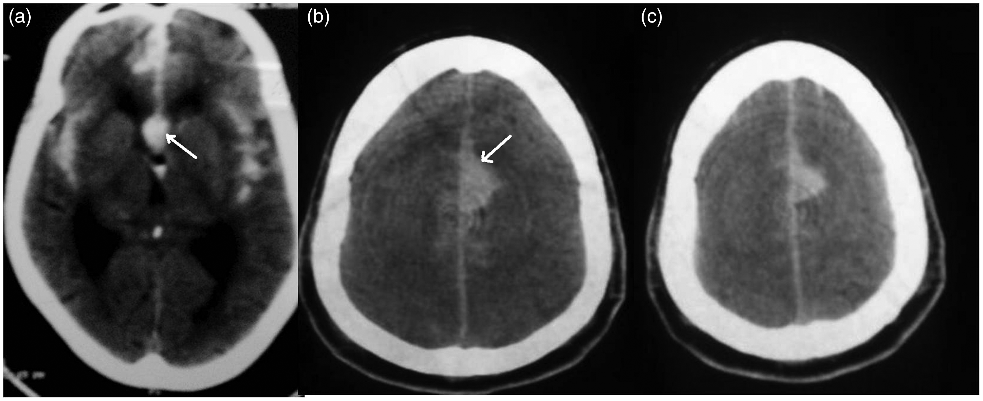
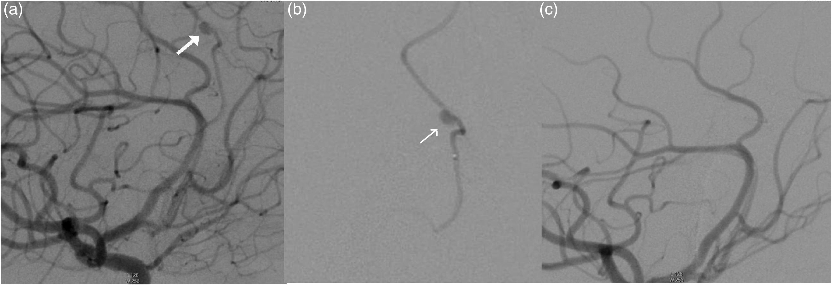
Endovascular occlusion was selected as the treatment choice owing to the nature and location of the aneurysm. Via a transfemoral route, a 7 F shuttle sheath was placed in the right internal carotid artery (ICA). A 6F Envoy guide catheter was placed in the petrous ICA. A 1.2 f magic (Balt) flow-guided microcatheter was negotiated under roadmap guidance into the frontopolar artery. A superselective angiogram confirmed the position of the microcatheter proximal to the aneurysm (Figure 2(b)). n-Butyl cyanoacrylate (33%) was injected into the aneurysm (Figure 3(a)). A post-procedure angiogram showed exclusion of aneurysm from the circulation (Figure 2(c)). Procedure was uneventful. Patient gradually improved over the next few days and was discharged in a stable state. On one- and six-month clinical follow-up, he did not have any clinical symptoms with normal neurological examination. A control angiogram done after six months showed stable occlusion with normal cerebral circulation (Figure 3(b)).
Fluoroscopic image showing the glue cast in the aneurysm and the parent vessel (a). Control angiogram showing stable exclusion of the aneurysm from the circulation (b).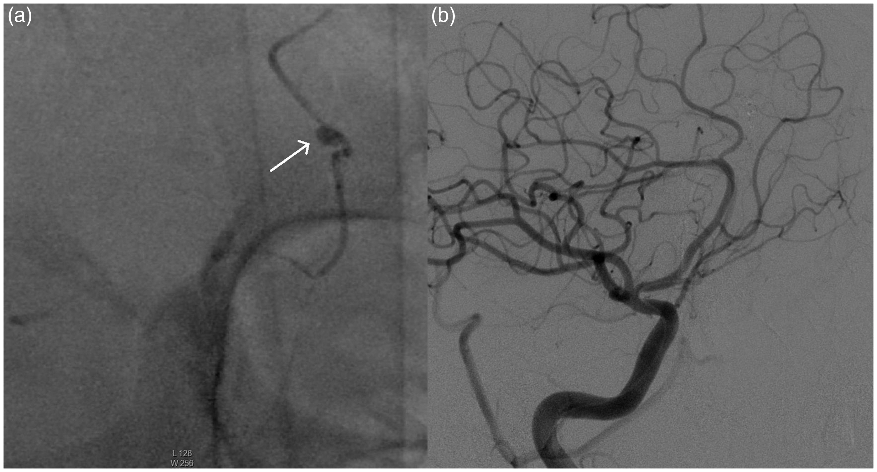
Discussion
Traumatic intracranial aneurysms are fragile lesions, 1 and hence challenging to treat either by surgical or endovascular modality. Delayed rupture of traumatic A2 aneurysms is associated with mortality of >50%. 3 Hence treatment should be done at the earliest opportunity to exclude these aneurysms from the cerebral circulation. Traumatic aneurysms contain hematoma with disruption of all the layers of the vessel. Because of the close proximity of the distal anterior cerebral artery, the free edge of the falx vessel wall may become disrupted in cases of closed head injury, as in the present case.
Aneurysms of the distal anterior cerebral artery have higher rates of surgery-related morbidity and mortality.4–6 Compared to other supratentorial aneurysms, because of the deep location with traumatic aneurysms, clip placement is difficult.5,6 Hence endovascular occlusion is an effective alternative approach to treatment of these aneurysms in order to prevent morbidity and mortality due to these aneurysms. Difficulty can also be encountered in embedding the dome, preserving veins, and prone tendency to rupture in these aneurysms. Intracerebral hemorrhage (ICH) has a negative implication on prognosis because of the limited subarachnoid space adjacent to the aneurysms. 7 Distal infarction is usually prevented by good and adequate leptomeningeal and pial collaterals in these cases. 8
We chose endovascular parent occlusion in our case, which is reportedly the best treatment option in traumatic pseudoaneurysms. The risk of distal ischemia is reported to be between 0% and 4.6%. 9 Distal ischemia usually does not occur in these cases due to sufficient leptomeningeal and pial collaterals. Parent vessel preservation with selective aneurysm occlusion will still have risk of continuing growth of dissecting aneurysm causing recurrent hemorrhage. 10 Because of the technological advancements in hardware, the availability of flow-guided microcatheters favoring access to distal locations makes the endovascular method a successful feasible option in these cases.
Conclusion
In the present report, we describe a rare case of traumatic frontopolar artery aneurysm treated successfully with endovascular embolization. Our case is unique because of the infrequent location of the aneurysm and small caliber of the artery posing challenge in catheter manipulation. It is important that the embolic agent reaches the pseudoaneurysm, failing which it is likely to fill retrograde from the collaterals. Very few cases of frontopolar artery aneurysms are reported in the literature and endovascular embolization is the treatment of choice in these aneurysms.
Footnotes
Declaration of conflicting interests
The authors declared no potential conflicts of interest with respect to the research, authorship, and/or publication of this article.
Funding
This research received no specific grant from any funding agency in the public, commercial, or not-for-profit sectors.