
Abstract
Biopolymer-based bead hydrogels present promising systems for the controlled delivery of therapeutic agents, enabling prolonged and site-specific drug action. Alginate and pectin, naturally occurring polysaccharides extracted from plant sources, have been widely valued for their non-toxicity, gelling ability, and compatibility with biological environments. In this study, hydroxypropyl methylcellulose was incorporated into the pectin–alginate bead formulation to enhance structural integrity and regulate drug diffusion. The composite beads were ionically crosslinked with calcium or iron ions and characterized by Fourier-transform infrared spectroscopy and scanning electron microscopy. Swelling behavior was investigated in simulated gastric and intestinal fluids to assess pH responsiveness. After 400 min, Ca2+ ion crosslinked beads reached ∼60% swelling in simulated gastric fluid and ∼202% in simulated intestinal fluid, whereas Fe3+ ion crosslinked beads reached ∼38% in simulated gastric fluid and ∼128% in simulated intestinal fluid, indicating that calcium-crosslinked systems swelled to a greater extent compared to their iron-crosslinked counterparts. Drug release studies further revealed that in simulated gastric fluid, Ca- and Fe-crosslinked beads exhibited cumulative amoxicillin releases of ∼15% and ∼11%, respectively, while in simulated intestinal fluid, the cumulative releases reached ∼90% and ∼75%. These results demonstrate that crosslinking density and polymer composition significantly affect both the release kinetics and the structural stability of the beads, supporting their potential use as pH-sensitive, biopolymer-based carriers in antimicrobial therapy.
Introduction
Biopolymer-based hydrogels have emerged as essential materials in the design of controlled drug delivery systems, offering versatile and biocompatible platforms for the sustained release of therapeutic agents. Composed primarily of natural polymers, these hydrogels can be engineered to regulate drug release kinetics in response to environmental stimuli such as pH, ionic strength, and temperature.1,2 In pharmaceutical applications, they are increasingly utilized in localized treatment, wound healing, and controlled drug release, providing precise dosage control, prolonged therapeutic efficacy, and reduced side effects.3–5 Among the various forms of hydrogels, bead structures have gained particular interest due to their ease of preparation, tunable size, and high drug encapsulation capacity.
As natural polymers, alginate and pectin have attracted significant attention due to their suitability for biomedical applications, particularly in drug delivery systems. Alginate, derived from brown seaweed, is a widely studied biopolymer that forms stable gels in the presence of cations such as divalent calcium (Ca2+) and zinc (Zn2+), or trivalent iron (Fe3+).6,7 Its gel-forming behavior under physiological conditions has made alginate a popular choice for controlled drug delivery, especially in oral systems where it responds to pH variations in the gastrointestinal tract.7–10 Pectin, a polysaccharide abundant in plant cell walls, is also well-known for its gel-forming ability and biocompatibility, making it ideal for drug encapsulation and release. 11 Its high water retention and swelling capabilities are beneficial for wound healing applications, while its pH-lowering effect can help inhibit bacterial growth.12,13 Importantly, both alginate and pectin can be processed into ionically crosslinked beads, providing a versatile platform for controlled release studies.14,15 These characteristics have facilitated their use in bead-based drug delivery systems.
To further improve the mechanical properties and biocompatibility of these hydrogels, hydroxypropyl methylcellulose (HPMC) has been incorporated into the systems. HPMC is a semi-synthetic, non-ionic cellulose ether that plays a pivotal role in modern pharmaceutical formulations. Its versatility as a film-former, binder, thickener, and release-controlling agent makes it one of the most commonly used excipients in oral drug delivery systems. In addition to modifying gel structure, HPMC significantly enhances the mechanical strength, mucoadhesiveness, and stability of hydrogel networks. Its high swelling capacity and excellent compatibility with other polymers enable the design of responsive matrices that ensure consistent and prolonged drug release.16–20 When combined with alginate and pectin, HPMC acts synergistically to improve the integrity and performance of hydrogels, making it particularly valuable for tailoring the release profiles of therapeutic agents.21,22 Previous studies have demonstrated that ionically cross-linked pectin–alginate beads can provide sustained drug release and tunable physicochemical properties.23,24 The gelation of alginate occurs through ionic crosslinking, where divalent cations such as Ca2+ coordinate with the carboxyl groups of guluronic acid blocks to form “egg-box” junctions; multivalent ions such as Fe3+ can produce stronger, more densely crosslinked networks, thereby altering mechanical strength and drug release behavior. 6
Oral drug delivery remains the preferred route of administration due to its convenience and patient compliance. 25 However, controlling drug release in the gastrointestinal tract remains challenging, particularly for antibiotics such as amoxicillin, which require sustained release to maintain therapeutic levels and prevent bacterial resistance. 26 The development of pH-sensitive systems for modulating drug release in the small intestine is therefore critical to improving bioavailability and overall therapeutic efficacy.27,28
Hoffman et al. highlighted the pharmacokinetic and pharmacodynamic rationale for designing controlled-release formulations of amoxicillin. 26 Several studies have since developed various biopolymeric systems.29–32 Naiel et al. reported the construction of gastroretentive beads composed of alginate/i-carrageenan and aminated chitosan for prolonged release of amoxicillin trihydrate. 33 Similarly, Güncüm et al. developed PVA/sodium alginate-based nanoparticles capable of sustained amoxicillin drug release. 34 Nia et al. formulated pH-sensitive carboxymethylcellulose–layered double hydroxides hydrogels, enabling targeted delivery of amoxicillin to the colon. 35 Collectively, these studies highlight the potential of biopolymeric carriers for site-specific and sustained amoxicillin delivery, improving therapeutic outcomes while reducing dosing frequency and minimizing the risk of bacterial resistance.
In line with these advancements, the present study investigates a novel hydrogel formulation based on alginate, pectin, and HPMC, crosslinked with Ca2+ and Fe3+ ions for effective amoxicillin delivery. The research focuses on evaluating the influence of these ions on the hydrogel’s morphology, chemical interactions, and network characteristics. In vitro swelling and release studies were conducted in pepsin and pancreatin supplemented simulated gastric and intestinal fluids to mimic gastrointestinal conditions. Scanning electron microscopy and Fourier transform infrared spectroscopy were employed to characterize the surface morphology and chemical interactions of the drug-free hydrogel beads. The ultimate aim is to optimize the formulation for sustained drug delivery, thereby enhancing the therapeutic efficacy of amoxicillin, as reflected by its encapsulation efficiency and controlled release behavior.
Experimental
Materials
All chemicals used in this study were obtained from commercial sources. Citrus pectin, sodium alginate, and pancreatin from porcine pancrease (≥3 × USP specifications) were both purchased from Sigma-Aldrich, USA. According to the manufacturer’s documentation, citrus pectin has a molecular weight range of 10–300 kDa, with typical values between 23 and 71 kDa. Sodium alginate, with a molecular weight of 12,000–80,000 Da and a viscosity of 15–25 cP at 1% in H2O, was obtained from Sigma-Aldrich. Pepsin (1:10000 USP ≥ 10,000 Nf Units/mg (Pepsin Activity)) was provided from AFG Bioscience LLC, USA. Sodium chloride, and anhydrous calcium chloride was acquired from J.T. Baker, USA. Hydrochloric acid (37%), used to adjust the pH to acidic conditions, was supplied by Merck, Germany, which also provided iron (III) chloride hexahydrate, sodium hydroxide for pH adjustments, and sodium dihydrogen phosphate as a buffering agent. All solutions were prepared using ultrapure water (18 MΩ·cm).
Preparation of ALG-PEC-HPMC beads
The process of preparing pectin–alginate beads began by dispersing 400 mg of alginate (ALG), 100 mg of pectin (PEC), and 100 mg of hydroxypropyl methylcellulose (HPMC) in distilled water. The mixture was allowed to hydrate for 2 h to ensure complete dissolution, and the volume was adjusted to 20 mL to obtain a solution with final concentrations of 2% (w/v) ALG and 0.5% (w/v) each of PEC and HPMC. The preparation of the polymer solutions was carried out at room temperature (25 ± 2°C). The solution was then transferred into a syringe equipped with a needle of 0.85 mm inner diameter and gently dripped from a height of 20 cm into a 50 mL gelling solution containing either 10% (w/v) calcium chloride (CaCl2) or 10% (w/v) ferric chloride (FeCl3) in an 80:20 ethanol–water mixture. This controlled dripping allowed uniform bead formation and proper interaction with the crosslinking ions. The beads were kept in the gelling solution for 24 h to complete the crosslinking process, ensuring sufficient gelation and structural stability. The crosslinking process was conducted at room temperature. After 24 h, the beads were separated from the gelling solution by gentle filtration and rinsed with distilled water to remove excess ions, and finally dried in an oven (Weightlab WF-HT45, Weightlab Instruments, Turkey) to obtain dried Ca-ALG-PEC-HPMC and Fe-ALG-PEC-HPMC named beads ready for further characterization.
Swelling behaviors of ALG-PEC-HPMC beads
The swelling behavior of the beads was evaluated by immersing accurately weighed samples of Ca-ALG-PEC-HPMC and Fe-ALG-PEC-HPMC in 30 mL of simulated gastric fluid (SGF, pH 1.2) and simulated intestinal fluid (SIF, pH 7.4).
SGF was prepared by dissolving 0.2% (w/v) NaCl and 0.32% (w/v) pepsin in distilled water, and the pH was adjusted to 1.2 using concentrated HCl. SIF was prepared by dissolving NaH2PO4 and 1% (w/v) pancreatin in distilled water, with the pH adjusted to 7.4 using NaOH. All pH measurements were performed using a DLAB DPH100 pH/ORP meter (DLAB Scientific Co., Ltd, China).
The bead samples were incubated in these media for about 6 h to observe their swelling characteristics under different pH and enzymatic conditions. During this period, the increase in bead weight was recorded at specified time intervals.
The degree of swelling was calculated using the following formula (Equation (1)):

Each experiment was carried out in triplicate, and the results were expressed as the mean ± standard deviation (SD) to ensure data accuracy and reproducibility.
Drug loading and encapsulation efficiency of ALG-PEC-HPMC beads
Drug-loaded beads were prepared following the aforementioned procedure, with 2% (w/w) drug added to the ALG-PEC-HPMC solution. The solution was transferred into a syringe equipped with a needle of 0.85 mm inner diameter and dripped from 20 cm into 50 mL of 80:20 ethanol–water solution containing 10% CaCl2 or 10% FeCl3 (Scheme 1). The beads were allowed to remain in the crosslinking solution for 30 min at room temperature without stirring to ensure proper gelation and stabilization. They were then collected, washed with distilled water to remove unencapsulated drug, and dried at room temperature. Encapsulation efficiency (EE%) was determined by measuring the concentration of unencapsulated drug in the crosslinker and washing solutions using a UV–VIS spectrophotometer (PG Instruments, T70, UK) at 228 nm, according to equation (2):

In vitro drug release study of ALG-PEC-HPMC beads
The drug release behavior of Ca-ALG-PEC-HPMC and Fe-ALG-PEC-HPMC beads was evaluated in simulated gastric fluid (SGF, pH 1.2) and simulated intestinal fluid (SIF, pH 7.4). Approximately 200 mg of beads were suspended in 100 mL of release medium and incubated at 37°C in a Biosan WB-4MS water bath (Biosan, Latvia) for 6 h to evaluate pH-dependent release. At predetermined intervals, 1 mL samples were withdrawn and replaced with fresh buffer to maintain sink conditions. Amoxicillin concentration was measured by UV–VIS spectroscopy. Calibration curves in SGF and SIF were prepared using standard amoxicillin solutions. Cumulative release (%) was calculated and plotted versus time to assess release kinetics. Drug release studies were carried out in triplicate, and data are expressed as mean ± SD to ensure reproducibility. Schematic illustration of the drug loading process into the ALG-PEC-HPMC gel beads.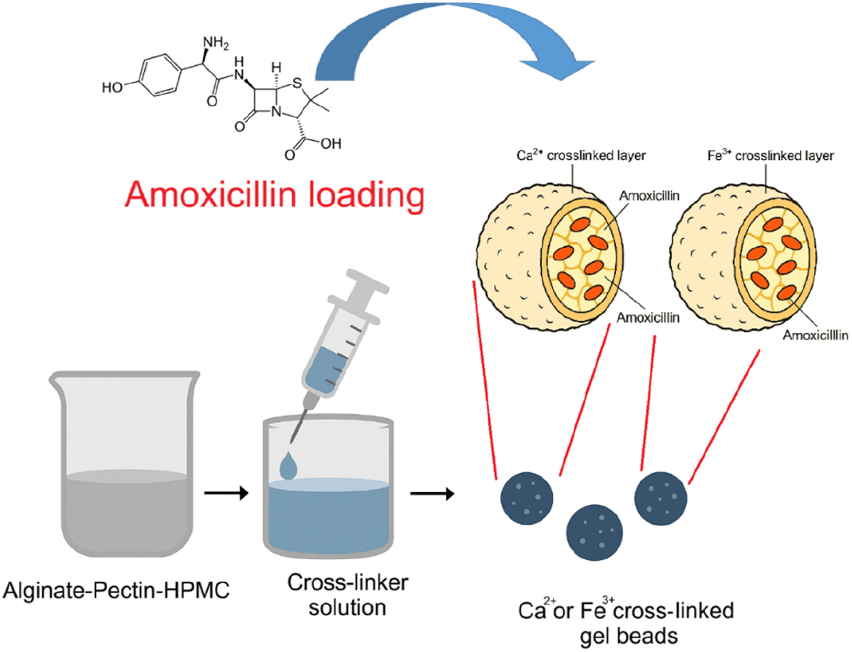
Characterizations of ALG-PEC-HPMC beads
Fourier transform infrared (FTIR) spectra of the beads were recorded using a FT/IR-4700 model FTIR spectrometer (Jasco Inc., Japan) with 32 scans at a resolution of 4 cm−1, within the spectral range of 4000–400 cm−1. For FTIR analysis, solid and dry crosslinked ALG–PEC–HPMC beads were directly placed onto the sample holder without further modification or grinding. Scanning electron microscopy (SEM) analysis of the alginate beads was performed using a scanning electron microscope (Zeiss EVO LS10, Zeiss, Germany). Before SEM imaging, the samples were coated with Au/Pd by using a Quorum SC7620 Mini Sputter Coater (Quorum Technologies, UK).
Results and discussion
Visual imaging and surface morphology analysis of ALG-PEC-HPMC beads
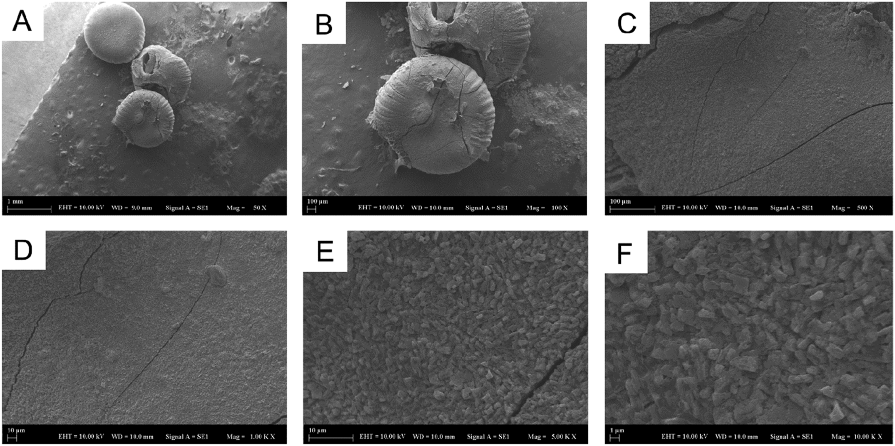
Photographs of the Ca-ALG-PEC-HPMC and Fe-ALG-PEC-HPMC beads in wet and dry states were captured using a mobile phone camera and are presented in Figure 1(a)–(d). Specifically, Figure 1(a) and (c) show the Ca-ALG-PEC-HPMC beads in wet and dry states, respectively, while Figure 1(b) and (d) show the wet and dry states of the Fe-ALG-PEC-HPMC beads. The calcium ion crosslinked beads exhibited a light beige color and a relatively smooth, spherical appearance (Figure 1(a) and (c)). In contrast, the iron ion crosslinked beads were noticeably darker, with a reddish-brown hue attributable to ferric ion incorporation (Figure 1(b) and (d)). The diameter of individual beads was measured using a digital caliper on five representative beads. The average diameter of Ca-ALG-PEC-HPMC and Fe-ALG-PEC-HPMC beads was 2.50 ± 0.05 mm before drying and 1.00 ± 0.03 mm after oven drying, reflecting shrinkage due to dehydration. SEM images also provide visual confirmation of single bead size and surface morphology. Due to the limitations of mobile phone imaging in capturing fine surface features, detailed morphological differences were further examined using scanning electron microscopy as presented in Figures 2(a)–(f) and 3(a)–(f). Photographs of Ca-ALG-PEC-HPMC beads (a and c) and Fe-ALG-PEC-HPMC beads (b and d) in wet (a and b) and dry (c and d) states. SEM images of Ca-ALG-PEC-HPMC beads (a–f) at different magnifications: (a) ×50, (b) ×100, (c) ×500, (d) ×1000, (e) ×5000, (f) ×10000. SEM images of beads of Fe-ALG-PEC-HPMC (a–f) at different magnifications: (a) ×50, (b) ×100, (c) ×500, (d) ×1000, (e) ×5000, (f) ×10000.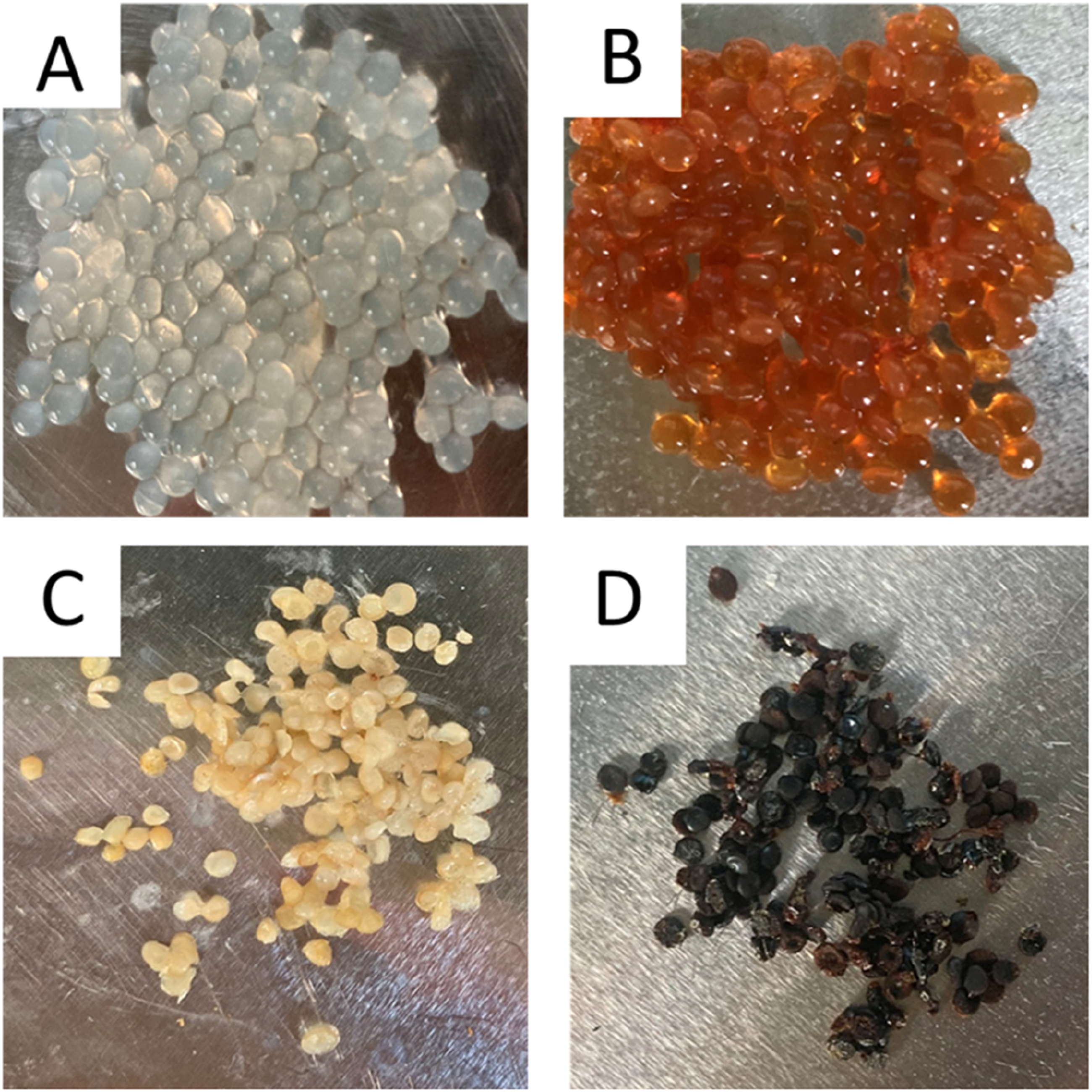
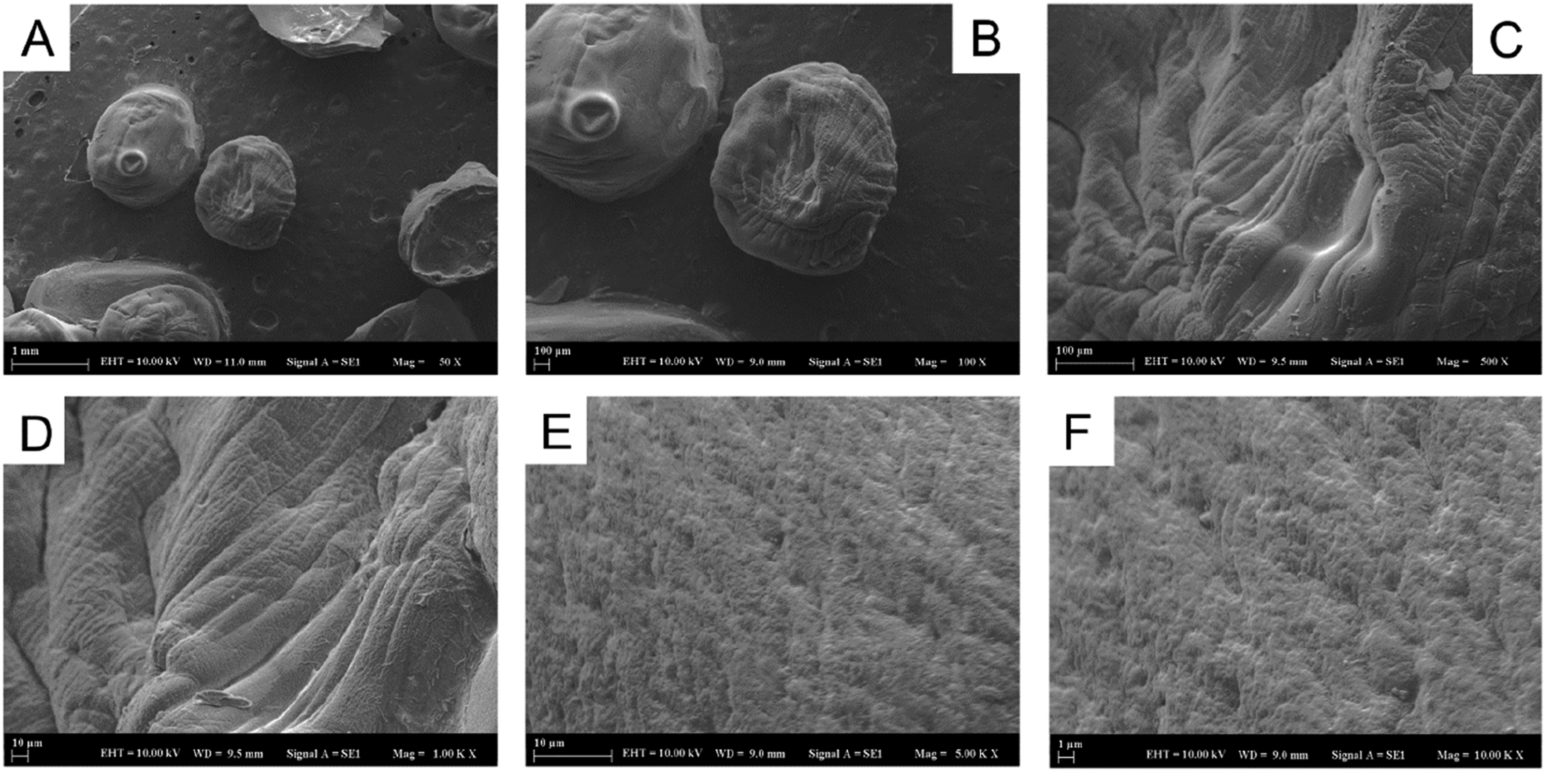
SEM images of alginate–pectin–HPMC beads crosslinked with calcium ions (Figure 2(a)–(f)) and iron ions (Figure 3(a)–(f)) were obtained at increasing magnifications to investigate surface morphology and structural integrity.
Calcium ion crosslinked beads imaged at ×50, ×100, and ×500 (Figure 2(a)–(c)) exhibit a generally spherical shape with moderately wrinkled surfaces and intact contours. At low magnification (Figure 2(a)), the beads show a rounded morphology with consistent size distribution and a slightly roughened outer layer (scale bar = 1 mm). The ×100 image (Figure 2(b)) reveals shallow surface cracks and concentric folds, likely formed during drying, yet overall integrity is preserved. At higher magnification (Figure 2(c), ×500), the surface appears dense and compact with limited porosity and minor crevices (scale bar = 100 µm), indicating a homogeneously gelled structure. The incorporation of HPMC further enhanced bead quality by smoothing the surface, reducing small surface indentations, and preserving structural integrity. These effects minimized minor deformations and prevented fragmentation, which could otherwise compromise bead performance.36–38 Moreover, the improved surface uniformity and low porosity are expected to contribute to a more controlled and predictable release profile, highlighting the practical advantages of HPMC inclusion in bead formulations. Overall, these observations indicate that the calcium ion crosslinked beads possess a compact, low-porosity morphology with stable surface characteristics, supporting their suitability for controlled release applications.
In Figure 3(a)–(f), the surface morphology of Fe3+ ion crosslinked ALG–PEC–HPMC beads was examined using SEM. Low-magnification images (Figure 3(a)–(c)) show overall bead shape, while high-magnification images (Figure 3(d)–(f)) reveal detailed surface texture and granularity. At ×1000 magnification, small surface cracks and a rough texture were clearly visible, while at ×5000 magnification, dense particle packing and fragmented morphology became visible (Figure 3(d) and (e)). In Figure 3(d)–(f), at ×1,000, ×5,000, and ×10,000 magnifications, the surface appeared rougher and more granule-like. At the highest magnification (Figure 3(f), at ×10,000 magnification), flake-like domains were observed, indicating localized variations in surface texture due to Fe3+ ion crosslinking. From Figure 2(a)–(f) and Figure 3(a)–(f), compared to the calcium ion crosslinked samples, Fe3+ion crosslinked beads exhibited lower smoothness, higher surface roughness, and greater rigidity, which may influence swelling behavior and drug release kinetics. These findings are consistent with previous studies: Kadji et al. (2022) reported compact but rough surfaces in Fe-alginate beads, 39 and Sharma et al. (2019) demonstrated that alginate–pectin beads displayed roughened surfaces with small surface variations, highlighting the role of pectin in modulating bead morphology. 40 Collectively, these results suggest that Fe3+ ion crosslinking, in combination with pectin and HPMC, produces beads with compact yet textured surfaces, potentially affecting their functional properties.
SEM observations provided clear insights into the effect of multivalent cation type on the microstructure of ALG-PEC-HPMC beads, revealing that calcium produces smoother and more elastic surfaces, whereas iron leads to denser, particulate-rich morphologies. Such structural distinctions play a crucial role in determining the swelling response, mechanical stability, and release characteristics of the beads in simulated body fluids. In agreement with literature data, Fe3+ ion crosslinking imparts higher mechanical strength and elastic modulus to alginate-based beads compared to Ca2+ ion crosslinking, likely due to the formation of a more compact ionically crosslinked network. 41
FTIR analysis of crosslinked ALG-PEC-HPMC beads
Figure 4(a)–(c) presents the FTIR spectra of ALG, PEC, HPMC, and their ionically crosslinked composites, which were analyzed to identify the main functional groups and to elucidate potential interactions among the biopolymer components and metal ions. In the ALG spectrum (Figure 4(a)), the broad absorption bands at 1594 cm−1 and 1405 cm−1 correspond to the asymmetric and symmetric stretching vibrations of carboxylate (–COO−) groups, while the peak at 1027 cm−1 is attributed to C–O stretching vibrations.21,42,43 In addition, a broad O–H stretching band was observed around 3400–3450 cm−1, consistent with previous reports on ALG.42–44 The spectra of PEC and HPMC are shown in Figure 4(b). PEC exhibits a broad O–H stretching band at 3363 cm−1, an ester carbonyl (C = O) stretching peak near 1732 cm−1, and a carboxylate band at 1606 cm−1, confirming the presence of galacturonic acid residues.
45
In contrast, HPMC displays a distinct O–H stretching peak at 3444 cm−1, along with characteristic bands corresponding to C–H stretching (∼2901 cm−1), C–O stretching (∼1050 cm−1), and –CH3 bending vibrations (1300–1460 cm−1), consistent with previous observations.43,46 FTIR spectra of ALG (a), PEC (b), HPMC (b) powders, and Ca-ALG-PEC-HPMC (c), and Fe-ALG-PEC-HPMC (c) beads.
The spectra of the ionically crosslinked composites, namely Ca–ALG–PEC–HPMC and Fe–ALG–PEC–HPMC, are presented in Figure 4(c). In the Ca–ALG–PEC–HPMC and Fe–ALG–PEC–HPMC spectra (Figure 4(c)), the broad O–H stretching bands, originally observed in alginate at 3336 cm−1, shifted to 3432 cm−1 and 3391 cm−1, respectively, indicating enhanced hydrogen bonding interactions among hydroxyl and carboxyl groups within the blended matrices.42–44 Similarly, the asymmetric and symmetric –COO− stretching vibrations, originally observed at 1593–1594 and 1405–1406 cm−1 in alginate, shifted to approximately 1620 and 1450 cm−1, respectively, in the composites, reflecting hydrogen bond formation and ionic coordination.43–47 The C–O stretching vibrations near 1030 cm−1 became broader and more intense, particularly in the Fe3+ ion crosslinked beads, suggesting the involvement of glycosidic linkages and hydroxyl groups in intermolecular associations. 46 These systematic shifts in –OH and –COO− vibrations confirm the establishment of hydrogen bonding and electrostatic interactions among ALG, PEC, and HPMC, further stabilized by coordination with Ca2+ and Fe3+ ions. 45 These findings highlight the structural integrity of the ionically crosslinked hydrogel beads and verify the successful integration of the biopolymer components.
Swelling behavior of ALG-PEC-HPMC beads in SGF and SIF
Figure 5(a) presents the swelling behavior of Ca-ALG-PEC-HPMC and Fe-ALG-PEC-HPMC beads over a 400 min incubation period in simulated gastric fluid (SGF, pH 1.2) at 37°C. Similar experiments with ionically crosslinked anionic polymer beads have shown that these systems generally swell more at neutral to mildly basic pH than under acidic conditions, due to the ionization of carboxyl groups and increased water uptake.48–50 Seeli et al. specifically reported this behavior in guar gum succinate–sodium alginate (GGS-SA) beads crosslinked with barium ions, which swelled more at pH 7.4 than at pH 1.2, reflecting the pH-dependent nature of anionic polymer chains.
51
Swelling profiles of ALG-PEC-HPMC hydrogel beads in (a) simulated gastric fluid (SGF, pH 1.2, with pepsin) and (b) simulated intestinal fluid (SIF, pH 7.4, with pancreatin).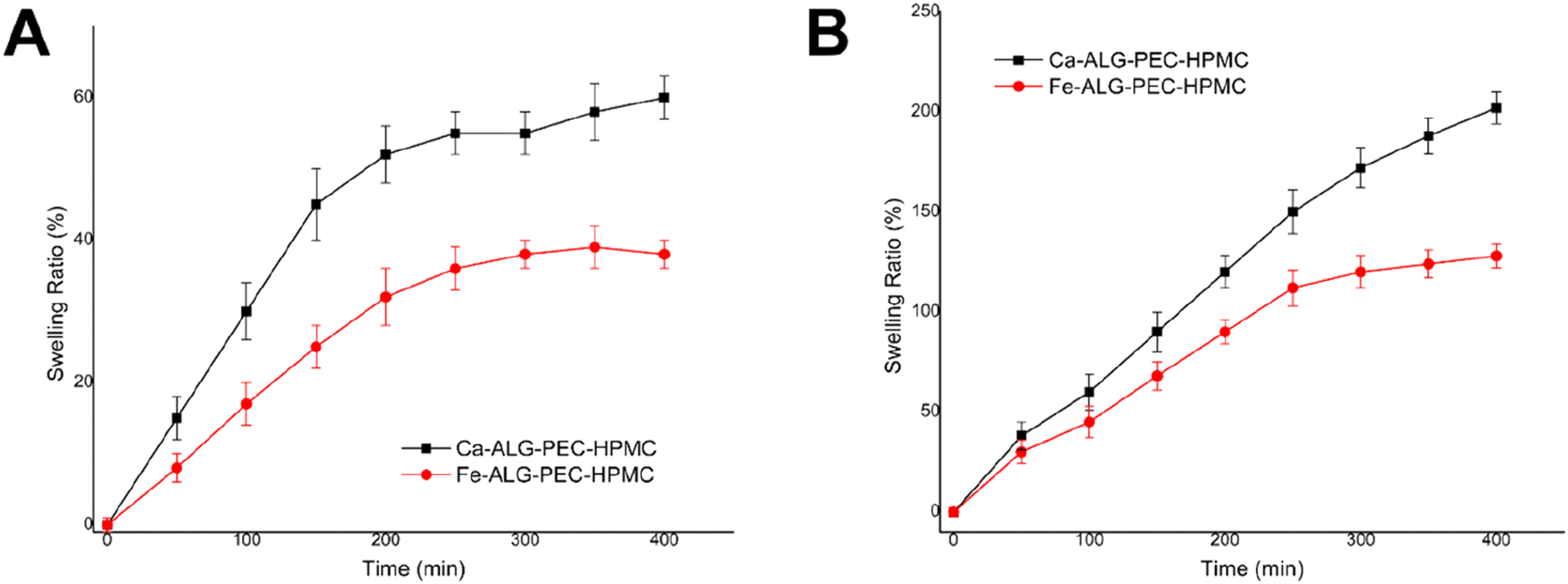
The SGF used in this study was supplemented with pepsin, the primary proteolytic enzyme in the stomach, to better replicate physiological gastric conditions. As illustrated in the graph in Figure 5(a), both types of beads exhibited a rapid increase in swelling during the initial phase (0–100 min), likely due to the fast absorption of the acidic medium. During this period, Ca-ALG-PEC-HPMC beads displayed a more pronounced increase, reaching approximately 30% swelling at 100 min, while Fe-ALG-PEC-HPMC beads reached around 17%. Between 100 and 200 min, swelling continued at a reduced rate, with Ca-ALG-PEC-HPMC and Fe-ALG-PEC-HPMC beads reaching approximately 52% and 32%, respectively. After 200 min, the swelling ratios of both bead types began to plateau. By 400 min, Ca-ALG-PEC-HPMC beads maintained a higher swelling ratio (∼60%) than Fe-ALG-PEC-HPMC beads (∼38%), suggesting a greater water uptake capacity in calcium ion crosslinked systems under acidic conditions. Similar observations were reported by Choi et al., who systematically compared CaCl2, FeCl2, FeCl3, and FeSO4 as crosslinking agents in alginate films and demonstrated that Fe3+ produces denser structures, while Ca2+ remains the most effective in improving water-related properties. 52
Calcium, being divalent, forms planar two-dimensional crosslinks with alginate in accordance with the classical “egg–box” model, resulting in a more loosely crosslinked network that allows greater water uptake.42,51,53,54 In contrast, trivalent Fe3+ ions promote stronger crosslinking, resulting in a denser network that restricts water penetration. 55
Figure 5(b) presents the swelling profiles of Ca-ALG-PEC-HPMC and Fe-ALG-PEC-HPMC hydrogel beads over a 400 min incubation period in simulated intestinal fluid (SIF, pH 7.4), supplemented with pancreatin to better replicate physiological conditions of the small intestine at 37°C. During the initial phase (0–100 min), both hydrogel systems exhibited a marked and rapid increase in swelling, indicative of significant fluid uptake and polymer network relaxation.56–58 Between 100 and 250 min, the swelling ratios continued to increase, but at a slower rate. At 250 min, Ca-ALG-PEC-HPMC beads attained a swelling ratio of approximately 150%, whereas Fe-ALG-PEC-HPMC beads exhibited a lower value of swelling around 112%. Beyond 250 min, Fe-ALG-PEC-HPMC beads reached a plateau, stabilizing at a final swelling ratio of approximately 128% at 400 min. In contrast, Ca–ALG–PEC-HPMC beads maintained a gradual upward trend, ultimately achieving a swelling ratio of 202%. This extended swelling phase suggests that the calcium ion–crosslinked network provides a more open and hydrated structure, thereby facilitating prolonged fluid diffusion and matrix expansion.41,59 Moreover, divalent cations such as Ca2+ generally produce less densely crosslinked and more flexible networks than trivalent ions, which explains the superior swelling behavior of Ca-ALG-PEC-HPMC beads. 6 The observed swelling trends at pH 1.2 (low swelling, Figure 5(a)) and pH 7.4 (enhanced swelling, Figure 5(b)) are likely related to the partial ionization of carboxyl groups under neutral conditions, leading to increased electrostatic repulsion and water uptake.48–50
The swelling experiments (Figure 5(a) and (b)) further confirm that Ca-ALG-PEC-HPMC beads exhibit significantly higher swelling ratios in SIF (pH 7.4, with pancreatin), indicating their enhanced suitability for sustained intestinal drug release.
The results highlight the pH-responsive nature of the ALG–PEC–HPMC beads and emphasize the role of ionic crosslinking in modulating water uptake. Such swelling characteristics are critical for the design of oral drug delivery systems, where water absorption directly influences drug release and therapeutic performance. In particular, Ca-ALG-PEC-HPMC beads exhibited superior swelling behavior compared to Fe-ALG-PEC-HPMC beads, reflecting the more open and hydrated network structure formed by calcium crosslinking. These findings indicate that Ca-ALG-PEC-HPMC systems hold strong potential for intestinal drug delivery applications, where extended swelling and sustained release are essential for effective targeted therapy.
pH-responsive controlled drug release from ALG-PEC-HPMC beads
The design of pH-responsive hydrogel systems relies on the combined effects of polymer composition, crosslinking ions, and matrix stabilizers. Integrating findings from multiple studies helps to illustrate how each factor contributes to controlled drug release. Previous studies have highlighted the role of pectin-based hydrogel matrices in controlling drug release,
12
while the presence of HPMC within the hydrogel matrix provides structural stabilization, complementing the role of pectin in regulating drug diffusion and contributing to sustained release.
20
Guo and Kaletunç, demonstrated that alginate–pectin hydrogel particles display pH-dependent dissolution kinetics, directly affecting drug diffusion rates.
60
Urbanová et al., reported that dual crosslinking with Ca2+ and Zn2+ ions strengthens the gel network and modulates release profiles, supporting the concept of ion-dependent control of drug delivery.
61
Based on these insights from previous studies, the pH-responsive drug release behavior of Ca-ALG-PEC-HPMC and Fe-ALG-PEC-HPMC beads was evaluated in both simulated gastric fluid (SGF, pH 1.2) and simulated intestinal fluid (SIF, pH 7.4) at 37°C, as shown in Figure 6(a) and (b). The results demonstrate a pH-responsive release profile, with significantly lower drug release in SGF compared to SIF for both formulations. In SGF, as illustrated in Figure 6(a), Ca-ALG-PEC-HPMC beads exhibited a cumulative release of approximately 15% after 400 min, whereas Fe-ALG-PEC-HPMC beads released about 11%. The initial release in the first 60 min remained below 10% for both, indicating strong resistance to acidic degradation. Cumulative release profiles of ALG-PEC-HPMC hydrogel beads in (a) simulated gastric fluid (SGF, pH 1.2, with pepsin) and (b) simulated intestinal fluid (SIF, pH 7.4, with pancreatin).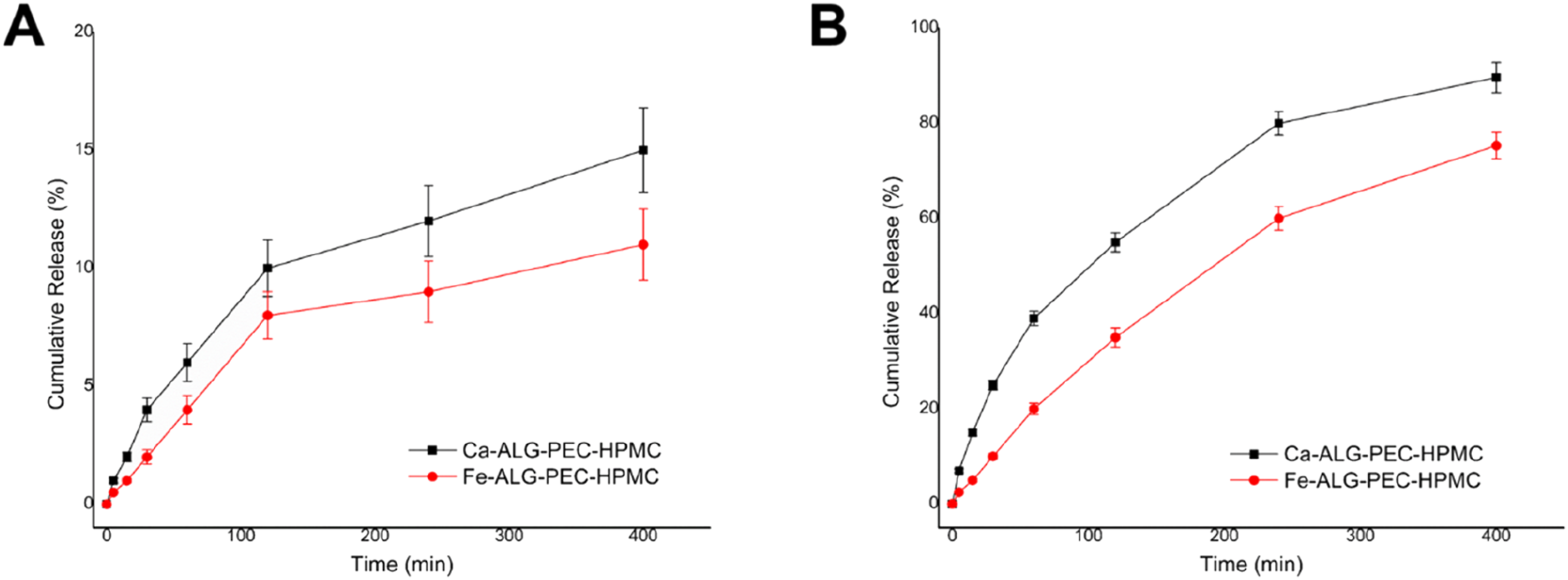
Upon placing the beads in SIF (Figure 6(b)), a significant increase in drug release was observed for both systems. The Ca-ALG-PEC-HPMC beads reached approximately 90% cumulative release within 400 min, while the Fe-ALG-PEC-HPMC beads achieved around 75%. The enhanced release in SIF can be attributed to swelling and matrix loosening induced by the ion exchange between the crosslinker ions (Ca2+ or Fe3+) and the monovalent Na+ ion present in the buffer.
Fe3+ ion crosslinked beads demonstrated more controlled release, retaining more drug in acidic conditions and gradually releasing it in the intestine, which is desirable for oral delivery. In contrast, Ca2+ ion crosslinked beads, with their weaker network, allow faster drug diffusion, suitable for applications requiring more immediate release.
Additionally, SIF contains pancreatin, which aids in digestion and could contribute to the breakdown of the matrix, facilitating the release process. This exchange weakens the gel network, promotes polymer relaxation, and facilitates drug diffusion. pH-sensitive gelation mechanisms, such as hydrogen-bond interactions in pectin-based hydrogels, contribute to the controlled release behavior under varying pH conditions. 62 Moreover, the stabilizing effect of pectin and HPMC has been shown to modulate both initial burst and sustained release phases, supporting controlled delivery under varying pH conditions.63,64
Although both formulations followed a biphasic release trend, with an initial burst followed by a more gradual sustained release, Fe-ALG-PEC-HPMC beads displayed a more controlled release behavior. This observation aligns with reports that Fe3+ crosslinking produces denser and more stable networks, enhancing resistance to premature erosion. 65 Overall, these results highlight the potential of both formulations for pH-targeted oral drug delivery, particularly for protecting the drug in the gastric environment and releasing it in the intestine, in agreement with general design principles established for pH-responsive hydrogels.66,67
Encapsulation efficiency and its impact on swelling and drug release
Encapsulation efficiency, swelling ratio, and cumulative drug release of Ca-ALG-PEC-HPMC and Fe-ALG-PEC-HPMC beads.
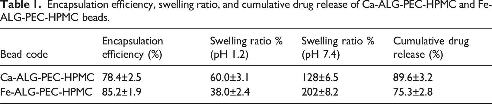
The encapsulation efficiency of Fe-ALG–PEC–HPMC beads was higher than that of Ca-ALG–PEC–HPMC beads, as shown in Table 1. This is likely due to the stronger coordination of Fe3+ ions with the carboxyl groups of alginate and pectin, forming a denser 3D crosslinked network that limits drug diffusion during gelation.55,65 As mentioned previously, the average diameter of Ca-ALG-PEC-HPMC beads was about 2.50 mm, whereas that of Fe-ALG-PEC-HPMC beads was around 2.00 mm. Assuming spherical geometry (V = 4/3πr3), this corresponds to a volume ratio of approximately 1.95:1, further reflecting the less compact network structure of calcium ion crosslinked beads. Malektaj et al. (2025) reported that among multivalent cations (Fe3+, Cu2+, Sr2+, and Ca2+), Fe3+-crosslinked alginate hydrogels showed the highest encapsulation efficiency and slowest release, underscoring the key role of cation type in tuning release behavior. 65 These observations highlight how both the crosslinker type and bead volume collectively influence encapsulation efficiency, swelling, and drug release profiles. The denser Fe3+-crosslinked network allows higher drug retention and more controlled release, whereas the more open Ca2+ network supports faster diffusion and greater swelling, making it suitable for applications requiring quicker therapeutic action. This emphasizes the tunability of the ALG–PEC–HPMC bead matrix for targeted oral drug delivery.
Conclusion
This study demonstrates that ionic crosslinking significantly modulates the structural and functional properties of ALG-PEC-HPMC beads for controlled drug delivery. Ca2+-ion crosslinked beads exhibited greater swelling and faster drug release, whereas Fe3+-ion crosslinked beads maintained stronger network integrity and provided more sustained release, highlighting the tunable performance of these biopolymeric systems. Structural analyses using FTIR and SEM confirmed bead integrity and the presence of characteristic ionic crosslinking interactions, supporting the observed differences in behavior. These results demonstrate the potential of pH-sensitive beads for tailored oral drug delivery applications, enabling controlled and targeted release of therapeutic agents and illustrating how different crosslinking strategies can be employed to achieve specific release profiles.
Footnotes
Acknowledgments
The author gratefully acknowledges the financial support provided by the Research Foundation of Istanbul Technical University through projects THD-2024-45406 and THD-2025- 46766 for this study.
Author contributions
Funding
The author disclosed receipt of the following financial support for the research, authorship, and/or publication of this article: Bilimsel Araştırma Projeleri Birimi, İstanbul Teknik Üniversitesi (THD-2024-45406, THD-2025-46766).
Declaration of conflicting interests
The author declared no potential conflicts of interest with respect to the research, authorship, and/or publication of this article.