






Abstract
Coir-nanocellulose (CNC) infused with antibiotics, is presented as a promising candidate for antimicrobial wound care applications. Nanocellulose was extracted from coconut husk fibers through alkali treatment and acid hydrolysis along with steam explosion. The obtained cellulose nanofibers exhibited diameters in the range of 50-100 nm with a zeta potential −37.2 ± 2.9 mV indicating the electrostatic stability of the dispersion and exhibited characteristic peaks corresponding to nanocellulose. The fluid absorption capacity of CNC was in the range of 1.45-3.08 g/g. Two broad-spectrum antibiotics; ciprofloxacin and gentamicin were independently infused into CNC to prepare ciprofloxacin loaded coir-nanocellulose (C-CNC) and gentamicin loaded coir-nanocellulose (G-CNC) respectively. The cumulative drug release profile indicated a burst release of 70.8 ± 3.4% for C-CNC and 64.3 ± 2.9% for G-CNC, both of which got stabilized within 10 h and the drug release kinetics followed Korsmeyer-Peppas model. Significant inhibitory effect on the tested bacterial strains was observed for both C-CNC and G-CNC which exhibited zone of inhibitions in the range of 2.3 ± 0.011 to 2.8 ± 0.045 cm for C-CNC and from 2.4 ± 0.031 to 2.8 ± 0.054 cm for G-CNC respectively and it was found proportional with the drug concentration in the sample. CNC with and without drug incorporation is found to be cytocompatible as indicated by MTT assay with viability >80% at 48 h. Results indicate that antibiotics functionalized CNC would be a viable approach for developing innovative wound care products with a unique combination of antibacterial functionality and inherent cytocompatibility.

Research highlights
• Nanocellulose with excellent fluid absorption capacity, was successfully extracted from coconut husk fibers by steam explosion, with the involvement of minimal chemical treatment methods. • The extracted coir-nanocellulose (CNC) was characterized by SEM, FT-IR, and XRD analyses along with fluid absorption studies which revealed excellent absorption characteristics. • The incorporation of two broad-spectrum antibiotics, Ciprofloxacin and Gentamicin, imparted sufficient antimicrobial properties on to CNC and demonstrated its potential antibacterial activity against Staphylococcus aureus, Escherichia coli, Bacillus cereus, and Pseudomonas aeruginosa. • The CNC was found to be cytocompatible as evident from MTT assay performed on L929 cell lines. • The combination of biodegradability, biocompatibility, and effective antimicrobial action positions CNC as a promising solution for addressing wound healing challenges.
Introduction
Wound management has become a complicated challenge predominantly in the context of growing antimicrobial resistance and mounting prevalences of diabetic ulcers, traumatic wounds, and post-surgical complications. 1 To obtain the best outcomes, a combinational strategy utilizing appropriate wound care, treating underlying medical conditions, and suitable wound healing approaches is essential. Wound healing outcomes can be improved by revolutions in the dressing methodologies used 2 and therefore, effective wound management involves choosing the appropriate dressing material that meets the demand for advanced functional wound care based on considering factors such as maintenance of a moist environment, absorption of wound exudate, protection against microbes, and provision for pain relief with good biocompatibility and biodegradability. 3 Biopolymers are a promising class of materials for wound healing applications because of their numerous inherent properties along with nominal toxicity and ease of access. Chitosan, κ-carrageenan, cellulose, gelatin, collagen, alginate, hyaluronic acid, silk fibroin, etc. are widely used biopolymers for wound healing applications in the form of hydrogels, nanofibers, microspheres, nanoparticles, hydrocolloids, foams, sponges, bandages, gauze, films and membranes. 4
Cellulose has great potential as cost-efficient innovative material for biomedical applications owing to its biocompatibility, biodegradability and low cytotoxicity. Since cellulosic materials offer a sustainable, cost-effective and versatile option, they are crucial for various therapeutic uses such as tissue engineering, wound healing, and drug delivery. 5 The amount of cellulose found in various categories of natural fibers varies arbitrarily, and coir fiber can be regarded as an excellent source of cellulose, constituting approximately 32-43% of coir fiber. 6 Coir fibers principally obtained from the husk of the coconut, delivers a sustainable strategy to tackle ecological challenges. 7 Coir fibers contribute to about 30 wt% of the coconut husk and hold distinct properties which makes them appropriate for an extensive array of diversified applications. The coir fibers derived from the outer mesocarp of coconut is globally identified for its typical features like longevity, adaptability and biodegradability in the manufacturing of geotextiles, reinforced composites, biofuel and biomedical applications. 8 Even though coir fibre is a renewable and sustainable source of cellulose, which is obtained in plenty as an agricultural by-product from the coconut industry, it is comparatively under-utilized. 9 This lignocellulosic biomass derived from an agricultural waste and can be replenished through coconut cultivation, making it a treasured resource for waste valorisation and circular bioeconomy initiatives. 10 Coir fiber can be differentiated from other established cellulose sources, owing to its relatively high lignin and cellulose content which in turn delivers remarkable contributions for the evolution of functional biomaterials. 11 The concentration of cellulose obtained may vary depending on several parameters such as the source and method of extraction employed. 12 With a cellulose concentration of approximately 38.01 weight percent, coir fiber has high potential for cellulose extraction among other natural lignocellulosic biomasses. 13
Owing to its important biological features like, structural similarity with the extra cellular matrix (ECM) and tunable physiochemical properties, nanocellulose has drawn a gradually growing interest in innovative wound care, in addition to their biocompatibility and biodegradability. However, despite its structural properties, the lack of inherent antimicrobial properties limits the utilization of cellulose nanofibers for the development of wound dressings. 14 The implementation of antibacterial agents and bioactive compounds augments their efficiency, making them suitable for an extensive series of wound healing applications. 15 Controlled or sustained drug delivery systems can help deliver effective drug concentration directly to the wound site limiting microbial infection. 16 In order to help prevent infection at wound site, broad spectrum antibiotics such as ciprofloxacin and gentamicin are actively involved. 17 Ciprofloxacin helps in the inhibition of bacterial DNA gyrase and gentamicin interferes with bacterial protein synthesis, making their incorporation effective against bacterial colonization and biofilm formation at wound site. 18
The most common pathogens that infect the wounds are Staphylococcus aureus, Pseudomonas aeruginosa and Escherichia coli. 19 Ciprofloxacin is a widely used bactericidal antibiotic, which acts either by inhibiting the activity of DNA gyrase or DNA topoisomerase IV. 20 Gentamicin is a broad-spectrum antibiotic which is essentially used against various infections ranging from skin disorders to pneumonia, bacterial meningitis, and even respiratory or urinary tract infections. 21 Both gentamicin and ciprofloxacin could be used against S. aureus, P. aeruginosa and E. coli and there has been even reports showing better synergistic activities of gentamicin-ciprofloxacin combinations against E. coli and P. aeruginosa strains. 17 Due to these complementary mechanisms, C-CNC and G-CNC can be effectively utilized for wound-management applications owing to their potential to limit bacterial colonization and reduce the formation of biofilms at the wound site.
By suppressing invading microorganisms and limiting infection, these antibiotics help maintain conditions that favour the natural wound-healing process. The different steps in wound healing, mainly collagen formation and tissue growth, are not directly promoted by antibiotics. In a study conducted by Agarwal et al., it was found that ciprofloxacin and gentamicin are effective antibiotics against free-living forms of P. aeruginosa. 22 The research conducted by Wang et al. revealed that the combination of fosfomycin and gentamicin is most effective against the biofilm of E. coli strains. 23 Research evaluating clinical strains identified in individuals with infections related to prosthetic joints discovered that the combination of gentamicin and ciprofloxacin demonstrated synergism more frequently against the biofilm formation of P. aeruginosa strains. 24
Although research exploring the potential of cellulose nanofibers for wound healing appear promising, investigations concerning the integration of antibacterial agents in to cellulose nanofibers seems limited. 25 In order to overcome this limitation, studies involving antibiotic incorporation into nanocellulose matrix has gained huge interest in recent years, as a strategy for localized and sustained drug delivery. The novelty of the work has been explicitly articulated as the transformation of coir biomass in to nanocellulose and its biomedical application for the development of a drug delivery system through the integration of clinically relevant antibiotics for wound management. The study exclusively integrates sustainability, material revolution and biomedical application which has been inadequately explored in existing literature.
Considering recent advances in wound management, the present study focuses on the viability of using Coir-Nanocellulose based antibacterial wound dressings for infected and chronic wound management, predominantly those that are susceptible to bacterial infections. 26 Based on the demonstrated sustained drug release behavior and antibacterial activity, the material is relevant for chronic, infected, non-healing wounds; where prolonged antibacterial action is indispensable. 27 For that, incorporation of antibiotics, ciprofloxacin and gentamicin were performed individually on the extracted Coir-Nanocellulose and conducted antimicrobial susceptibility studies on different microbial strains, that are the most common causative agents of skin and soft tissue infections and surgical wound infections. Cytocompatibility studies were also performed to evaluate the effectiveness and suitability of CNC and drug loaded CNC for wound dressing applications.
Materials and methods
Materials
Raw coir fiber was sourced from the Central Coir Research Institute, Coir Board, Kerala, India. All reagents used namely sodium hydroxide, sodium chlorite, oxalic acid, copper (II) sulfate pentahydrate and ethylenediamine obtained from Merck (Mumbai. India) were of analytical grade. Ciprofloxacin (≥98%, HPLC) and gentamicin solution (10 mg/ml) procured from Sigma Aldrich (India) were used for drug loading and antimicrobial studies. Staphylococcus aureus (MTCC#96), Escherichia coli (MTCC#68), Bacillus cereus (MTCC#1307), and Pseudomonas aeruginosa (MTCC#1688) were purchased from MTCC, Chandigarh. Nutrient agar, nutrient broth, Luria Bertani Agar and MacConkey Agar procured from Hi-Media (Mumbai, India) were used for bacterial studies. Standard antibacterial discs of ciprofloxacin (CIP 10 mcg, Hi-Media) and gentamicin (GEN 10 mcg, Hi-Media) were used as positive controls for agar disc-diffusion studies.
Methods
Preparation of coir-nanocellulose (CNC)
Coir Nanocellulose (CNC) was extracted from coir fibers according to a previous method reported by Abraham et al., with slight modifications.
28
Briefly, coir fiber was soaked in 2% NaOH overnight, then subjected to steam explosion by autoclaving at 20 lbs and 121o C for 1 h, which was further treated with 2-3% sodium chlorite (NaClO2) and then exposed to the second step of steam explosion for bleaching. This bleaching process was repeated until the material got completely lightened and the resultant sample was acid hydrolyzed with 5% oxalic acid during the last phase of steam explosion. The acid-treated sample was then subjected to ultrasonication, followed by rinsing with distilled water until the pH reached 7.0. The washed neutral sample was then air-dried, and ground in a mixer grinder to obtain nanocellulose fibers.

Chemical composition analysis
Composition analysis of raw coir fiber, alkali treated fiber, steam exploded fiber, bleached fiber, acid hydrolyzed fiber and CNC were analyzed according to ASTM standards viz. ASTM D 1103-55T for cellulose and ASTM D 1106-56 for lignin. Moisture content was analyzed using a Moisture Analyzer (RADWAG, MA 50.R).
Morphological evaluation
Scanning Electron Microscopy was used to observe the morphological changes that occurred during various stages of processing of the coir fibers until the formation of CNC. Scanning electron microscopy images were captured at a resolution of 0.05 μm and 100 μm using a JEOL Model JSM-6390LV.
Zeta potential measurement
Zeta potential measurements of CNC were performed using the Malvern Zetasizer (Malvern Panaytical, UK) based on the electrophoretic light scattering (ELS) measurements employing M3-PALS technique. Samples were analyzed at 25 ± 0.1°C post equilibration and the measured electrophoretic mobility was converted to zeta potential by using the Smoluchowski approximation.
Fourier transform infrared (FT-IR) spectroscopy
A Perkin Elmer Spectrum two FT-IR spectrometer was used to record the Fourier transform infrared (FTIR) spectra of the fibers at various stages of treatment as well as post-drug loading. The spectrophotometer uses LiTaO3 MIR detector and Attenuated Total Reflection (ATR) contact sampling method. Fibers (2 mg) were crushed in liquid nitrogen and mixed with KBr (1:100) to form an 1 mm thick disc, which was used for the analysis.
X-ray diffraction (XRD)
RIGAKU MINIFLEX 600 Benchtop Powder X-Ray Diffractometer (XRD) instrument with Cu tube (as the X-ray source, λ = 1.54 Ao) and a Hybrid pixel array detector (HPAD), was used to get X-ray diffraction profiles of the CNC, C-CNC and G-CNC using CuK radiation at operating voltage and current of 30 kV and 20 mA, respectively and the diffraction strengths were measured from 3° to 80° (2θ range). The Crystallinity Index values (CrI) has been calculated from the XRD Spectrum using the equation:

Degree of polymerization (DP)
The degree of polymerization of nanocellulose samples was determined using intrinsic viscosity measurements in cupriethylenediamine (CED) solution by following TAPPI T 230 standard protocol.
29
Freshly prepared CED solution (0.5 M) was used for determining DP and for that, approximately 40 mL of 5 M NaOH was added to 12.5 gm of CuSO4.5H2O dissolved in 100 mL D.H2O under constant stirring. A light blue precipitate of Cu (OH)2 is formed and addition is continued until precipitation is completed. The precipitate is filtered and washed multiple times with D.H2O and 25 mL of ethylenediamine (EDA) is added to the precipitate under constant stirring. The solution turned deep blue indicating the formation of CED complex and the final volume is made up to 100 mL by adding D.H2O. Finally, the extracted nanocellulose sample was dissolved in CED solution at four different concentrations 0.05, 0.1, 0.15 and 0.2 g/dL and viscosity measurements were performed using Ubbelohde viscometer at 25 ± 0.1°C. The intrinsic viscosity
Fluid absorption studies
The fluid absorption characteristics of CNC were measured according to a previously reported method.
30
The dressing components were cut into 1 cm2 squares and weighed, denoted as the dry weight (Wd). Phosphate Buffer Saline (pH 7.4) was used for conducting fluid absorption studies, in which, the sample was immersed for different time periods (0.5 h, 2 h, 4 h, 8 h, 12 h and 24 h) while being covered to stop evaporation. After the specific time period, samples were taken out from PBS and allowed to drip for 30 s, and the wet weight (Ww) was measured.

Preparation of antibiotic loaded CNC and determination of percentage drug loading
A stock solution of ciprofloxacin, was prepared by dissolving 150 mg ciprofloxacin hydrochloride in 150 mL distilled water to obtain a final concentration of 1 mg/mL. Similarly, 1 mg/mL stock solution of gentamicin was prepared by dissolving 15 mL gentamicin sulfate solution (10 mg/mL) in 150 mL distilled water. To each of these stock solutions, 300 mg CNC, was introduced, maintaining a sample-to-drug ratio of 2:1, and the mixtures were stirred at 60 rpm for 48 h. The products obtained were washed with distilled water 2-3 times in order to remove any unbound drug, and the spent liquid was analyzed using UV-Visible spectrophotometry, to calculate the drug loading efficiency. 31 To obtain C-CNC and G-CNC, the samples were dried overnight in a hot air oven at 70°C.
The ratio of the difference between the total amount of drug used to prepare the sample and the amount of drug contained in the aqueous medium to the mass of the polymer was used to determine the percentage of drug loaded in the nanocellulose sample.
32

The encapsulation efficiency was determined by calculating ratio of the difference between the total drug added and the free drug, to the total drug added.

In vitro drug release studies
The drug-loaded CNC samples of dimension 1 cm2 were taken in 250 mL conical flasks containing 250 mL phosphate-buffered saline (pH 7.4). These flasks were incubated to maintain dynamic conditions in a shaker at 100 rpm and temperature of 37°C. From this, 2.5 mL of release media was taken at pre-determined intervals (2, 4, 6, 8, 24, 48 and 72 h) and restocked with an equal volume of fresh PBS. The absorbance of the samples was measured using UV-Visible Spectrophotometer and the in vitro drug release were calculated from the standard curve. Ciprofloxacin and Gentamicin stock solution in PBS was prepared at an initial concentration of 1 mg/mL, serially diluted to 100 μg/mL to obtain the standard curve. Absorbance was measured at 270 nm (ciprofloxacin) and 202 nm (gentamicin). A plot of absorbance against concentration and fitting the result to the equations y = 0.633 x + 0.0067 and y = 0.0252 x + 0.2261, with an R2 value of 0.998 and an R2 value of 0.997 generated the standard curves of ciprofloxacin and gentamicin, respectively. The actual levels of ciprofloxacin and gentamicin in the dissolution medium at different time points, were back-calculated from the plotted standard curve. 33
Drug release kinetics
Drug release profile of C-CNC and G-CNC were fitted on to different models to find out the best fit for explaining the release mechanism. Different kinetic models such as Zero order, First Order, Higuchi and Korsmeyer-Peppas kinetic were considered and the release kinetics parameters such as rate constant (k), correlation coefficients (R2) and release exponent (n) were determined and those with the highest R2 value is considered as the best fit explaining the release mechanism. 34
Effect of C-CNC and G-CNC on the microbial growth
Separate cultures of the test organisms S. aureus, B. cereus, E. coli, and P. aeruginosa, were grown in 50 mL of sterilized nutrient broth maintained at 37°C for overnight. The following day, conical flasks containing 250 mL of fresh medium were inoculated with 10 mL of overnight-grown culture, to obtain a bacterial concentration of 1.5 × 10⁸ CFU/mL, equivalent to 0.5 McFarland standard. 35 The microbial cultures were mixed with the samples (CNC, C-CNC, G-CNC, control) and kept in a shaking incubator at 37°C and 180 rpm. The control group contained microbial cultures without any samples. The concentration of each flask was recorded every 30 min by measuring optical density at 600 nm, using a UV-Visible Spectrophotometer. The growth rate of microbial cells was calculated from a plot of the log of optical density vs. time. 36
Antimicrobial susceptibility studies
Four different microbial strains, gram-positive and gram-negative, viz. S. aureus, B. cereus, E. coli, and P. aeruginosa, which are among the leading causes of skin, soft tissue, and surgical wound infections, were employed for the Kirby-Bauer test. 37 Petri dishes with a diameter of 14 cm were prepared using 25 mL Mueller-Hinton (MH) agar. The test cultures were prepared with a bacterial concentration of 1.5 × 10⁸ CFU/mL, equivalent to the McFarland standard of 0.5, evenly dispersed on the surface of MH agar plates using a sterile cotton swab. Then, along with the positive (CIP-10 mcg disc and GEN-10 mcg disc, Hi-Media) and negative (CNC sample without drug) controls, sample discs containing C-CNC and G-CNC at concentrations of 1 mg/mL, 0.5 mg/mL and 0.25 mg/mL cut at around 6 mm in diameter, were placed under sterile conditions on the culture plates. After 24-h of incubation at a temperature of 37°C, the diameters of the zone of inhibition of each plate were measured.
Cytocompatibility studies
Sterile CNC (0.1 mg) was immersed in 1 mL of DMEM F12 media (Hi-Media-AL127) supplemented with 10% fetal bovine serum (FBS, Gibco - 10270106) and 1% penicillin-streptomycin (Penstrip, Hi-Media-A001 A) for 24-h at 37°C and 5% CO2. Cytocompatibility evaluation was performed using L929 cells procured from the National Center for Cell Science (NCCS, Pune). The cells were grown in DMEM supplemented with 10% fetal bovine serum and 1% penicillin-streptomycin antibiotic solution and maintained at 37 ᵒC and 5% CO2 in an incubator. After reaching 90% confluency, the cells were detached from the flask using trypsin-EDTA, and cytocompatibility studies were performed. L929 cells were seeded in a 96-well plate (Tarson- 980040) at a density of 2000 cells per well and cultured in the same medium under identical conditions for 24 h. The cell culture medium was then replaced with conditioned medium extracted from the scaffold, and the cells were further cultured for 48 h. To each well, 10 µL of MTT reagent at a concentration of 10 mg/mL was added to evaluate the cell viability and proliferation, followed by incubation for 4 h at 37°C. Subsequently, 100 µL of 10% sodium dodecyl sulfate (SDS) solution was added to each well to solubilize the formed formazan crystals, and the plate was incubated overnight at room temperature. Finally, a Tecan multimode plate reader was used to measure the absorbance of the solubilized formazan crystals at 570 nm.
Statistical analysis
All experiments were performed in triplicate (n = 3) and the results are represented as mean ± standard deviation (SD). The statistical analysis was conducted using GraphPad Prism version 10.4.1 (GraphPad Software, Boston, MA, USA). Variances amongst experimental groups were assessed using one-way analysis of variance (ANOVA) and for growth curve studies involving multiple treatment groups measured over time, two-way ANOVA was employed. Statistical significance was calculated at a confidence level of 95%, and the differences obtained were considered as significant when p < 0.05.
Results and discussion
Preparation of coir-nanocellulose (CNC)
Coir fibers were initially treated with sodium hydroxide to make the fibers more susceptible to bleaching and acid hydrolysis. Sodium chlorite aided in the elimination of residual lignin in steam-exploded, alkali-treated fiber. Bleaching is a critical step for removing lignin and other non-cellulosic components from lignocellulosic biomass. Bleaching parameters such as the time and number of steps help in the modulation of the relative lignin content in the fibers. 38 Because acid hydrolysis was performed in a heterogeneous system, the yield was affected by a variety of factors such as pre-treatment, acid concentration and hydrolysis time. The intention of the pre-treatment method was to allow the hydrolysate to permeate the cellulosic fiber more easily. When oxalic acid is employed as a hydrolyzing agent, it combines with the cellulose surface hydroxyl groups, and pure cellulose is formed when oxalic acid reacts with the sodium derivative of the fiber. Under acid hydrolysis, the amorphous portions of cellulose present in coir fibers behave as structural defects and cause the microfibrils to split into nanofibers. 39 In terms of particle morphology, the use of oxalic acid during hydrolysis appeared to be a superior option compared to strong mineral acids, to avoid extreme conditions that result in total cellulose hydrolysis.
For every 100 g of coir fiber, 37 g of coir-nanocellulose was obtained after chemical treatment and steam explosion. The yield of CNC from coir fiber is influenced by a multitude of factors, including fiber composition, presence of contaminants, pre-treatment methods, process parameters and purification techniques. A yield of 37% highlights the possibility of using coir fibers as an environmentally friendly resource for CNC production. Owing to its unique properties, including high strength, stiffness and cytocompatibility, CNC can be considered as a nanomaterial with a broad range of applications. The ability to obtain a relatively high yield of CNC from coir fiber makes it a promising feedstock for the production of this valuable material. A flow-chart of CNC production is depicted in Figure 1. Flow-chart of synthesis of Coir-Nanocellulose (CNC).
There have been rigorous studies for extracting nanocellulose from various lignocellulosic biomass such as sugarcane bagasse, corn stover, almond shells and other available substrates. 40 In a review by Hu et al., the yield of cellulose from different lignocellulosic biomasses under different processing conditions were considered and it was found that the yield varied from 50% in tea stalk with 65% H2SO4 to 32.2% in bleached bagasse pulp with 80% citric acid to 8.2% in sweet potato peels with 72% malic acid. 41 The yields reported in our study align with the reports from Silva et al., where coconut fibre residue yields around 30% of nanocellulose after delignification and bleaching. 42 Some studies on coconut cellulose also reports that the yield of nanocellulose is around 20%, depending on the amount of cellulose in the substrate used, the type and severity of the pretreatment used. 12
Chemical composition analysis
Chemical composition at different stages of CNC production.
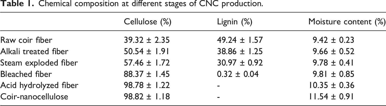

Chemical composition and moisture content at different stages of CNC production viz. Raw Coir fiber (RCF), alkali treated fiber (ATF), sodium chlorite treated fiber (SCT), Oxalic acid treated fiber (OAT) and Coir-Nanocellulose (CNC).
When treated with NaOH and subjected to steam explosion, the cellulose proportion increased. Lignin reacts with NaClO2 to form lignin chloride. 43 The percentage increase in pure cellulose content and decrease in lignin and hemicellulose contents with each processing step was another main observation, which in turn confirmed the effectiveness of the extraction process.
Scanning electron microscopy (SEM)
Scanning electron microscopy images at different stages of processing (Figure 3) evidences the transformation of coir fibers to nanocellulose having an average diameter of 50-100 nm with apparent variations in the surface structures and with individual random fibers presenting approximately 88 nm diameter. The coir fibers initially observed as thick individual strands, were converted to dense intricate networks, meticulously interwoven, forming an inter-connected mesh-like structure with numerous pores, clearly confirming the formation of microfibrils. The increase in the surface area and porosity aids in acquiring the unique properties of CNC, such as fluid absorption, which can also help serve as an appropriate framework for the incorporation of antibiotics. Scanning Electron Microscopic images of: (a) Raw coir fiber, (b) Alkali treated fiber, (c) Bleached & Acid hydrolysed fiber, (d) Coir-Nanocellulose.
The surface roughness of the raw coir fiber can be attributed to the presence of polyphenolic chemicals, which cause its coarse granular morphology. 44 It was found that the fibers got softened after steam explosion using sodium hydroxide. It has been previously demonstrated that majority of the lignin and hemicellulose can be eliminated by mercerization. The depolymerization and defibrillation that occurred during steam explosion, further eliminated the remaining amorphous elements of lignin, hemicellulose, tannin, pectin, etc. from the core of the fiber. 45 When steam-exploded fibers were bleached with sodium chlorite, substantial changes in morphology were observed, the product became fibrillated, and the size was reduced to the nanoscale range, which reached a range of 80 ± 10 nm after acid hydrolysis and grinding, as evident from the scanning electron microscopic image. 46
Zeta potential measurement
The surface charge and colloidal stability of pristine and drug loaded CNC were determined by zeta potential measurements. CNC exhibited a zeta potential of −37.2 ± 2.9 mV confirming the electrostatic stability of the dispersion, with absolute value higher than 25 mV. The highly negative surface charge is attributed to the presence of surface hydroxyl groups in CNC. 47 For C-CNC, a significant reduction in zeta potential was observed following drug incorporation with a value of −26.7 ± 2.3 mV which may be due to the interaction between CNC matrix and ciprofloxacin causing partial neutralization of surface charges. The strong cationic nature of gentamicin further reduced the zeta potential of G-CNC to −15.4 ± 1.8 mV with its strong ionic interactions with CNC. The reduction in zeta potential for C-CNC and G-CNC confirms effective drug loading and surface modification, indicating moderate stability. Thus, the zeta potential measurements reveal that CNC-drug interactions substantially alter the surface characteristics of CNC which in turn modulate drug release and dispersion properties. 48
Fourier transform infrared (FT-IR) spectroscopy
FTIR spectra of CNC along with coir fiber at various stages of treatment are shown in Figure 4. The -OH and C–O stretching from the ether groups were responsible for the prominent spectral bands at 3327 and 1024 cm-1 respectively. C-H stretching of CH2 groups resulted in a peak in the region of 2900 cm-1. The vibration at 2850 cm-1 due to C–H stretching in lignin is absent in the treated samples, confirming the removal of this fraction.
49
The peak at 1641 cm-1 represents adsorbed water (H–O–H deformation), in the extracted cellulose samples, while the minimal spectral intensity ensures the elimination of hemicellulose fractions during processing. The band at 1432 cm-1 indicates C–H deformation in lignin, as well as deformation of this bond in carbohydrates in the presence of C = O as it moves down from its normal location. Similarly, after chemical and physical modifications, the peak at 1024 cm-1 became sharper, and this band was associated with CH2 rocking vibrations in cellulose. FT-IR spectra of coir fiber during different stages of processing (a) Raw Coir fiber (RCF), (b) alkali treated fiber (ATF), (c) sodium chlorite treated fiber (SCT) (d) Oxalic acid treated fiber (OAT) and (e) Coir-Nanocellulose (CNC).
Because the treated material was first neutralized with NaOH, the acid-extracted product did not show bound sulfate groups at 1210 cm-1, but showed absorption peaks at 1160, 1024, and 736 cm-1, indicating enhanced asymmetric stretching of the ring and vibration of C–O in cellulose. 49 In the majority of the spectral bands, the extracted CNC were identical to the previously reported reference cellulose samples, indicating a strong correlation.
FTIR analysis of drug-loaded-CNCs is shown in Figure 5. A distinct characteristic peak at 3301 cm-1 was identified in the FTIR spectrum of C-CNC and was attributed to the OH stretching vibration. Another band at 2894 cm-1 corresponds to aromatic C-H stretching and alkenes, and the peak at 1627 cm-1 corresponds to the -C = O vibration.
50
The peaks at 1319 cm-1 reveal the bending vibration of the O-H group, which suggests the existence of carboxylic acid, while the bands at 1498 cm-1 are related to C-H stretching. In addition, the C-F group showed a significant absorption peak at 1053 cm-1 G-CNC
51
shows peaks at 1630 cm-1, and 1316 cm-1 corresponding to amide I and amide III of gentamicin respectively. A new peak at 1050 cm-1 has emerged in C-CNC and G-CNC which may be attributed to the C-F stretching in ciprofloxacin and C-O stretching in gentamicin. The irregular peaks around 1400 cm-1 indicates the cross linking of nanocellulose with the antibiotics.
52
FT-IR spectra of (a) Coir-Nanocellulose (CNC), (b) Ciprofloxacin loaded Coir-Nanocellulose (C-CNC) and (c) Gentamicin loaded Coir-Nanocellulose (G-CNC).
The N-H bending vibration at 1631 cm-1 can be attributed to the aromatic amine of gentamicin. 53 Distinct N-H and alkyl C-H stretching vibrations are reflected as bands at 3394 and 2906 cm-1, respectively. The S-O bending and stretching vibrations were observed at 660 and 1054 cm-1 respectively, which are related to the sulfur content of gentamicin sulfate, whereas the HSO4 vibrational band was observed at 1104 cm-1.54
X-ray diffraction (XRD) analysis
The coir fibers have crystalline and amorphous regions, as indicated by the XRD graph. Alkali treatment removed cementing components such as lignin, hemicellulose, and pectin, causing the percentage crystallinity of the fiber to increase. The cellulose components tend to arrange themselves in an organized manner after lignin was gradually removed by various treatments. The lignin disintegrated during the bleaching process became lignin chloride, leaving the intact cellulose component behind.
In the XRD spectra of CNC, C-CNC, and G-CNC, three characteristic peaks at 2θ = 16.8°, 22.7°, and 34.8° were observed. These diffraction peaks are characteristic of native cellulose (cellulose I) and correspond to the (110), (200) and (004) crystallographic planes, respectively, indicating the presence of a highly crystalline cellulose structure.
55
Zimowska et al. had also reported that these peaks are dominant in monoclinic crystal lattice structure with well-ordered cellulose.
56
These peaks present in CNC, also exist in C-CNC and G-CNC which suggest that loading antibiotics does not change the crystallinity substantially. However a small increase in peak intensity has been observed after loading CNC with gentamicin, which aligns with the increase in crystallinity index calculated. The crystallinity index values for CNC, C-CNC and G-CNC are 51.85, 51.89 and 57.44 % respectively. The results are also in agreement with the reports by Costa et al., where nanocellulose particles showed a crystallinity index value of 55%.
57
(Figure 6). X-Ray diffraction pattern of (a) Coir-Nanocellulose (CNC), (b) Ciprofloxacin loaded Coir-Nanocellulose (C-CNC) and (c) Gentamicin loaded Coir-Nanocellulose (G-CNC).
Degree of polymerization (DP)
The intrinsic viscosity [η] of CNC was calculated to be 273 ± 8 mL.g-1 and Degree of Polymerization (DP) of CNC were computed to be 205 ± 6. The characteristics of derived nanocellulose predominantly depends on the polymeric chain length in addition to the degree of polymerization. The different pretreatment and extraction methods cause isolation of pure and crystalline cellulose by confirming the breaking of bonds between cellulose and non-cellulosic components viz. lignin and hemicellulose. 58 The different treatments have removed amorphous regions and retained the crystalline structure as evident from the crystallinity index of the synthesized CNC. These removal of amorphous regions is caused by the breakage of the polymeric chains present in the material, which ultimately causes reduction in the degree of polymerization which in turn increases the porosity, surface area and reactivity of nanocellulose. 59
Fluid absorption studies
Although there are various processes for managing fluid via absorption, passive fluid uptake is the most frequently evaluated characteristic and is an important consideration for wound dressings. It regulates wound infection and aids in preventing excessive build-up of exudate at the wound site.
60
The absorption measurement obtained using a standard immersion technique revealed that with time, the fluid absorption increased for a period of up to 4 h, and then remained constant, indicating a maximum absorption of 3.08 g/g in 4 h. Thus, the CNC sample absorbed up to 3.08 times its own weight during swelling in phosphate-buffered saline (Figure 7). (a) Fluid absorption studies at different time periods (A) 0.5 h, (B) 2 h, (C) 4 h, (D) 8 h, (E) 12 h and (F) 24 h; (c) Cross-sectional view of CNC pressed mats (a) before and (b) after immersing in 0.9% saline depicting the faster wettability and swelling characteristic of CNC. (c) Time-dependent fluid absorption characteristics of CNC.
Fluid absorption studies performed on CNC dressings revealed the efficient absorption of excess fluid, which can help prevent maceration and promote a healthy wound environment, as excessive exudate can create a favorable environment for bacterial growth. By minimizing the fluid buildup, CNC dressings can help reduce the risk of infection. Therefore, a well-managed wound environment can facilitate healing and reduce the risk of complications. Rapid and substantial absorbency and moisture management are major requirements in the development of appropriate dressing for exuding wounds. A fast-wetting quality enables dressing to quickly absorb exudate and prevent maceration of the surrounding skin, which was observed in the case of CNC. Furthermore, high absorbency is preferred for all liquids released by wounds. The frequency of changing the wound dressing can be significantly reduced with high absorbency, thereby minimising the chances of disturbing the wound. 61
The ability of a dressing material to absorb wound exudate while maintaining a moist environment is one of the most desirable and critical property of a wound dressing material. The abundant surface hydroxyl groups present on CNC might have contributed to the fluid absorption capacity which augments water absorption through hydrogen bonding. In a study conducted by Minsart et al., the swelling capacities of 11 commercially available wound dressings were reported and compared and was found to be ranged in between 1.5 and 23.2 g/g, depending on material type and morphology. 62 The obtained value of CNC falls within the clinically relevant range particularly intended for light to moderately exuding wounds. The relatively moderate swelling behaviour of CNC is due to the compact nanofibrillar structure and absence of any surface modifications. In most cases, moderate fluid absorption is preferred as excessive absorption may lead to mechanical disintegration of wound dressings and drying of wound environment.
For fluid absorption studies one-way ANOVA has been performed which revealed a significant effect of immersion time on fluid uptake and obtained a p-value of 0.0154, which indicates that the fluid absorption increased significantly with time, further implying the progressive swelling behaviour of CNC matrix. The initial rapid increase in absorption occurred due to the availability of hydroxyl groups in cellulose, which followed by a saturation of the same, resulted in a subsequent plateau. Thus, the fluid absorption profile exhibited by CNC indicates that it holds suitable moisture absorption capacity maintaining structural stability, making it appropriate for wound healing applications, specifically for light to moderately exuding wounds.
Drug loading studies
Drug loading and entrapment efficiency are essential metrices for drug delivery systems. Drug loading and entrapment studies were performed for ciprofloxacin and gentamicin. The percentage drug loading for ciprofloxacin and gentamicin is 17.68 ± 0.63% and 19.27 ± 0.96% respectively with drug entrapment efficiency as 42.97 ± 1.26% for ciprofloxacin and 47.76 ± 1.93% for gentamicin respectively. In the drug loading and drug entrapment studies, although gentamicin demonstrated slightly better results, both antibiotics were successfully loaded and entrapped within the delivery systems. The variability or uncertainty in the results is indicated by the standard deviations linked to each measurement. According to the work done by Zahra Sobhani et al., ciprofloxacin hydrochloride-loaded chitosan nanoparticles with 1:0.5 mass ratio, the encapsulation efficiency was found to be 23%. 63 Another study indicated that the best encapsulation performance for gentamicin sulfate which is 45%, was achieved using PLGA 50:50 at 2% nominal loading and a buffer at pH 6.0. 64
In a recent study by Roy et al., it was demonstrated that ciprofloxacin can be successfully loaded on to nanocellulose owing to the strong electrostatic and hydrogen bonding interactions among the cellulose functional groups and drug molecules and the percentage loading can be enhanced through surface functionalization like dialdehyde modification. 65 Similarly, recent research which focused on the development of bacterial nanocellulose incorporating gentamicin-loaded chitosan nanoparticles as a promising multifunctional wound dressing biomaterial revealed a sustained release pattern with strong antibacterial activity and good biocompatibility. 66
The interaction between cellulose and drug molecules occurs mainly through hydrogen bonding and surface adsorption and not through true encapsulation in to a porous nanocellulose matrix. The highly crystalline nature of cellulose limits free sites for drug loading and the hydrophilic nature of ciprofloxacin and gentamicin cause partial drug loss during purification and washing steps. Consequently, the loading efficiency got restricted to 17-19% and entrapment efficiency to 42-47%. Recent studies reported that the bonding between ciprofloxacin and unmodified CNC is through weak intermolecular interactions and surface modification can provide additional binding sites to improve ciprofloxacin loading through strong electrostatic interactions. 65 Likewise, previous studies report that gentamicin loading on to cellulose carriers happens through adsorption and partial penetration and hence the drug release is mainly governed by a balance between absorbed and adsorbed drug fractions. 67 In a study by Gulsu et al., it was reported that oxidised cellulose enhances the loading efficiency of gentamicin by the utilization of reactive functional groups. 68 Unmodified CNC systems exhibit moderate loading but deliver sustained drug release over extended period of time, making it beneficial for wound healing applications. The dense hydrogen bonding network in CNC considerably reduces drug diffusion and enhance prolonged antimicrobial activity in spite of moderate loading efficiencies. 69 Hence, the drug loading and drug entrapment efficiencies recorded in this study indicates the inherent characteristics of unmodified CNC system.
In vitro drug release studies
The rate of drug release from the nanofiber surface had a significant impact on the effectiveness of the wound dressing material. The cumulative release of ciprofloxacin and gentamicin from CNC under in vitro conditions at various time points during a 72 h period was determined (Figure 8). Drug dissolution occurs when an aqueous medium enters the CNC structure, causing pores between the fibers to allow diffusion of the dissolved drug out of the membrane.
70
Both ciprofloxacin and gentamicin loaded CNC samples exhibited burst release of 70.8 ± 3.4% and 64.3 ± 2.9% respectively, within a period of 2 h. The release rate became stable after 10 h which was further maintained at ∼80-85% and in case of both C-CNC and G-CNC, 92 ± 2.6% of the drug got released by 72 h. In-vitro drug release profile of CIP and Genta loaded CNC for a period of 72 hrs.
There observed a significant difference in the release behaviour of C-CNC and G-CNC during the initial period up to 12 h, where the p-value obtained was 0.0167 (1 h), 0.0142 (2 h), 0.0106 (4 h), 0.0084 (6 h), 0.0121 (8 h), and 0.0465 (12 h). This indicates that the physicochemical properties of two antibiotics influenced their diffusion from the CNC matrix. In contrast, no significant difference was observed after 24 h with p-value >0.05, suggesting sustained release equilibrium caused by the diffusion of drug molecules from the internal porous CNC matrix.
The drug release rate plays a crucial role in determining the effectiveness of wound dressings. Previous research has demonstrated that an antibiotic, when delivered rapidly, helps in controlling the infection at the wound site, and when delivered gradually over the course of a couple of days, can regulate wound healing in a better way. 71 In our study, the initial rapid release might have resulted from the diffusion of drug molecules that were superficially bound to the outer surface of the C-CNC and G-CNC samples. Thus, an initial burst can provide a high concentration of antibiotics at the wound site to combat existing bacteria, whereas sustained release offers long-term protection. Controlled drug release can also help minimize the development of antibiotic resistance. Thus, the combination of burst release and sustained release can be beneficial for wound healing, as it allows for the rapid control of infection and long-term protection. As the envisioned application was for the development of antimicrobial wound dressings, it might be considered suitable for treating wounds and preventing infection at the wound site based on the drug release rate.
Drug release kinetics
The drug release data were fitted in to different models viz. Zero order, First order, Higuchi and Korsmeyer-Peppas for C-CNC (Figure 9(a)) and G-CNC (Figure 9(b)) and the results are consolidated in Table 2. Both C-CNC as well as G-CNC followed a two-stage release profile featuring an initial burst release followed by a sustained release pattern. The initial burst release may be associated with release of drug molecules loosely bound to the surface of nanocellulose.
72
The lower correlation coefficient values for Zero order, First Order indicate that the release mechanism does not follow constant-rate or a concentration dependent process.
73
Drug release kinetics modelling performed on C-CNC (a) and G-CNC (b). Drug Release Kinetics Modelling by comparing values of correlation coefficient (R2) and kinetic rate constant (k).
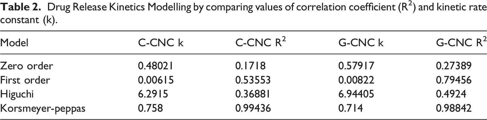
The zero order, first order and Higuchi models exhibited low correlation coefficient values for both C-CNC and G-CNC indicating that the drug release do not follow a constant rate or is not based on diffusion alone. Comparing the different release models, the Korsmeyer-Peppas model exhibited highest correlation coefficient having R2 values of 0.99 for C-CNC and 0.98 for G-CNC confirming the best fit for the drug release mechanism to be
The release exponent (n) calculated from Korsmeyer-Peppas model for both systems were significantly below 0.45 and was approximately 0.11462 for C-CNC and 0.05316 for G-CNC confirming a Fickian diffusion-controlled release mechanism. 74 The low n values indicate the presence of a highly restricted dense nanocellulose matrix and strong intermolecular interactions. Recent studies further states that nanocellulose based drug delivery systems permits tunable and sustained drug release making them promising candidate for advanced drug delivery applications. 75 Thus, the initial burst release followed by the sustained release highlights the potential of C-CNC and G-CNC for prolonged antibacterial activity in wound healing applications.
Effect of C-CNC and G-CNC on the growth of microorganisms
A series of phases in the development of the bacterial culture that were characterized by differences in growth rate were observed in all four strains of tested bacteria: B. cereus, S. aureus, P. aeruginosa and E. coli (Figure 10(a)–(d)). A normal bacterial growth curve with lag, log, and stationary phases was observed for bacteria alone and with CNC inoculated nutrient broth. In contrast, the growth declined for both C-CNC and G-CNC sample treated cultures, from 0.5 h onwards. This indicates that the C-CNC and G-CNC samples have a growth-inhibiting effect on microbes even during their lag phase, suggesting that multiplication is averted.
76
Growth curve of microbes under the influence of CNC, C-CNC and G-CNC compared to the normal growth curve, depicting the microbiocidal nature of drug loaded CNC for the tested strains of (a) B. cereus, (b) S. aureus, (c) P. aeruginosa and (d) E. coli.
The bacterial growth curves were analyzed using two-way ANOVA and it was observed that compared with untreated bacterial control, both C-CNC as well as G-CNC exhibited significant inhibition of bacterial growth for all the tested strains of microorganisms across all time points with a p-value <0.0001 and also a statistically significant difference was observed between C-CNC and G-CNC with a p-value of 0.0016, indicating drug formulation dependent antimicrobial activity.
Antimicrobial studies
The presence or absence of inhibition zones were used to evaluate the antibacterial activity of all the samples, including PC (CIP-10 mcg disc), PC (GEN-10 mcg disc), NC (CNC), C-CNC (1.0, 0.5, 0.25 mg/mL) and G-CNC (1.0, 0.5, 0.25 mg/mL), against the four tested bacterial strains. Pure CNC used as the negative control, did not show any antimicrobial activity against the tested organisms. In contrast, the incorporation of different concentrations of Ciprofloxacin (1.0, 0.5 and 0.25 mg/mL) (Figure 11) and Gentamicin (1.0, 0.5 and 0.25 mg/mL) (Figure 12) on to the CNC resulted in significant inhibition of growth against the tested strains of E. coli, B. cereus, P. aeruginosa and S. aureus and was found to be proportionate with the concentration of the drug used with the highest ZoI measuring 2.7 ± 0.024 cm for E. coli, 2.3 ± 0.011 cm for S. aureus, 2.6 ± 0.037 cm for B. cereus and 2.8 ± 0.045 cm for P. aeroginosa for C-CNC with a concentration of 1 mg/mL which was comparable with positive control. Similarly, ZoI measuring 2.6 ± 0.042 cm for E. coli, 2.4 ± 0.031 cm for S. aureus, 2.7 ± 0.073 cm for B. cereus, and 2.8 ± 0.054 cm for P. aeruginosa was observed for G-CNC at a concentration of 1 mg/mL. Inhibitory effect of C-CNC on Muller-Hinton agar plates at varying concentrations (a-Negative Control, b- Positive control, c- C-CNC 1.0 mg/mL, d- C-CNC 0.5 mg/mL and e- C-CNC 0.25 mg/mL), (a) E. coli, (b) P. aeruginosa, (c) S. aureus, (d) B. cereus. Inhibitory effect of G-CNC on Muller-Hinton agar plates at varying concentrations (a-Negative Control, b- Positive control, c- G-CNC 1.0 mg/mL, d- G-CNC 0.5 mg/mL and e- G-CNC 0.25 mg/mL) for (a) P. aeruginosa, (b) E. coli, (c) S. aureus and (d) B. cereus.

Analysis of the data (Figure 13) indicated that both C-CNC and G-CNC samples had significant inhibitory effects against the tested strains of gram-positive and gram-negative bacteria, and the maximum growth reduction was observed at a concentration of 1 mg/mL.77,78 Quantitative analysis of Zone of Inhibitions of (a) C-CNC and (b) G-CNC at different concentrations (1, 0.5.0.25 mg/mL) along with positive control (PC) and negative control (NC) tested against E. coli, S. aureus, P. aeruginosa and B. cereus.
The healing process is frequently slowed down by wound infection caused by bacteria and other microbes, which can also result in potentially fatal consequences. To avoid infection and promote wound healing, the wound must be covered with a suitable dressing material that hinders microbial growth on the wound surface. 79 CNC loaded with Ciprofloxacin and Gentamicin exhibited antibacterial activity against the tested bacterial strains, which are common causatives of skin and soft tissue infections, and the activity was proportional to the concentration of the incorporated antibiotic. By inhibiting bacterial growth, antibiotic-loaded CNC can help prevent wound infections and promote healing. The ability to adjust the antibiotic concentration allows for tailoring of the antibacterial activity to specific wound needs.
Cytocompatibility studies
MTT assay performed with L929 cells cultured in conditioned media extracted from CNC, C-CNC and G-CNC evaluated the impact of CNC and antibiotic incorporation on to CNC in altering cellular viability. The results exhibited good cytocompatibility with significant viability and proliferation. Compared to the control group, which received fresh DMEM F12 media, cells exposed to the conditioned media exhibited an approximately 80% increase in cell survival after 48 h as shown in Figure 14, which was further confirmed by the absorbance measurements at 570 nm. Morphology of L929 cells (a) control, (b) treated with CNC, (c) treated with C-CNC and (d) treated with G-CNC for 48 h.
The statistical analysis performed using one-way ANOVA revealed that both C-CNC and G-CNC exhibited larger zones of inhibition compared to control with p-value <0.05 and also the inhibition was more significant with higher drug concentrations. This also confirms that the antimicrobial effect has initiated from the loaded antibiotics rather than the nanocellulose carrier.
Several studies have explored the biocompatibility of nanocellulose for various applications. Cellulose nanofibers extracted from lemongrass were found to be nontoxic in the L132 cell line and all other cell lines studied, when exposed to 0-1000 µg/mL. 80 In another study, the cytotoxicity of CNC was tested in GM07492 A cells, and it was observed that CNF showed more than 70% viability. 81 Cell viability and proliferation of L929 cells cultured in CNC in comparison with C-CNC and G-CNC conditioned media revealed no significant difference in cell viability. This results in comparison with the control demonstrated the possibility of using CNC-based antibiotic loaded wound dressings to deliver a favorable microenvironment for cell proliferation, where prolonged contact with healthy tissue is essential.
Conclusion
The present study involves successful extraction of coir-nanocellulose from coconut husk fibers and demonstrates the potential of antibiotic infused coir-nanocellulose as a viable material for innovative wound treatment. The good fluid absorption capacity of the dressing ensures a moist environment that promotes healing and incorporation of antibiotics reduces the risk of bacterial colonization at the wound site. This strategy seeks to combine the targeted antibacterial activity of encapsulated antibiotics with the advantageous fluid absorption potential of nanocellulose. The initial burst release of antibiotics at the wound site delivers a high concentration of the drug, which helps protect against existing bacterial load at the wound site, and sustained release over an extended period of time aids in the maintenance of a minimal concentration of drug required to avert further infection at the wound site. Overall, the current study delivers thoughtful insights into the potential of coir-nanocellulose based antibiotic-infused wound dressings, which could become a valuable tool in wound care with further research and development. Further research focusing on the in vivo aspects are required to confirm the efficacy of the wound-dressing material.
Footnotes
Acknowledgements
The author acknowledges the support and facilities extended by the Chairman, Coir Board, Director, Central Coir Research Institute, Coir Board, Senior Scientific Officers, and Research Scholars of the Institute.

Author contributions
Geena M.G.: Conception and Design, Methodology, Data curation, Analysis and Interpretation of the data, Drafting of the Paper, Visualization, Investigation.
Ajith Sudhakaran: Data curation, Analysis and Interpretation of the data, Visualization, Investigation.
Akhila K.P.: Methodology, Data curation.
Anita Das Ravindranath: Supervision.
Rahul Rajan: Methodology.
Athira Johnson: Drafting of the paper.
Sailaja G.S.: Supervision, revising critically for intellectual content, Final approval of the version to be published.
Funding
The authors disclosed receipt of the following financial support for the research, authorship, and/or publication of this article: Ministry of MSME, Coir Board.
Declaration of conflicting interests
The authors declared no potential conflicts of interest with respect to the research, authorship, and/or publication of this article.
Statements and declarations
The authors confirm that this manuscript has neither been published, nor is under consideration for publication elsewhere, and that all authors have approved submission of the manuscript.