
Abstract
We present a rare image documenting direct transit of a thrombus originating from the venous circulation and passing through a patent foramen ovale linked to a malpositioned cannula in the setting of temporary mechanical circulatory assistance in a young patient who underwent a peripheral extra corporeal membrane oxygenation as a circulatory support.
Case presentation
A 21-year-old male patient was referred to our intensive care unit for cardiogenic shock. He was diagnosed with infective endocarditis on a prosthetic aortic valve implanted four years ago. Preoperative left ventricular (LV) ejection fraction was normal. Postoperative course was complicated by severe LV dysfunction. Both aortic prosthesis valve examination and coronary angiogram were normal. Peripheral extra corporeal membrane oxygenation (ECMO) along with intra aortic balloon pump were implanted as a circulatory support. Routine bedside transthoracic echocardiography (TTE) revealed undiagnosed malposition of the venous cannula into the left atrium, crossing through the patent foramen ovale (PFO; Figure 1(a); Supplementary Material online Video 1). Under TTE control, we corrected the position of the cannula by withdrawing it into the inferior vena cava. This operation left a vermicular thrombus floating in the right atrium and stuck into the PFO (Figure 1(b) and (c); Videos 2 and 3). Control TTE performed few days later revealed that the thrombus had completely resolved under anticoagulant therapy without any manifestation, suggesting neither pulmonary nor systemic embolization. As he did not recover under ECMO, the patient was transplanted on day 28 with uneventful postoperative course. He is doing well at 12 month follow-up. Images documenting direct transit of a thrombus originating from the venous circulation and passing through a PFO are scarce.1,2 We report a rare case of a floating thrombus in the setting of temporary mechanical circulatory assistance linked to a misplaced venous cannula and disappearing under well-managed anticoagulant treatment. Surgical embolectomy might not always be the rule in these critical patients, free from embolic events. 3
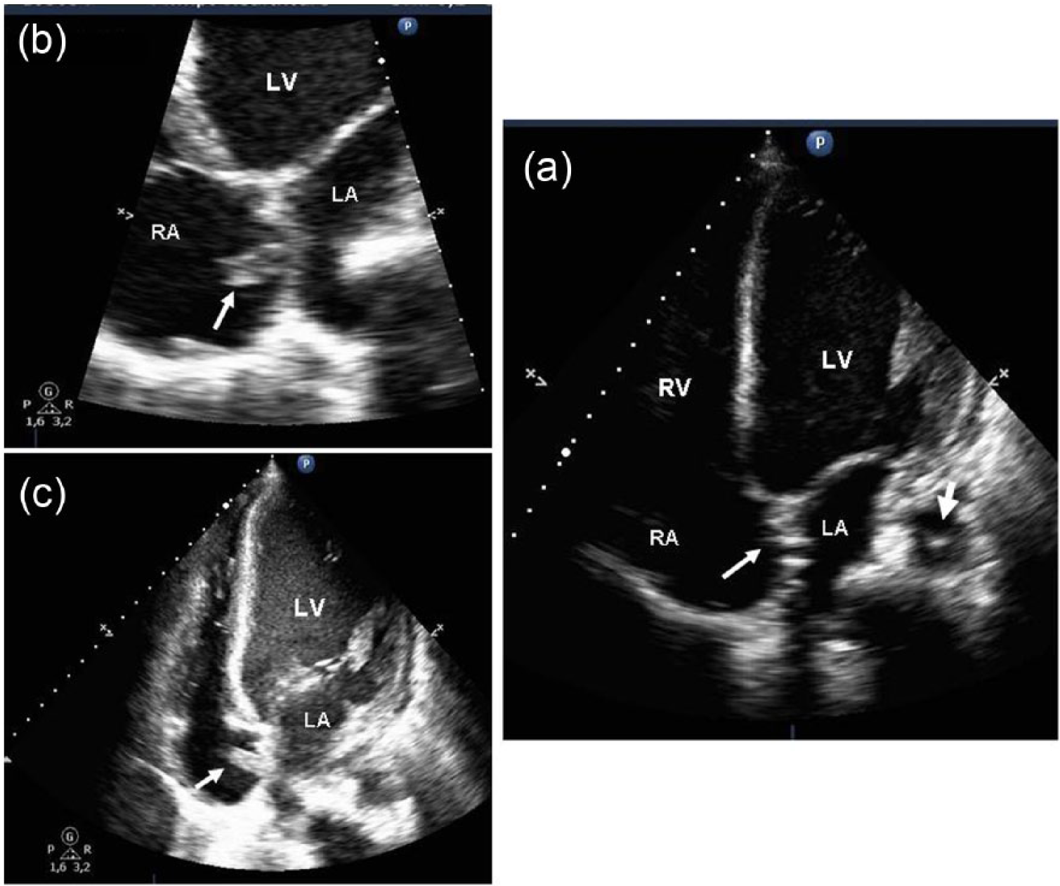
(a) Bedside transthoracic echocardiography, apical 4-chamber view showing the malpositioned venous cannula passing through the patent foramen ovale (PFO, small arrow) and reaching the left atrium. The large arrow refers to intra aortic balloon pump in the descending aorta, (b) after the correction of cannula’s position. Bedside transthoracic echocardiography, apical 4-chamber view, zoom on the interatrial septum showing the entrapped thrombus into the PFO, (c) after the correction of cannula’s position. Bedside transthoracic echocardiography, apical 4-chamber view showing the entrapped thrombus into the PFO (arrow).
Footnotes
Conflict of interest
The authors declare that there is no conflict of interest.
Funding
This research received no specific grant from any funding agency in the public, commercial, or not-for-profit sectors.