





Abstract
Tooth wear is increasing in prevalence within the United Kingdom. Treatment of tooth surface loss can be daunting for both the clinician and patient. However, use of additive resin composite restorations is a minimally invasive treatment modality. This case illustrates the treatment of tooth surface loss in both the maxillary and mandibular arches with direct composites restorations using putty indices generated from a diagnostic wax-up recorded in centric relation. The tooth surface loss had resulted in reduced restorative space on the right hand side. The restorative treatment involved increasing the anterior vertical dimension, enabling the provision of a cobalt-chrome partial denture. This case shows the restoration of form, function and aesthetics using a reorganised occlusal approach.
Learning Objectives
To understand treatment of tooth surface loss using additive, minimally invasive, direct resin composite restorations using a diagnostic wax-up to increase the patient’s vertical dimension
To get an overview of restoring the lower partially edentulous arch with a cobalt-chrome partial denture
To be able to discuss the treatment plan and evidence base
Introduction
The Adult Dental Health Survey (ADHS 2009), highlighted that 94% of the population in England, Wales and Northern Ireland were dentate, having at least one tooth. 1 Adults had on average 25.7 teeth, with 17.9 sound untreated teeth. 1 Nearly 20% used a removable prosthesis, with 13% relying on both dentures and natural teeth. 1 The survey also reported the prevalence of tooth surface loss (TSL) into dentine, with over 75% of dentate adults exhibiting anterior tooth wear. 1 Moderate to severe tooth wear was found in 15% and 2% of adults respectively. 1 Moderate TSL had increased from 11% in 1998 to 15% in 2009. 1 Greater TSL was associated with increased ages. 1 The average number of teeth associated with moderate tooth wear was 3.1. 1 Tooth wear is a multifactorial process involving attrition, abrasion and acid erosion, 2 with growing evidence that dietary habits increase the risk of erosion occurrence. 3
Several techniques exist for the restorative management of TSL including the use of direct and indirect restorations as well as fixed and removable prostheses. TSL is often an asymptomatic condition and its management may cause concern for both the patient and treating clinician. 4 TSL can also reduce the occlusal vertical dimension (OVD), which is the distance between mandibular and maxillary teeth when they are occluding. 5 Furthermore, tooth loss can also lead to over-eruption of unopposed teeth, further complicating space management in a TSL scenario.
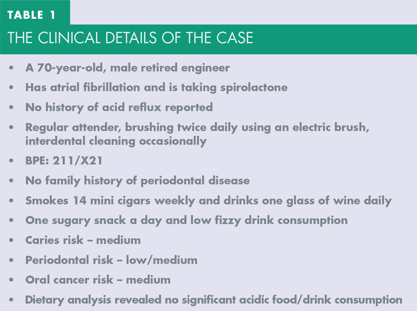
The following clinical case (Figures 1-11) illustrates how TSL can be treated, using additive and minimally invasive composite restorations to increase the OVD in a patient who has lost restorative space posteriorly due to the over-eruption of maxillary posterior teeth. Treatment of the TSL enabled the restoration of the mandibular arch using a removal prothesis to re-establish posterior support. Table 1 provides the clinical details of the patient.
The clinical details of the case

Pre-operative anterior retracted view showing a shortened dental arch and evidence of TSL

Occlusal view of the maxillary teeth showing amalgam restorations in UR6 and UL4, a metal-ceramic crown UL5, as well as incisal and slight palatal TSL

Patient demonstrating the use of Green Tepe™ interdental brushing following supragingival cleaning with the aim to reduce calculus deposits

Composite restoration LL3 distal caries (A) Preoperative view, (B) Immediate postoperative view

Diagnostic wax-up of maxillary anterior teeth

Diagnostic wax-up of mandibular anterior teeth.

Restoration of the maxillary anterior teeth; (A) shade match completed prior to rubber dam isolation using Venus Pearl composite, (B) rubber dam placement retained using clamp and wedgets, (C) use of palate stent and PTFE tape to restore every other tooth, (D) immediate postoperative view, (E) immediate postoperative view following polishing

Restoration of UL4 using Venus Pearl composite; (A) placement of pre-wedges to protect neighbouring teeth, (B) immediate postoperative view

Isolation of LL3 buccal abrasive cavity using cotton wool roll and PTFE tape in the gingival sulcus; (A) preoperative and (B) postoperative

Restoration of LL7 using rubber dam isolation and Venus Pearl resin composite; (A) preoperative and (B) postoperative

Partial chrome denture restoring the lower edentulous regions; (A) design of prosthesis, (B) fit of prothesis
Figures 1-2 show the pre-operative presentation, an anterior retracted view and a maxillary view of the patient who exhibited TSL with attrition, abrasion and erosion elements. The loss of the lower right posterior teeth had also resulted in over-eruption of the opposing teeth and loss of restorative space within the sextent. The extra-oral and intra-oral examinations revealed no hard or soft lesions, and no discomfort or notable clicks were recorded from the temporomandibular joints (TMJ). The maxillary arch was recorded as a Class 1 Kennedy classification with free end saddles distal to UR6 and UL5. The mandibular arch was recorded as Class 2 Modification 1 Kennedy classification with a free end saddle distal to LR3 and a bounded saddle between LL47. Edentulous regions had sufficient alveolar width and height. There was evidence of over-eruption of the upper right maxillary teeth (UR654) opposing the mandibular free end saddle associated with a loss of restorative space. When the patient occluded, there was 3-4 mm of restorative space between UR7 cusp tips and the mandibular edentulous region.
Overall his gingival health was very good, although some plaque and calculus deposits were noted anteriorly, which he struggled to maintain even after routine cleaning and tailored oral hygiene instructions.
Caries was noted on the lower left canine (LL3) distally and the lower left second and third permanent molars (LL78) were unopposed and none-functional. The patient was functioning with a shortened dental arch (SDA). No teeth were tender to percussion (TTP) and all responded positively to ethyl chloride. No apical pathology was noted on radiographic examination.
An occlusal assessment was completed prior to any confirmation of the treatment plan. A Lucia jig was made using Trim (Bosworth Company) and the patient was deprogrammed and manipulated into centric relation (CR). The first contact on the retruded arc of closure appeared to be the UL4 against the opposing LL3. A minimal shift from CR to centric occlusion (CO) was noted, no major vertical and minimal horizontal components were recorded. Dynamic functions were assessed which identified anterior protrusion with posterior disclusion (UL45 region), canine guided occlusion during right lateral excursion, no obvious working/non-working side interferences and early canine guidance during left lateral excursion shifting to involve LL12 as well.
The treatment plan involved general supragingival cleaning and tailored oral hygiene instructions using TepeTM brushes (Figure 3). The caries (LL3) was restored using Venus Pearl composite (Kuzler) (Figure 4).
The TSL was restored using additive, resin composite (Venus Pearl Kuzler) restorations based on a diagnostic wax-up taken from articulated study models (Figure 5-6) on which the vertical dimension has been increased by 2-3 mm. The restorations were placed in the maxillary arch (Figure 7), one tooth at a time, using rubber dam isolation using an index of the wax-up with a slight bevel preparation. Polytetrafluoroethylene (PTFE) was used as a physical barrier. PTFE can help create tight contacts and it does not interfere with indices. The UL4 was restored at a subsequent visit (Figure 8) using pre-wedges (Palodent V3 Wedgeguards).
The lower incisors were restored using the same approach employed for the upper anteriors, whilst the lower left canine (LL3) buccal cavity was restored with cotton wool roll and PTFE tape (Figure 9) and the LL7 was restored as shown in Figure 10.
Rubber dam isolation during the placement of resin composites is not routinely carried out. 6 One of the most commonly cited reasons for not using rubber dam is time constraints. 7 However, it is considered the optimal technique for providing sufficient moisture control during adhesive dentistry. 8 Resin composites placement is influenced by several contaminants including blood, cervical fluid and saliva and isolation does influence the longevity of adhesive restorations. 9
A Cochrane review investigated the importance of rubber dam isolation for restorative treatment in dental patients, 10 it assessed randomised controlled trials (RCTs) or quasi-randomised controlled trials (qRCTs) involving both direct and indirect restorative procedures. Patients were in either the intervention (rubber dam isolation) or comparison (cotton wool roll) groups. Primary outcomes assessed included survival rates at six-month, one, two, five and ten-year intervals as well as adverse events. Clinical evaluation of the restorations quality, cost considerations and patient acceptance/satisfaction. After removal of duplicated references, 781 studies were identified and only four studies were included in the review. 10 The authors concluded there is very low-quality evidence from single studies that rubber dam isolation may enable higher survival rates for restorative treatments. 10 However, it is important to highlight the studies did not address the primary outcomes sufficiently; the treatments provided included non-carious cervical lesions, fissure sealants and atraumatic restorative techniques in primary teeth. The studies reviewed did not involve the restoration of carious adult teeth. The quality of the evidence is not robust enough to make definitive conclusions about the influence of rubber dam isolation on survival rates of restorative treatments. 10 Even with this limited data, however, this does not mean rubber dam use should not be used, as numerous other advantages when isolating with rubber dam exist, such as airway protection, better access, less mirror fogging, better visual access and easier soft tissue management.
Studies have reported the suitability of resin composite as a material for restoring worn dentitions with hybrid composite materials being recommended. 11 When using resin composites the following should be considered:
Enamel bonding: To be completed using a separate etch and bonding system followed by resin composite infiltration. The use of self-etch resins with a single step is not advised. 11
Dentine bonding: More challenging due to its heterogenous composition. Dentine bonding involves conditioning, priming and bonding. Conditioning effectively is acid etching and either removes or modifies the smear layer. The use of a separate etch and rinse of enamel and dentine is recommended for treating worn teeth. 11
Surface preparation: Both enamel and dentine preparation is advised with a bevelled enamel margin and roughened dentinal surface. In this case, a small amount of incisal preparation was completed and the teeth were sand blasted with aluminium oxide prior to bonding.
Application of composite build ups: A recent European consensus on the management of severe tooth wear highlighted a conservative minimally invasive, additive approach should be used. 12 Survival rates of composites in the restoration of wear have been reported of between 50-95% for up to ten years 11 with survival being affected by patient age and gender. Lower survival rates have been reported in older patients, especially in patients with limited posterior support. 13
Changing vertical dimension: Studies have failed to identify any negative effects of increasing the occlusal vertical dimension using direct composite restorations. 14
A 2013 prospective randomised, controlled, split-mouth clinical trial, reported a seven-year follow-up of direct composite restorations in the worn mandibular anterior dentition. Fifteen patients were involved, 107 direct composites were placed and their performance alongside patient satisfaction were evaluated. 15 Survival was defined as a restoration which had not been lost, repaired or replaced and the seven-year survival rate was 85%, with 53% of patients having all their restorations present. 15 No biological complications were associated with the anterior build ups in this study, which considered tooth, periodontal and TMJ issues. Restoration of the worn mandibular anterior teeth was associated with improvements in aesthetics, and sensitivity as well as reduced concern for longevity. 15 The most common clinical complication associated with the direct composite restorations was marginal breakdown. The split-mouth design involved restorations being placed with or without circumferential preparation, which was not associated with any significant differences in results. 15 However, the study was limited in patient and restoration number.
A 2016 systematic review evaluated the treatment performance/longevity of dental materials/techniques indicated to restore teeth with severe wear. 16 This study analysed retrospective and prospective studies and investigated the annual failure rates (AFR %). From 511 initial articles, 12 were included in the review which highlighted ARF ranges from 0.4% and 26.3% for microhybrid and microfilled direct resin composites respectively and 0% to 14.9% for indirect resin composites, and 2.7% for porcelain veneers. 16 The authors concluded no strong evidence exists that any material is superior and both direct/indirect materials may be used to treat severe wear. 16
Another systematic review evaluated the survival rates of anterior composites in managing tooth wear. 17 From 666 articles only five studies were included in the review, which involved 772 direct and indirect anterior composite restorations. 17 The follow-up periods ranged between five months and ten years. 17 However, no meta-analysis was completed due to heterogeneity in the data. The survival rates for anterior composites were >90% and 50% at 2.5 and five years 17 and the authors concluded that evidence does support the use of composite restorations in the treatment of anterior tooth wear at an increased vertical dimension. However, the literature provides only short to medium-term outcomes and more standardised studies are required to provide long-term outcomes. 17
With respect to the clinical case discussed above and following the restoration of the TSL, additional restorative space was generated on the right side, enabling the mandibular arch to be restored with a cobalt-chrome partial denture (Figure 11) using techniques advocated previously. 18 A 2012 Cochrane review investigated the effects of different prostheses for the treatment of partially absent dentition in terms of long-term success, function, morbidity and patient satisfaction. 19 The review considered only RCTs, and 21 RCTs were included in the analysis, which concluded that insufficient evidence exists in the literature to recommend one prosthetic option over an alternative for partially edentate patients. 19
Conclusion
Tooth surface loss is increasing in prevalence within the United Kingdom. Understanding and identifying the aetiological factors in the management of TSL is vital. Where treatment is required, additive, minimally invasive treatment approaches should be employed prior to the use of more invasive options. As demonstrated in this case, space can be generated for removable prostheses by the placement of direct composite resin restorations at an increased vertical dimension.
Footnotes
Acknowledgements
This case was submitted as a documented case study as partial fulfilment of the Diploma in Restorative Dentistry awarded by the Faculty in General Dental Practice (UK). Thanks is given to the patient who consented to being used as a case study and to Mr Nicholas Lewis who assisted in the treatment planning.