





Abstract
Effective communication between clinicians and the dental laboratory is paramount in the realm of dental implant restorations, where precision and meticulous planning are key for success. This article explores the transformative shift towards digital workflows, highlighting the collaborative dance between the clinician and the dental laboratory. The power of visual communication is emphasised, with examples illustrating its impact on the precision of treatment plans. Celebrating these digital tools, this article envisions a new era of collaboration, transcending traditional communication methods for more predictable and successful implant restorations.
Learning Objectives
To provide an overview of the importance of communication between clinicians and the dental technician in the fabrication of an implant borne restoration
To highlight specific aspects of the relationship between clinician and technician and how to identify and avoid particular pitfalls
To demonstrate the level of communication required to achieve a predictable outcome using the example of a case presentation of a failed incisor tooth
Introduction
Effective communication between dental team members is the cornerstone in achieving optimal outcomes in the intricate field of dental implant restorations, where precision is paramount and success hinges on meticulous planning. Nowhere is this more evident than in the collaborative dance between the dental surgery and the dental laboratory, mainly when planning and restoring implants. Thus, it is imperative to recognise that the fundamentals – planning and communication – provide the groundwork for success and prevent simple mistakes from becoming major stumbling blocks.
In recent years, the implant field has witnessed a transformative shift towards digital workflows, revolutionising how implant surgeons and dental technicians communicate and plan implant restorations. The advent of three-dimensional (3D) viewers and other visual communication platforms has ushered in an era of enhanced collaboration, facilitating a seamless exchange of information between the surgical suite and the dental lab. This digital evolution allows for the easy sharing of virtual models, enabling surgeons to convey intricate details of implant positions, angulations, and restorative space with unparalleled clarity. Visualising the treatment plan in three dimensions instead of relying solely on written descriptions or phone calls minimises the risk of misunderstandings and ensures a shared perspective on the desired outcome.
The human brain’s remarkable ability to process visual clues plays a pivotal role in the effectiveness of this digital communication revolution. Research findings suggest that a substantial portion of the brain is specialised in processing visual information, highlighting the potency of combining visual communication with clear written text. This effectiveness is underscored by the ground-breaking work that led to Allan Paivio’s Dual Coding Theory. 1
Richard Mayer, renowned for his cognitive theory of multimedia learning, delves into this concept in his book Multimedia Learning. 2 Within this work, Mayer examines twelve principles of instructional design, all rooted in experimental research studies and a comprehensive theory of how individuals comprehend information presented through words and pictures. Noteworthy among these principles is the Multimedia Principle, which suggests that individuals comprehend information more deeply when presented with a combination of words and pictures, as opposed to words alone. 2
In the context of implant restorations, the ability to share and view 3D representations of the planned restoration fosters a deeper understanding and alignment between surgeons and dental technicians. It represents an opportunity to tackle potential challenges at the earliest phase of the planning stages.
To illustrate the power of this visual communication paradigm, consider a straightforward example. In a traditional workflow, a surgeon might describe the desired emergence profile of a restoration in written form, leaving room for misinterpretation. In a digital workflow, the same surgeon can share a 3D scan showcasing the precise contours and dimensions, providing the dental technician with a clear visual reference for crafting the restoration. This is very important when we consider the rapid rise of bone level implants that sit just below the bone level: too broad an emergence could lead to bone engagement where the bone prevents passive seating to the implant, leading to complications such as screw loosening or fracture as the bone resorbs, and the once tight abutment starts to move.
Figure 1 provides an example of this potential issue with the clinician’s notes accompanying the case, in addition to the expected information, including “Implant deep, please only flare near gingival margin. Risk of bone engagement, especially mesially.” This information influenced the design provided by the laboratory, which was confirmed by the clinician, and all before a commitment to manufacturing the abutment. At this stage any changes can easily be made without additional costs or issues for either clinical or technical team.

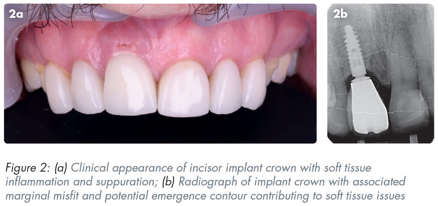
Figure 2 shows the clinical and radiographic presentations of an implant-supported crown replacing a central incisor, with associated soft tissue complications. This could be attributed to several factors, including poor marginal fit of crown to abutment. However, the abutment choice and design resulting in an excessive emergence profile of the restoration may also have played a role. These issues may arise when there is minimal clinical input regarding the restoration material and design. This is an example of the importance of abutment choice and design for a more predictable biological outcome.
The application and design of titanium custom abutments can be used as another example of the close collaboration needed between the technical and clinical teams, to provide a predictable outcome. A narrow emergence from the bone level can be provided with a custom design or with a standard titanium base. This can lead to significant differences in the support of the overlying material, often zirconium. Thin sections of zirconium are liable to fracture which can be typically seen with standard abutments. 3
Figure 3 shows a molar implant crown on a standard titanium abutment, where there is fracture and loss of zirconium at the cervical abutment interface. This is where the loading forces are greatest 4 and where the forces become more exaggerated due to an off-centre placement, creating a cantilever force. Similarly, failures of ceramic at the abutment interface can also occur with replacement of anterior teeth (Figure 4).


Figure 5 shows an alternative approach for a molar replacement restoration, with the abutment custom-designed to allow sufficient support for the restoration and soft tissues, and also compensating for a slightly distal placement of the implant head. Modern technology and materials now allow for more robust and biologically compatible restorations where technicians can provide results with far fewer issues when the guidelines on materials, loading and emergence profiles are followed.

As we continue to explore the intricacies of implant restorations, let us not only recognise the importance of planning and communication but also celebrate the digital tools that empower us to communicate with unprecedented clarity. By embracing these technological advancements, the dental community can foster a new era of collaboration that transcends the limitations of traditional methods of analogue communication, ultimately leading to more predictable and successful implant restorations.
Effective communication between the dental surgery and laboratory is crucial for successful implant placement and restoration. This can take place in the form of emails, large file transfers using tools such as WeTransfer (WeTransfer B.V., Amsterdam, The Netherlands), custom cloud-based lab portals and software, or platforms such as Slack (Salesforce Inc., San Francisco, CA, USA) or Teams (Microsoft, Redmond, WA, USA) (Figure 6).

Below are some key principles and potential pitfalls, explained in plain entry-level language:
Principle: Ensure the surgeon and the lab are on the same page regarding the treatment plan. Pitfall: Assuming everyone knows the plan without discussing it thoroughly. Clear communication upfront prevents misunderstandings later.
Principle: Provide comprehensive details about the patient’s condition and the desired outcome. Pitfall: Being too brief or unclear about expectations. The lab needs as much information as possible to create the best restoration.
Principle: Accurate impressions are vital for a well-fitting implant restoration. Pitfall: Rushing through the impression process. Take the time to ensure a precise analogue or digital impression, as errors here can affect the entire restoration at the point of fit.
Principle: Convey the desired shade and surface characteristics for the final restoration. The topic of shade assessment and communication is complex and falls outside of the remit of this article. However, photographs taken using a digital single-lens reflex (DSLR) camera are increasingly considered as a valuable method of communicating shade characteristics rather than shade tabs alone. Utilising polarised photography and exploring the use of eLab system (eLab Prime, Freiburg im Breisgau, Germany) (Figure 7) are becoming increasingly popular in overcoming the limitations of photographs alone. They allow the laboratory to analyse the shade information in more detail and use their software to carry out virtual try-ins. Ultimately, the aim is to eliminate the need for the patient to have to visit the dental laboratory for a shade-taking appointment. It should be reiterated that shade and characteristic matching remain some of the biggest challenges in indirect restorative dentistry, particularly in complex aesthetic cases, such as restoration of the single tooth adjacent to a natural dentition. To date, the author is not aware of any methods that are available for a clinician to consistently and predictably communicate all the required detailed information to technician colleagues for them to replicate the characteristics of a natural tooth. Pitfall: Assuming the lab knows the exact colour. Communicate preferences explicitly to avoid discrepancies in the final result.
Principle: Regularly update each other on progress and consult on any changes. Pitfall: Delaying communication until the last minute. Timely updates allow for adjustments and prevent rushed decisions.
Principle: Encourage an open discussion about any challenges or concerns. Pitfall: Ignoring problems in the hope they will resolve themselves. Address challenges promptly to find collaborative solutions.
Principle: Verify all details before finalising the restoration. Pitfall: Assuming details are accurate without cross-checking. Double-checking prevents errors that may lead to non-fitting implants.
Principle: Work together to solve any unexpected issues during the process. Pitfall: Blaming each other for problems. Approach challenges as a team to find the best solutions.

Good communication involves being thorough, transparent, and collaborative at every step, from the initial planning stages to the completion of the implant restoration. Communicating effectively helps avoid common pitfalls and ensures a successful outcome for both the surgical and laboratory teams. The case presented in Figures 8 and 9 highlight the workflow and communication involved in the management of a patient presenting with a failed maxillary lateral incisor. The technical team are involved at the outset to provide an efficient and predictable process and a successful outcome.


Conclusion
Effective communication is the linchpin for success in dental implant restorations, and the digital age has ushered in unprecedented clarity and precision. As the dental community embraces these technological advancements, the potential for collaboration between dental surgery and laboratory reaches new heights. This article underscores the importance of clear communication and celebrates the digital tools that empower dental professionals. The provided takeaways offer practical insights, emphasising the principles and pitfalls in implant treatment planning. By incorporating these principles and navigating potential pitfalls, the dental community can ensure a smooth journey from planning to the completion of implant restorations, fostering a collaborative approach that leads to predictable and successful outcomes.
Footnotes
Acknowledgements
The author would like to thank Martin Wanendeya for his contribution to Figures 6, 8 and 9, and to acknowledge Martin as the clinical partner in the case illustrated in Figures 8 and 9.