
Abstract
Ectopic ureters are a found in one of every 2000–4000 people. This abnormality can present with urinary tract infections, haematuria, and occasionally incontinence. Ectopic ureters traditionally follow the Weigert-Meyer Law, which describes the relationship of the lower and upper renal moieties. It states that the lower renal pole drains into a laterocranial ureteral orifice (and may reflux), while the upper renal pole drains into a mediocaudal ureteral orifice (and may be obstructed). If a duplicated ureter does not insert into the bladder, it by rule, originates from the upper pole. We present a case of a 42-year-old male who was incidentally found to have what appears to be an ectopic ureter draining from the lower renal pole into a structure contained in the scrotum, hence violating the Weigert-Meyer law.
Keywords
Case report
A 42-year-old male, with no significant past medical history, presented with an incarcerated left inguinal hernia. A computed tomography (CT) scan confirmed the presence of omentum in the inguinal canal along with a tubular structure arising from the lower pole of the left kidney, following the gonadal vein, and ending in the scrotum (Figure 1). This was noted to be arising from a kidney with a normal appearing interpolar renal pelvis (Figures 2–4). The hernia was manually reduced in the emergency department, and the patient was kept overnight for observation due to leucocytosis and persistent abdominal pain. The following morning his pain worsened, and due to concern for ischemic bowel, he was taken to the operating room for a diagnostic laparoscopy. A urology consult was obtained at this time to see if reconstruction was necessary. Due to absence of any history of urinary tract infection, epididymitis or other related clinical symptoms, intervention for this urologic abnormality was not pursued. After discussion, a definitive repair of the hernia with mesh was undertaken. During the operation, the ectopic ureter was visualised and traced through the inguinal canal. It was carefully preserved and the operation was completed without incident. The patient’s postoperative course and follow-up has been uncomplicated.
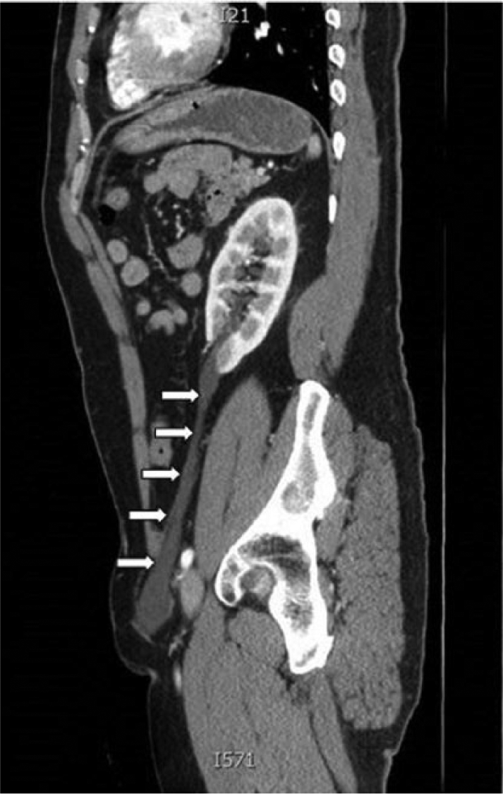
Ectopic ureter (white arrows) arising off lower pole of the left kidney and inserting into a scrotal structure.
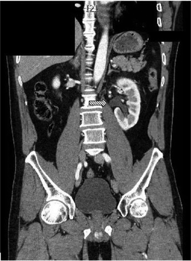
Interpolar ureter (hashed arrow) arising from interpolar pelvis of left kidney.
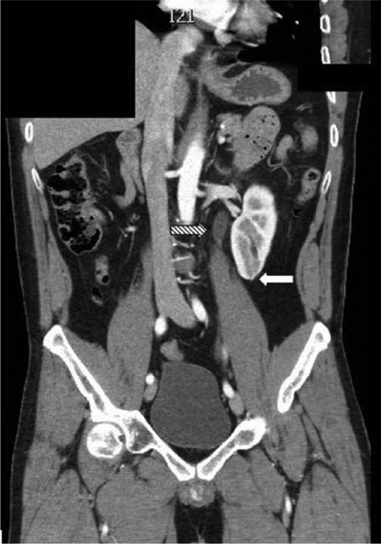
Left interpolar ureter (hashed arrow) and the separate ectopic ureter (white arrow) as it begins to come off the lower pole of the left kidney.
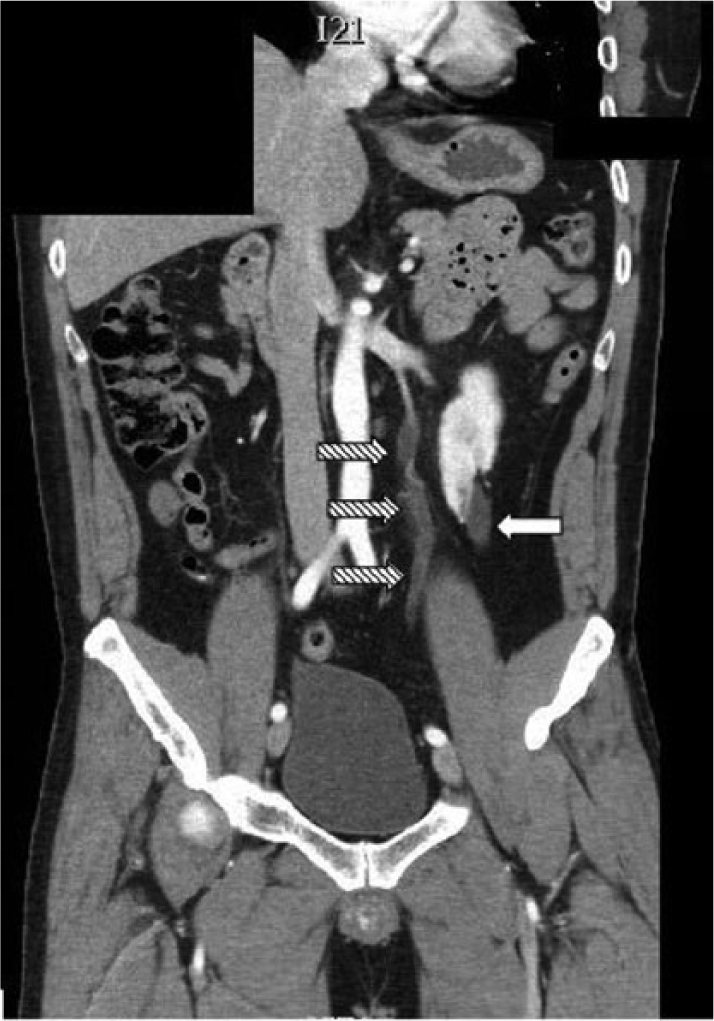
Left interpolar ureter (hashed arrow) and the separate ectopic ureter (white arrow).
A duplex collecting system is the most common renal abnormality. 1 Some individuals present with urinary tract infections, haematuria, and in rare female patients, continuous incontinence due to ureteral insertion distal to the urethral sphincter complex.2–5 Carl Weigert and Robert Meyer noted an invariable relationship between the ectopic and the orthotopic ureters, with the lower renal pole draining into the laterocranial ureteral orifice, and the upper pole draining into the mediocaudal ureteral orifice.1,4 Thus, if a duplex ureter does not insert into the bladder (and rather drains into a more caudal structure), it by rule should originate from the upper pole. Our case appears to be a rare example that violates the Weigert-Meyer law.2–5 It is the first case report to show this variant anatomy via CT reconstruction.
Footnotes
Acknowledgements
None.
Conflicting interests
The Author(s) declare(s) that there is no conflict of interest.
Funding
This research received no specific grant from any funding agency in the public, commercial, or not-for-profit sectors.
Ethical approval
Our institution does not require ethical approval for reporting individual cases or case series.
Informed consent
Verbal informed consent was obtained from the patient(s) for their anonymized information to be published in this article.
Guarantor
CSE.
Contributorship
KM and CSE researched literature and conceived the case report. KM wrote the first draft of the manuscript. KM and CSE reviewed and edited the manuscript and approved the final version of the manuscript.