
Abstract
Male circumcision is an extremely common urological procedure worldwide, with many variations in technique. Despite the large volume there is a low incidence of complications associated with circumcisions, with the majority being Clavien-Dindo I or II. In this study, we analyse the outcomes and complication rates associated with a continuous wound closure following a male circumcision.
Methods:
In a urology department from a single institution, 201 male circumcisions with a continuous wound closure were performed in a 4-year period. Outcomes were analysed retrospectively looking at postoperative complications and readmissions to hospital via our clinical portal.
Results:
No patients had complications that required admission or re-operation at our institution.
Conclusion:
No major post-operative complications were observed from our cohort. There were also no documented admissions back to our institution with wound healing complications. However, a limitation is that Clavien-Dindo I and II complications and treatment at general practitioner surgeries were not captured and may not accurately represent our complication rates quoted. Nevertheless, we can conclude from these data that closure for a circumcision using a continuous suture technique gives favourable outcomes with acceptable complication rates.
Level of evidence:
Not applicable for this multicentre audit.
Introduction
Male circumcision is an extremely common urological procedure that is frequently performed worldwide amongst many religions, races and cultures. 1 It has been reported that over a third of the male population are circumcised, with approximately 30,000 undergoing a circumcision every year in the United Kingdom.2,3 Despite the large volume, there is a low incidence of complications associated with circumcisions at 1–4%, with the majority being Clavien-Dindo I or II.4,5 Pain, bleeding and haematoma formation, wound infection and wound dehiscence are reported as most commonly seen in the emergency department. 4 Unsurprisingly, there are a plethora of approaches to performing a circumcision, along with variations in closure technique. 6 Interrupted sutures are most commonly used worldwide to approximate wound edges together after a circumcision. 4 Studies have shown that variations such as the sutureless technique using cyanoacrylate glue can provide better cosmesis, reduced post-operative pain as well as a reduction in operative time and cost.3,6 However, most of these studies only involved paediatric groups with very little information on adult cohorts. Reports on subcuticular closure have also quoted favourable outcomes; however, due to the thin prepucial skin, others have reported its closure inferior to more conventional techniques.7,8 It has been reported in the literature that closure of skin using a continuous suture may be superior due to its haemostatic properties and reduction of wound breakdown. 9 However, it is also reported that wound dehiscence may be higher with this technique. 9 Continuous suturing also extends to anatomy not involving skin such as bowel anastomoses.9,10 As far as we are aware there are no documented outcomes following continuous wound closure of a circumcision.
In this study, we analyse the indications, outcomes and complication rates associated with a continuous wound closure following a male circumcision. Indications for circumcision ranged from congenital or acquired phimosis, posthitis and balanitis xerotica obliterans (BXO). There were no exclusion criteria used on our cohort.
Materials and methods
In a urology department from a single institution, 201 male circumcisions with a continuous wound closure were performed in men aged 17–84 from June 2014 to November 2018. The circumcisions were performed under a single consultant in this period, or by direct supervision under him.
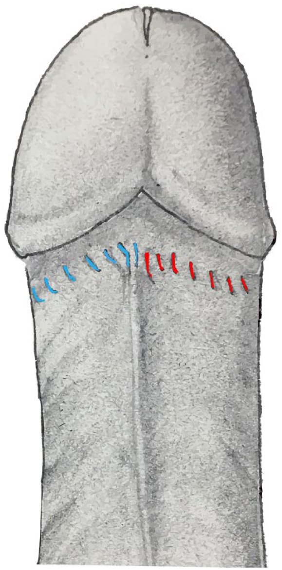
Patients are placed in the supine position and initially given a general anaesthetic, or a local penile and ring block using a 50/50 ratio of 1% lignocaine and 0.5% levobupivacaine. The skin is then prepped using chlorhexidine, with the borders draped to sterilize the remaining operating field. An outer cuff sleeve incision is made circumferentially proximal to the corona. A dorsal slit using McIndoe scissors is then made to the outer cuff incision line following a haemostatic clamp using artery forceps (Figure 1). The foreskin is then retracted and the glans thoroughly cleaned using chlorhexidine. An inner cuff incision is then made to the prepuce, approximately 3–4mm distal to the coronal ridge. The foreskin is subsequently removed using McIndoe scissors by joining the outer cuff incision to the inner cuff incision (Figure 2) and is sent off for histology. If necessary, the frenulum is reconstructed using a continuous suture with vicryl. Meticulous haemostasis is then performed using bipolar diathermy forceps until no further bleeding is observed. The foreskin is then sutured to the prepucial skin proximal to the corona in a continuous fashion (Figure 3). Knots are tied in the 12 to 6 (blue) and 6 to 12 o’clock (red) position using vicryl rapide sutures (Figures 4 and 5). The wound is finally dressed with Jelonet and blue gauze.
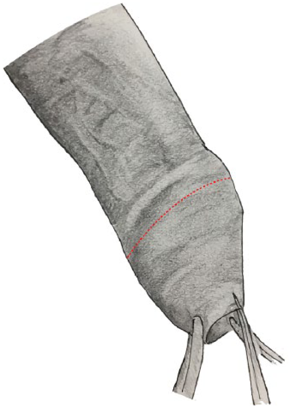
Commencement of dorsal slit to outer cuff incision.
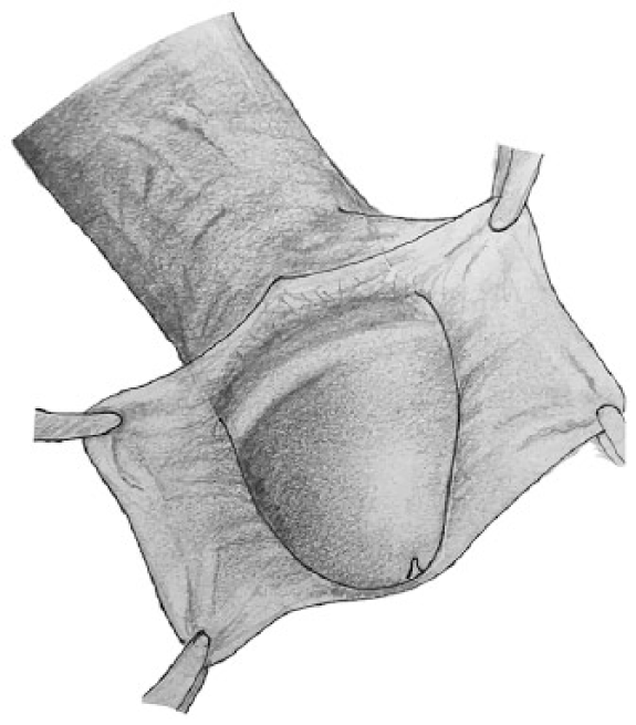
Excision of foreskin.
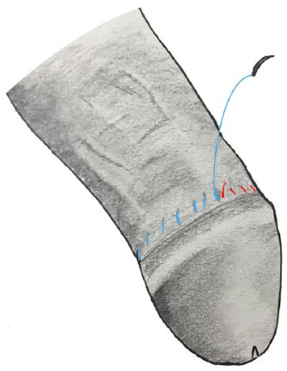
Continuous suture used to oppose prepucial skin to coronal mucosa.
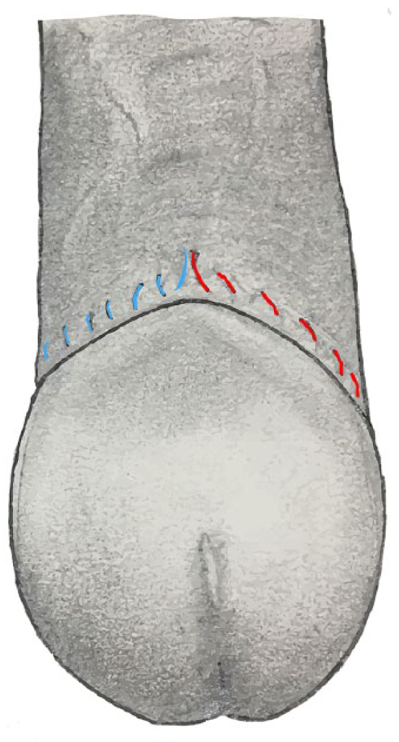
Dorsal view of circumcised penis.
Ventral view of circumcised penis.
Outcomes were analysed retrospectively by looking at operation notes and our online clinical portal. Readmissions due to post-operative complications and histology were subsequently analysed.
Results
The mean age of our cohort was 43 years (17–84). Men who underwent a general anaesthetic had a median performance score of 1.5 (1–3) as per the American Society of Anaesthesiologists (ASA) classification system, with 11.5% of the cohort having the procedure under local. A single consultant had performed 79% of the circumcisions, with the rest performed under his direct supervision. Phimosis secondary to clinical BXO was the most common indication for circumcision at 88%. The remaining indications included paraphimosis (1%), retention (1%), posthitis (2%), dyspareunia (4%) and chronic balanitis (4%). All patients underwent a circumcision, with 28% of them requiring a frenuloplasty. Penile biopsies were performed in two patients and a bilateral vasectomy was additionally performed in one man. The average operation time from anaesthetic induction to closure was 53 minutes (29–84).
Histology had revealed that 75% of specimens had evidence of BXO or lichenoid inflammation and 18% showed signs of chronic balanitis. The remainder of the histology had shown acute inflammation (4%), posthitis (2%), psoriasis in one patient and human papillomavirus-related dysplasia in another.
No patients were admitted with complications that required admission or re-operation.
Conclusion
There were no major post-operative complications (Clavien-Dindo I and II) observed from our cohort. There were also no documented admissions back to our institution with wound-healing complications. However, a limitation to this study is that presentations and treatment at general practitioner surgeries was not captured and therefore may not accurately represent our complication rates quoted. Nevertheless, we can conclude from these data that closure for a circumcision using a continuous suture technique can give favourable outcomes that do not require hospital admission or re-operation (Clavien-Dindo III and IV). Furthermore, it is a suitable method for those who are less experienced, as well as surgeons who are well practised with performing circumcisions.
Footnotes
Acknowledgements
We would like to thank Prof. H Leung for his assistance and guidance in this research.
Conflicting Interests
The authors declare that there is no conflict of interest.
Funding
This research received no specific grant from any funding agency in the public, commercial, or not-for-profit sectors.
Ethical approval
Not applicable.
Informed consent
Not applicable.
Guarantor
HF.
Contibutorship
MR and HF conceived the review. MR researched the literature. HF identified the cases and MR wrote the first draft of the manuscript. MR and HF reviewed and edited the manuscript and approved the final version of the manuscript. SA provided the illustrations for the manuscript.