
Abstract
Mild brain injuries are frequent in athletes engaging in collision sports and have been linked to a range of long-term neurological abnormalities. There is a need to identify how these potential abnormalities manifest using objective measures; determine whether changes are due to concussive and/or sub-concussive injuries; and examine how biological sex affects outcomes. This study investigated cognitive, cellular, and molecular biomarkers in male and female amateur Australian footballers (i.e. Australia’s most participated collision sport). 95 Australian footballers (69 males, 26 females), both with and without a history of concussion, as well as 49 control athletes (28 males, 21 females) with no history of brain trauma or participation in collision sports were recruited to the study. Ocular motor assessment was used to examine cognitive function. Telomere length, a biomarker of cellular senescence and neurological health, was examined in saliva. Serum levels of tau, phosphorylated tau, neurofilament light chain, and 4-hydroxynonenal were used as markers to assess axonal injury and oxidative stress. Australian footballers had reduced telomere length (p = 0.031) and increased serum protein levels of 4-hydroxynonenal (p = 0.001), tau (p = 0.007), and phosphorylated tau (p = 0.036). These findings were independent of concussion history and sex. No significant ocular motor differences were found. Taken together, these findings suggest that engagement in collision sports, regardless of sex or a history of concussion, is associated with shortened telomeres, axonal injury, and oxidative stress. These saliva- and serum-based biomarkers may be useful to monitor neurological injury in collision sport athletes.
Keywords
Introduction
Collision sports often results in participants sustaining mild brain traumas, and this has been linked to long-term neurological consequences.1–3 However, there is still a lack of understanding concerning how these changes manifest; whether they are specific to those who experience clinical concussions or if sub-concussive impacts also contribute; and whether men and women are differentially affected.1,2 Furthermore, considering the heterogeneous and subjective nature of the neurological symptomatologies reported by collision sport athletes, the identification of objective biomarkers that are sensitive to the effects of mild brain traumas has become a research priority in this field.4,5
The ocular motor (OM) system is a highly ramified neural network involved in the generation of eye movements; the most common type of eye movement is saccades, a quick ballistic movement from one point to another.6,7 While the primary nuclei for saccade generation are located in the brainstem (e.g. pontine premotor nuclei for horizontal saccades and midbrain premotor nuclei for vertical saccades), 8 the higher order saccades (i.e. cognitive saccades) – which are the primary focus of this paper – engage widespread cortical networks involved with cognitive processing pertinent to the situational demands and/or the intention of the individual.7,9 For example, a saccade to a visually salient target (visually guided) is primarily driven by a simple neural network, involving the midbrain superior colliculus, brainstem saccade nuclei, with minimal cognitive involvement (i.e., attention). 7 Alternatively, the introduction of a cognitive demand, such as the requirement to inhibit an automatic saccade generated by a suddenly appearing target in favour of a saccade to the diametrically opposite location (antisaccades), 10 or performance of a saccade to a remembered (memory guided saccades), 11 engage frontoparietal networks associated with inhibitory control, executive control function, and visuo-spatial memory.6,7 Given this disseminated network and implication of cognitive processing, it is not surprising that a variety of saccadic parameters have been shown to be sensitive to diffuse brain dysfunction following mild brain injuries such as concussion; most notably, latency, erroneously performed saccades, and visual spatial accuracy.6,7,12 We previously found OM saccade abnormalities in a cohort of male Australian footballers with a self-reported history of concussion (HOC) 13 ; however, whether these changes are dependent on an HOC, or differ between sexes is unclear. Therefore, in the present study we investigated OM performance on visually guided, antisaccade, and memory guided OM saccade tasks.
Telomeres are nucleo-proteomic complexes that consist of a repeating sequence of non-coding DNA that protect the ends of linear eukaryotic chromosomes from damage. 14 Shortened telomere length is associated with aging and neurodegenerative changes, and may represent a biomarker of neurological health. 15 We have previously found reduced telomere length in rats with mild brain injuries 16 ; however, whether this occurs in humans with mild brain injuries is unknown.
Circulating protein biomarkers have the potential to detect changes that reflect a range of neuropathophysiological processes that have been implicated in mild brain trauma. 17 For example, serum levels of tau and neurofilament light (NF-L), which are considered markers of axonal damage, have been found to be increased acutely after sports-related concussion. 18 In other studies, rats given mild brain traumas were found to have chronically elevated plasma protein levels of 4-hydroxynoneal (4-HNE), a marker for oxidative stress. 19 However, few studies have examined blood protein biomarkers in the chronic stages of mild brain trauma, and whether these outcomes are modified by biological sex, in humans.
The primary aim of this study was to investigate OM performance, telomere length, and serum protein biomarkers in male and female Australian footballers (i.e. Australia’s most participated collision sport) 15 in comparison to control athletes with no history of neurotrauma or participation in collision sports. Secondary to this, to elucidate the potential effects of sub-concussive versus concussive injuries, we also investigated whether there were any differences on these measures between Australian footballers with and without a HOC, sub-concussive impacts of engaging in collision sports and whether demonstrated abnormalities differed between footballers with and without a history of concussion. As both the men’s and women’s Australian football leagues follow similar full collision rules, another secondary aim of the study was to investigate potential sex differences within the same collision sport. Overall, we hypothesized that OM performance, telomere length, and serum protein biomarkers would differ between Australian footballers and controls, and that sex and HOC might modify these outcomes.
Methods
Participants
A total of 95 (male = 69, female = 26) amateur Australian football players were recruited from the Victorian Amateur Football Association to participate in this study. A control group of 49 athletes (male = 28 and female = 21), with no history of brain trauma or engagement in collision sports, was also recruited from local amateur sporting teams (i.e., basketball, tennis, cricket, track and field). Individuals with a history of neurosurgery or major psychiatric disturbances were excluded. To minimize any confounding effects of recent concussive and sub-concussive injuries, Australian football players who had sustained a concussion in the past 6 months were not enrolled into the study, and testing was performed during preseason. Study procedures were approved by the Melbourne Health Human Ethics institutional review board, were in accordance with The Code of Ethics of the World Medical Association (Declaration of Helsinki) for experiments involving humans, and all participants provided written informed consent prior to the study.
Clinical interview
A general medical history questionnaire was administered to each participant pertaining to demographics, HOC, sporting history, education history, as well as any learning difficulties. The Beck Depression Inventory (BDI) was used to measure self-reported depression and The National Adult Reading Task (NART) was used to measure premorbid intelligence. 13 The Sports Concussion Assessment Tool (SCAT3) was also incorporated as part of the clinical interview.20,21
OM assessment
62/95 Australian footballers (males = 37, females = 25) and 39/49 control participants (males = 18, females = 21) completed the OM assessment. The missing data from some participants was due to a combination of participants refusing OM testing and technical issues with OM equipment that prevented some of the participants from completing the assessment. The horizontal displacement of both eyes (i.e. saccades) was recorded using an Eyelink 1000 dark pupil, video-oculography system (SR-Research Ltd., Mississauga, Ontario, Canada). This is a high-resolution (noise limited at < 0.01 degree) and high-acquisition-rate (1000 Hz) system. Participants were seated in a dark room with their head supported by a chin rest 750 mm from a 60-Hz LCD monitor (resolution = 2048x1154). Task stimuli consisted of red and green crosses (0.5° visual angle) of equal luminance displayed on a black background. All task stimuli were generated using Experiment Builder (version 1.6.121). A 5-point calibration sequence was performed at the beginning of each task with in-vivo calibrations included to ensure the accuracy of the initial calibration. 13
The visually guided saccade task requires the performance of an eye movement to a suddenly appearing visual target. Participants fixated on a central green cross for 1250–1750ms, and performed a saccade to a suddenly appearing peripheral target (1250–1750ms), as it randomly stepped horizontally 5° or 10°, to the left or right of centre (24 trials). 13
The antisaccade task requires the inhibition of an automatically generated response elicited by the presentation of a salient peripheral target in favour of a saccade to the same location in the contralateral hemispace. Participants fixated on a central green cross for 1250–1750ms which disappeared concomitant to the appearance of a peripheral target at either 5° or 10°, left or right of centre. Participants were instructed to perform a saccade to the contralateral location (48 trials). 13
The memory guided saccade task assesses the ability to perform a saccade to a remembered location. Participants fixated on a central green cross, concurrently, a peripheral red cross briefly appeared (500 ms) at either 5° or 10°, left or right of centre. Participants were instructed not to look at the peripheral cross directly, but to remember its spatial location. Following the extinction of the central green cross (1500–2500ms), participants performed a saccade to the remembered spatial location of the previously illuminated red cross. 22
OM analysis
Monocular analysis was performed for OM output using a customised MATLAB program. For all OM tasks, saccade latency was calculated as the temporal difference between trial onset and saccade onset using a velocity criterion of 30° per second.
For the antisaccade and memory guided tasks, visual spatial accuracy was measured by calculating the ratio between the amplitude of the saccade at its final resting place (final eye position, FEP) and the target location, and is referred henceforth as FEP gain: 1 = on target, >1 = overshot target location, <1 = undershot target location.
For the antisaccade task, an error was defined as a saccade towards the peripheral target. For the memory guided saccade task, an error was defined as a saccade towards the peripheral red cross, or towards the same location within 100 ms of trial onset.13,22
Trials were removed from analysis of saccade latency and FEP gain where: 1) the task was not completed in accordance with task (error as explained below); 2) fixation was not maintained within 1.5̊° of the central target; 3) a blink occurred around trial onset that was thought to affect saccade onset; 4) no response was made within the trial period, and 5) a saccade made within 100 ms of target onset.
Telomere length analysis
A 5 mL saliva sample was collected from 90/95 Australian footballers (males = 65, females = 25) and 42/49 control participants (males = 24, females = 18) using Oragene DNA OG-500 collection tubes (DNA Genotek Inc., Ottawa, ON). Some participants were excluded due to discordance with the required procedure (e.g. ate or drank 30 minutes prior to testing) or refusal to partake. Genomic DNA was extracted from the saliva sample using QIAamp DNA Mini kit, according to the manufacturer’s protocol. The DNA quality and concentration were measured using the NanoDrop 2000 (Thermo Fisher Scientific, Waltham, MA). Samples were diluted to a concentration of 20 ng/μL and stored at –20°C in preparation for telomere length analysis. Primers were designed for telomeres and the single copy 36B4 gene.16,23,24 Each PCR reaction was run in duplicate and contained 1 μL of gDNA and a Master Mix of 1 x SYBR Green FastMix with Rox, DEPC-treated nuclease-free water, together with forward and reverse primers to a total volume of 20 μL per well. Two plates were performed for each DNA sample; using telomere then 36B4 primers. No-template control wells were also present on the plate in order to ensure reagents were not contaminated. RT-qPCR was performed to determine telomere length by comparing telomere to single-copy 36B4 gene ratio (T/S). Accordingly, a T/S of 1 indicates that the sample DNA is identical to the reference DNA, concerning telomere repeat number and single-copy gene number. The T/S ratio has been calculated to be approximately [2Ct(telomere)/2Ct(36B4)]−1 = −2−ΔCt. The linear regression equation, y = 1910.5x + 4157 (where y = telomere length and x = −2−ΔCt) was used to determine the telomere length of each sample.16,23,24
Serum protein markers
10 mL of whole blood was collected using standard phlebotomy procedures into a BD Vacutainer® SST™ II Advance tube from 51/95 Australian footballers (males = 43, females = 8) and 35/49 control subjects (males = 24, females = 11). The missing samples were due to participants refusing to donate blood and discordance from procedures. The tube was inverted several times and allowed to clot at room temperature for 30 minutes prior to being centrifuged at 1500 g for 10 minutes to separate serum. Serum was then transferred into 0.5 mL aliquots, flash-frozen and stored at -80°C.
Sample preparation, printing, scanning, and data analysis for reverse phase protein microarrays (RPPM) were performed as described previously.19,25 Serum protein levels of tau, phosphorylated tau (pTau) and NF-L (i.e. markers of axonal damage),17,26 as well as 4-HNE (i.e. a marker of oxidative stress),19,25,27,28 were analysed. Primary antibodies were diluted with 1x Azure Protein-free buffer in 1.5 mL Eppendorf Tubes to the desired concentration making a final volume of 250 μL with the following dilutions: 4-HNE (1:100; EMD Millipore, Prod # 393207), tau (1:100; Cell Signalling, Prod # 4019S), pTau (1:100; Sigma, Prod # SAB4504563), and NF-L (1:100; Protein Tech, Prod # 12998–1-AP). Primary antibodies were pipetted onto the slides, incubated overnight (8–12 hours) at 4 °C, and washed. Secondary antibodies were diluted in 1x Azure Protein-free buffer (1:10,000 dilution) as follows: goat anti-rabbit IgG (H + L) Superclonal™ Secondary Antibody, Alexa Fluor 790 (Thermo Fisher, Prod # A27041) and goat anti-mouse IgG (H + L) Superclonal™ Secondary Antibody, Alexa Fluor 790 (Thermo Fisher, Prod # A28182). Slides were incubated with their respective secondary antibody solutions at room temperature for 1 hour, washed, dried, and then loaded into the tray of the Innopsys InnoScan 710-IR scanner for XDR (extended dynamic range) signal acquisition at 785 nm. Scanner fluorescence data were imported into a Microsoft Excel-based bioinformatics program. After correcting for local background noise, points indiscernible from background were excluded (SNR <2, Net Fluorescence <10) and secondary-only signals were subtracted from corresponding slides. Net intensity vs. dilution was plotted on a log2-log2 scale; each local block of samples is fit individually, using inter-quartile range to exclude obvious outliers. The slope of the linear portion of the logistic curve was calculated and the line extrapolated back to zero (i.e. the y-intercept), assessing the amount of protein expressed.
Statistical analysis
SPSS 23.0 (IBM Corp., Armonk, USA) was used for statistical analyses. Two-way ANOVA was used for the statistical analysis of all variables. The NART was used as a covariate due to sex differences revealed on this variable (see Table 1). The primary analysis compared OM, telomere length, and serum biomarker measures between Australian footballers (i.e. collision athletes) and control non-collision athletes, with collision sport involvement and sex as the between-subject factors. To investigate the influence of an HOC on the above outcomes, a secondary analysis was performed to compare the difference between male and female Australian footballers both with and without a HOC. Last, receiver operating characteristic (ROC) and leave-one-out discriminant analyses (LDA) were conducted to determine the predictive utility of the significant biomarker measures either independently (ROC) or combined (LDA). Sample size calculations were based on our previous studies that have applied OM, telomere length, and serum protein measures in the context of mild brain trauma.13,16,19 Statistical significance was set at p < 0.05.
Participant demographics.

Note: The average age, NART score, and BDI score for non-collision sport controls and Australian rule footballers.
aSignificant difference in NART score between males and females irrespective of collision sport involvement. Results are presented as Mean ± SEM.
Results
Clinical history
Table 1 summarizes participant demographic information, including the average age, NART, and BDI score. There were no significant differences in any of these variables (p > 0.05), except on the NART (F1,140 = 6.57, p = 0.011), with males having a significantly higher NART score than females irrespective of collision sport involvement. The NART score was therefore used as a covariate for subsequent analyses.
OM performance in collision sport athletes
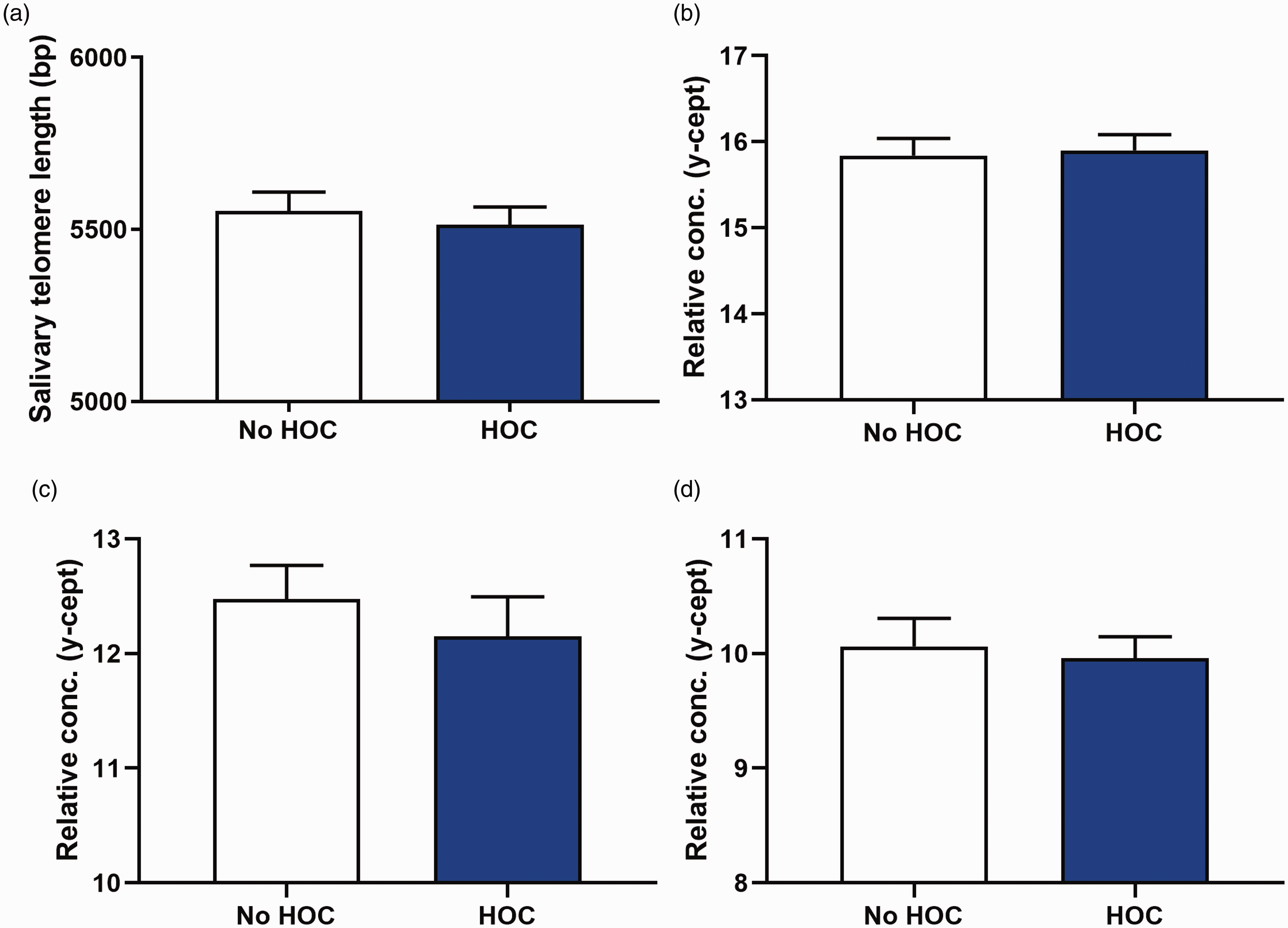
OM findings are presented in Table 2. There was no significant difference between Australian footballers and controls on any ocular motor variable. For latency, a significant main effect of sex was found on the visually guided (F1, 93 = 4.386, p = 0.039), antisaccade (F1,93 = 5.560, p = 0.02), and memory guided saccade tasks (F1, 88 = 16.119, p = 0.001), with males performing significantly shorter latencies than females on each task.
OM measures for controls and Australian footballers.
Note: OM outcome measures for the visually guided, antisaccade and memory guided saccade task.
aMales performed significantly shorter latencies than females irrespective of collision sports involvement. The NART score was used as a covariate. Results are presented as Mean ± SEM.
Shorter telomere length in collision sport athletes
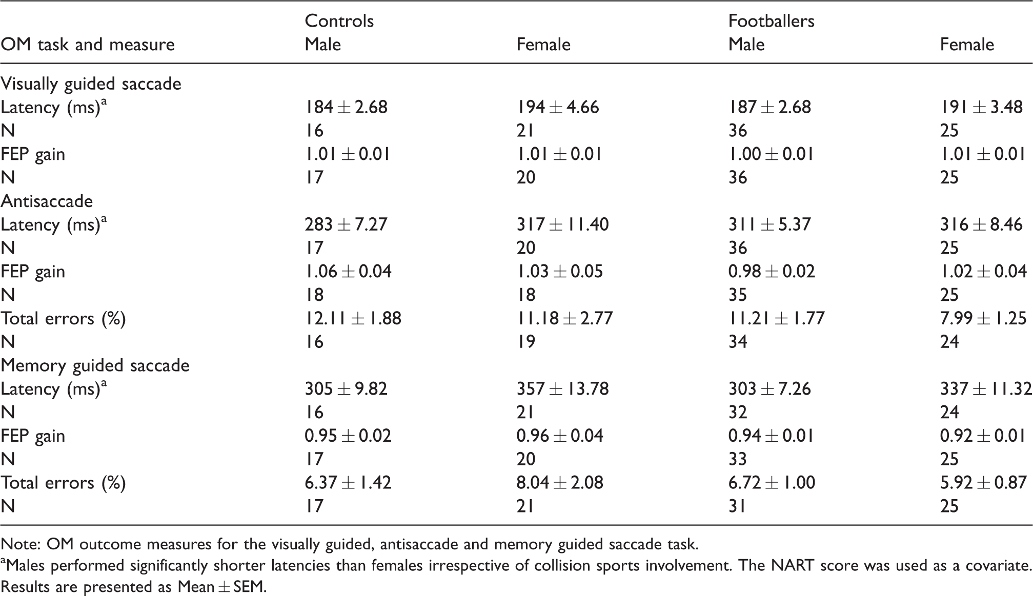
A significant main effect for collision sport was found on the measure of salivary telomere length (F1, 127 = 4.749, p = 0.031, Figure 1(a)), with Australian footballers having shorter telomeres than the non-collision sport athlete group. There were no significant findings related to sex (p > 0.05).
Telomere length and serum protein biomarkers in non-collision control athletes and Australian footballers. (a) Australian footballers had significantly shorter telomere length compared to non-collision sport control athletes. Australian footballers also had significantly increased serum protein levels of (b) 4-HNE, (c) tau, and (d) pTau. Bar graphs represent group Mean ± SEM. * = p < 0.05.
Serum protein levels of 4-HNE, tau, and pTau are increased in collision sport athletes
There were significant main effects found for collision sport participation on serum protein levels of 4-HNE (F1, 81 = 11.495, p = 0.001, Figure 1(b)), tau (F1, 81 = 7.586, p = 0.007, Figure 1(c)) and pTau (F1, 81 = 4.523, p = 0.036, Figure 1(d)), with Australian footballers having significantly increased levels. No significant differences related to NF-L or sex were found (p > 0.05).
OM performance, shortened telomeres, and serum protein findings are not dependent on an HOC
A secondary analysis was conducted to investigate whether OM performance, telomere length, and serum protein levels differed between Australian footballers with and without an HOC. Table 3 summarises the demographics and HOC of the Australian footballers. Australian footballers with an HOC had significantly greater BDI scores (F1, 89 = 5.211, p = 0.025) and more years of collision sport participation (F1, 91 = 6.066, p = 0.015) than footballers with no HOC. Male footballers were also found to have significantly higher NART scores (F1, 91 = 7.880, p = 0.006) and years of collision sport participation (F1, 90 = 13.8966, p = 0.001) compared to female footballers irrespective of an HOC.
Australian footballer demographics and HOC.

Note: The average age, NART score, BDI score and years of collision sport participation for all Australian footballers. The number of previous concussions and time since last concussion is reported for players with an HOC.
aMale footballers had a significantly greater NART and years of collision sport participation compared to female footballers irrespective of an HOC (p < 0.05). Results are presented as Mean ± SEM.
bFootballers with a history of concussion had a significantly greater BDI and years of collision sport participation than footballers without a history of concussion (p < 0.05).
There were no significant findings found in relation to HOC on any OM measures (p > 0.05, Table 4). There was however a significant main effect of sex, with male footballers having shorter memory guided saccade latency (F1, 51 = 4.978, p = 0.03) than female footballers irrespective of an HOC.
OM measure for Australian footballers with and without an HOC.

Note: OM outcome measures for the visually guided, antisaccade, and memory guided saccade task for Australian footballers with and without an HOC. No effect of an HOC was revealed for the FEP gain of the memory guided saccade task. The NART score was used as a covariate. Results are presented as Mean ± SEM.
aMales exhibited significantly shorter latencies on the memory guided saccade task than females.
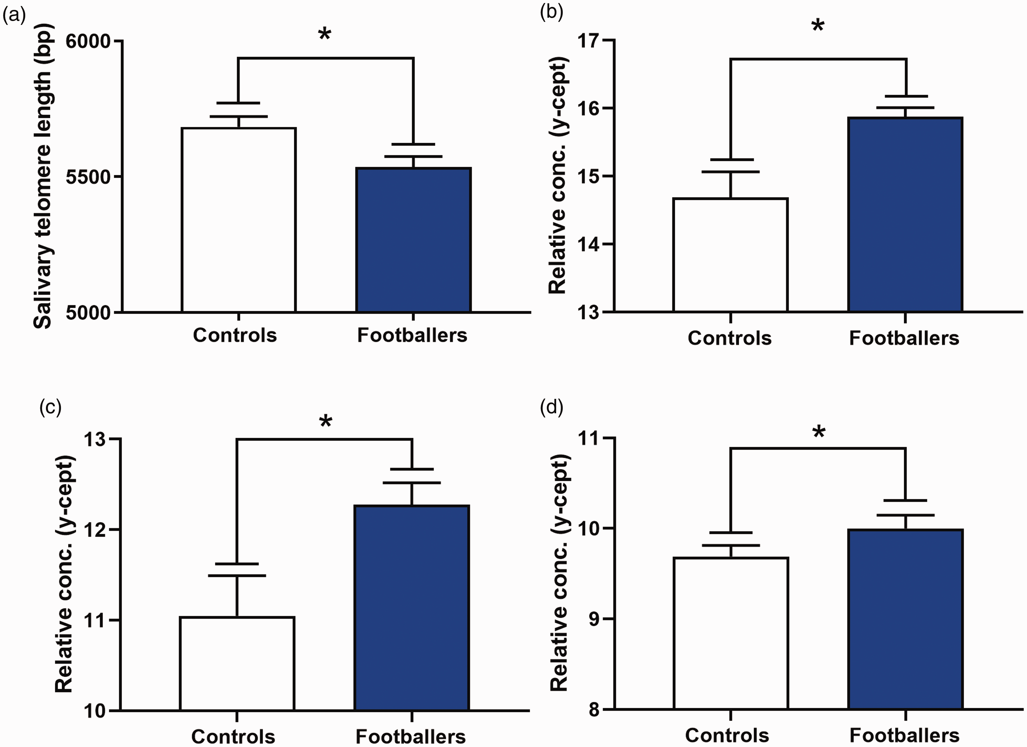
There were also no statistically significant findings related to HOC found for telomere length or the serum protein markers (p > 0.05, Figure 2). However, there was a statistically significant effect of sex on serum levels of 4-HNE (F1, 46 = 4.554 p = 0.038), with females having increased levels compared to males.
Shortened telomere length and serum protein levels among Australian footballers are not dependent on an HOC. There was no significant main effect of HOC on the measure of A) telomere length, or serum protein levels of (b) 4-HNE, (c) tau, and (d) pTau in Australian footballers. Bars represent group Mean ± SEM.
Ability of OM, telomere length, and serum protein measures to discriminate collision athletes
ROC analysis and LDA were conducted to examine whether the four biomarker measures found to be significantly different between Australian footballers and controls (i.e., telomere length, tau, pTau, and 4-HNE) were predictive of collision sport participation (Table 5).
Area under the curve (AUC) for ROC analysis in Australian footballers versus control athletes.

For the participants who had measures for all four of the biomarkers (n = 80; 50 Australian footballers, 30 controls), LDA correctly differentiated Australian footballers from controls in 72.5% of cases (Table 6).
Leave-one-out discriminant analysis showing ability of combined biomarkers to discriminate Australian footballers from control athletes.

Discussion
This study used a multimodal approach, including assessment of OM function, telomere length, and serum protein biomarkers, in male and female Australian football players to provide insight into the neurological health of collision sport athletes, and whether abnormalities are dependent on an HOC or biological sex. Relative to the non-collision control athletes, Australian footballers had significantly shorter telomeres, and increased circulating protein levels of markers for axonal injury and oxidative stress irrespective of sex or an HOC. Both female and male Australian footballers exhibited similar ocular motor performance to control athletes.
Telomere length as a marker of neurological health in collision sport athletes
Australian football players had significantly shorter telomeres than the non-collision sport athlete controls. This occurred independent of an HOC or sex, which again suggests cumulative damages associated with both concussive and sub-concussive injuries occurs in both male and female athletes. Whilst our laboratory has previously found shortened telomeres in rodents given mild brain injuries,16,24 this is the first evidence of shortened telomere length in humans in this context.
Reduction in telomere length is implicated in aging and neurodegenerative diseases (e.g. Alzheimer’s), 14 and may be an indicator of general neurological health as well as cellular biological age (as opposed to chronological age). 29 A recent systematic review suggested that the attrition of telomere length is 24.7 bp/year. 30 Our study identified a difference of ∼150 bp between Australian footballers and controls. Based on these figures, our findings suggest that Australian footballers in this study had an accelerated biological age of 6 years. This is of course speculative and should be interpreted with caution, particularly as the relationship between peripheral and central telomere length is not well understood.30,31 However, we previously identified a high correlation between telomere length quantified from an ear notch sample and brain tissue in rodents, 24 and similarly in humans, peripheral leukocyte telomere length has been correlated with cortical thickness, 32 both suggesting that peripheral telomere shortening may reflect central neurological health.
It is also important to consider that factors in addition to brain injury may influence telomere length. Aging, 14 stress, 33 a history of smoking, 34 alcohol consumption, 35 and poor sleep 36 have been associated with shortened telomeres, whereas exercise may attenuate these deficits. 37 In this study, the Australian footballer and control groups were comparable with respect to the other potential modifying factors that were measured (e.g. age and exercise). Furthermore, our previous rodent studies found shortened telomere length after a mild brain injury despite frequent exercise. 38 While future studies are required, our findings provide novel evidence of telomere shortening in male and female collision sport athletes.
Increased circulating markers of axonal injury and oxidative stress in collision sport athletes
Australian footballers had elevated serum protein levels of 4-HNE, a marker of oxidative stress,19,25,27,28 as well as tau and pTau, markers of axonal injury,17,26 when compared to the non-collision control group. As axonal injury and oxidative stress are considered common neuropathophysiological consequences of brain injury,39–41 these findings are somewhat expected. There is also evidence that oxidative stress may contribute to axonal injury after mild brain injury,40,41 which aligns with our finding of concomitant increases in markers for both of these processes. However, our finding that the elevated serum biomarkers in the collision sport athletes were not dependent on sex or HOC are novel, and further support the notion that sub-concussive impacts may have neurological consequences in both men and women.
Nature of OM findings
There were no differences in OM performance between Australian footballers and control athletes. This is in contrast to our previous work in which we found that male Australian footballers with a HOC had significantly shorter visually guided saccade latencies, reduced cognitive flexibility when required to switch from performing an antisaccade to a visually guided saccade, and increased antisaccade errors. 13 However, it is important to note these previous findings were only found under conditions of increased cognitive load. Specifically our previous study utilised a complex switch design (interleaved visually guided saccades and antisaccades) as opposed to the simpler block design (all same trials i.e, all antisaccades or all visually guided saccades). The OM tasks used in this study were the simpler block design visually guided and antisaccades, and may not have been sensitive enough to detect differences between groups. Furthermore, whilst the memory guided saccade task imposes an increased cognitive demand (i.e. to remember the target location) to potentially discriminate more subtle cognitive deficits, the previous utility of this task has only been demonstrated at acute or subacute timepoints post-concussion.42,43 Taken together, we recommend the use of cognitively complex saccade tasks when investigating the chronic consequences of mild brain injury on OM function.
Limitations and future directions
The mechanisms underlying the telomere shortening and serum protein findings, and whether any of these changes are linked, require further investigation. There are inherent challenges studying their mechanistic underpinnings, and whether they are brain-specific, in humans due to limited access to brain tissue and cerebrospinal fluid. Future studies could employ animal models of mild brain injuries as they provide direct access to paired brain and peripheral tissues.44,45
Our findings should be viewed in light of the self-reported measure of HOC used in our study. Moreover, a history of engaging in collision sports acts only as a surrogate measure of exposure to sports-related mild brain injury. Therefore, utilizing new clinical modalities such as mouth guard embedded telemetry to record impact biomechanics over an athletic season may be a means to quantify and delineate the mediating effects of sub-concussion and concussion. Alternatively, animal model studies that are able to investigate the effects of brain impacts at different forces in a highly controlled environment would be useful in determining the neurological burden of sub-concussive impacts.44,46–48
Despite finding statistically significant differences between collision versus non-collision athletes across the modalities examined in this study, the area under the curve (AUC) and LDA findings (e.g. all AUCs < 0.80) suggest that these measures may have limited clinical utility as classifiers in the context examined in this study (i.e. differentiating young adult amateur collision sport athletes from young adult amateur non-collision sport athletes). However, considering the young age of this cohort it is possible that the abnormalities in the Australian rule’s footballers may become more pronounced with time, or that the present changes predict the later manifestation of other neurological consequences. As such, longitudinal studies following large cohorts of collision sport athletes are required to determine the utility of OM, telomere, and serum protein biomarkers in a range of clinical settings.
Conclusion
This study found that amateur Australian footballers had shorter telomeres and increased levels of serum protein markers reflective of axonal injury and oxidative stress relative to a non-collision athlete control group. These abnormalities occurred independent of an HOC and sex which supports the emerging literature that collision sport athletes may be susceptible to neurological injury from not only concussive injury, but also from exposure to sub-concussive mild brain injuries. Although further research required, these findings have important implications and support the continued investigation of these methods as objective markers of neurological injury in collision sport athletes.
Footnotes
Acknowledgements
The authors thank Jack Germaine, Rafael Smith, Joshua Kimpton, and Wilfred Speagle for their contributions to this study. This study was funded by a grant and fellowship from the Australian National Health and Medical Research Council to SRS, and a grant from the Canadian Institute of Health Research to RM and SRS.
Authors’ contributions
SRS, RM, JF, DVD, OW, DKW, TOB, and LA conceptualized and designed the study. GFS, MC, WTO, JE, SS, DC, MS, RDB, SJM, JM, RA, IL, and SRS acquired and analyzed the data. GFS, MC, DVD, JF, RM, and SRS drafted significant portions of the manuscript. All authors contributed to editing the manuscript
Declaration of conflicting interests
The author(s) declared no potential conflicts of interest with respect to the research, authorship, and/or publication of this article.
Funding
The author(s) received no financial support for the research, authorship, and/or publication of this article.