






Abstract
A novel temporary aortopulmonary shunt, constructed between the aorta and main pulmonary artery with flexible cannulas, was used to facilitate right ventricular outflow tract reconstruction in one neonate and creation of a central aortopulmonary shunt in a second neonate. Although cardiopulmonary bypass is readily available in Turkey, the strategy described in the case report may prove especially useful in developing nations with limited access to cardiopulmonary bypass (CPB).
Keywords
Introduction
Neonatal operations to create an aortopulmonary shunt and perform right ventricular outflow tract (RVOT) reconstruction are frequently performed on cardiopulmonary bypass (CPB) due to perioperative progressive hypoxia. We report two patients in whom an RVOT reconstruction (patient 1) and an aortopulmonary shunt (patient 2) were performed without CPB, with stable hemodynamics facilitated via a provisional shunt constructed between the aorta and main pulmonary artery with flexible cannulas.
Case 1
A 20-day-old neonate (3.5 kg) was found to have RVOT obstruction, including infundibular and pulmonary annular hypoplasia and hypoplastic right and left pulmonary arteries, a thin patent ductus arteriosus (PDA), an atrial septal defect (ASD), and a right aortic arch, on echocardiography. Respiratory rate was 60 breaths per minute, oxygen saturation on pulse oximetry was 65%, hemoglobin (Hb) was 17 g/dL, and pH was 7.3. The neonate was taken into the operation theater with plans for a pulmonary valvotomy via transannular incision and pericardial patch reconstruction. Median sternotomy was performed and the pericardium was opened. A side-biting clamp was placed on the pulmonary annulus. The oxygen saturation decreased to 40% and the patient became bradycardic. The side clamp was removed, and cannulation purse string sutures were placed in the ascending aorta, the distal main pulmonary artery on the left side of the pulmonary bifurcation, and right atrial appendage. (The purse string suture was placed in the right atrial appendage, a precaution in case the patient needed to be placed on CPB emergently. Also, this purse string suture in the right atrial appendage can be used to retract gently the right atrial appendage and aid in exposure of the right pulmonary artery when this temporary aortopulmonary shunt is used to facilitate creation of a surgical aortopulmonary shunt to the right pulmonary artery.) After systemic heparinization with 300 U/kg of heparin, the ascending aorta and the distal main pulmonary artery were cannulated with 8F flexible aortic cannulas. Both cannulas connected to each other with connectors and tubing, and a provisional aortopulmonary shunt system was established. After the opening of the temporary shunt, arterial oxygen saturation was 70%, blood pressure was 75/35 mm Hg, and pulse rate 150 beats/min. The side-biting clamp was again placed onto the infundibulum (Figure 1 ). The pulmonary valvular tissue was excised via a transannular incision and transannular reconstruction was performed with a pericardial patch. The hemodynamic status was stable during the RVOT reconstruction. Then, the clamp was then removed. The provisional shunt was clamped and the arterial oxygen saturation became 90%. Intraoperative transesophageal echocardiography documented satisfactory patency of the RVOT. The sternum was closed after removal of provisional shunt cannulas. The neonate was extubated on first day after surgery and was discharged on seventh day after surgery. Echocardiographic evaluation of the RVOT has documented satisfactory reconstruction of the RVOT. Although directly opening and visualizing the RVOT using CPB offers better visualization of the RVOT in comparison to the exposure that can be achieved utilizing a side-biting clamp, adequate visualization of the RVOT may be achieved with this technique without CPB.
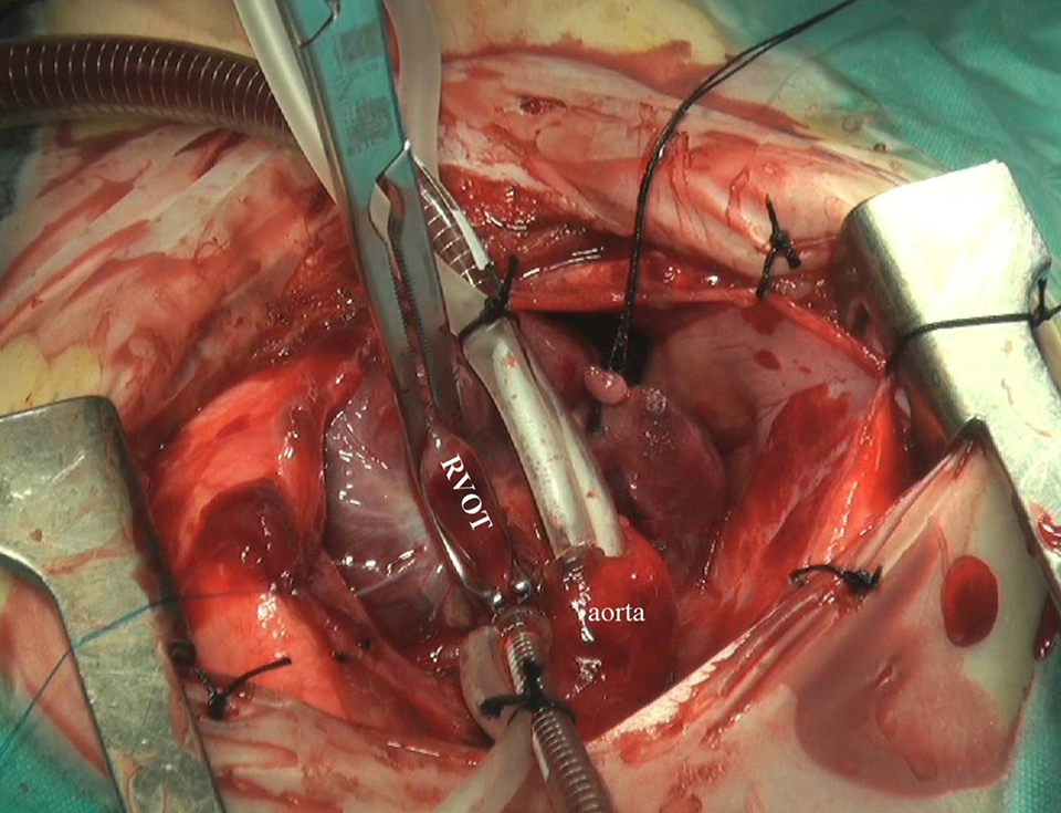
An intraoperative photograph (patient 1) that shows a side-biting clamp placed onto the right ventricular outflow tract (RVOT) after establishment of the temporary aortopulmonary shunt system.
Case 2
18-day-old neonate (4.0 kg) was found to have a ventricular septal defect, severe pulmonary stenosis and a PDA, on echocardiography. A stent was placed in the PDA in the cardiac catheterization laboratory. The child developed severe arterial desaturation and hemodynamic deterioration, and medical resuscitation was provided. Inotropic support was initiated and the neonate was immediately taken into the operation theater. Emergent median sternotomy was performed. Arterial oxygen saturation became 75% after cannulation with the same temporary shunt described in case 1 (Figure 2 ). After 30 minutes with the temporary shunt open, the blood pressure was 70/25 mm Hg, the acidosis on the arterial blood gas resolved, and myocardial contractility was significantly increased. A side-biting clamp was placed on the right pulmonary artery, and after 3 minutes, arterial oxygen saturation was 70%. With the hemodynamics stabilized, a 3.5 mm polytetrafluorethylene (PTFE) graft was placed between the right pulmonary artery and ascending aorta. After all clamps were removed and the temporary shunt was removed, the arterial oxygen saturation was 75%. The patient was transferred to the intensive care unit (ICU) with an open sternum, stable hemodynamics, and arterial oxygen saturations around 75%. The sternum was closed on the second day after surgery. Daily echocardiography documented an open shunt. The patient was discharged on the ninth day after surgery.
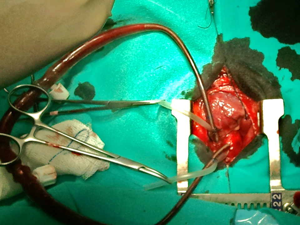
An intraoperative photograph (patient 2) that shows the provisional aortopulmonary shunt system that was established between the aorta and the main pulmonary artery.
Discussion
Aortopulmonary shunt operations performed via median sternotomy may require CPB due to hypoxia or hemodynamic instability. 1,2 The need for CPB may be unavoidable when pulmonary blood flow is ductal dependent and ductal flow is compromised. Cardiopulmonary bypass may also be necessary during neonatal RVOT reconstruction. Cardiopulmonary bypass can have adverse effects on many organ systems, especially in the neonates. 3–7 The postoperative recovery period may be adversely influenced by these negative aspects of CPB. 8 The provisional shunt system described in this article consists of connecting two 8F arterial cannulas after routine heparinization for CPB. The overall volume of the temporary shunt system is 9 mL, and the length of the tubing utilized is 38 cm. In both patients, the main pulmonary artery was cannulated at the bifurcation close to the left pulmonary artery. Echocardiographic evaluation of the branch pulmonary arteries has not revealed any stenosis of the branch pulmonary arteries.
Use of the temporary shunt described in this article may help avoid the need for cardiopulmonary bypass when performing aortopulmonary shunts and RVOT reconstructions in neonates. This temporary shunt may allow for the resuscitation of critically ill neonates, as demonstrated in the second case presented in this manuscript. This temporary shunt can help to achieve and maintain stable hemodynamics and oxygen saturations while performing neonatal aortopulmonary shunts and RVOT reconstructions. In cases where the method turns out to be insufficient, it is then possible to insert a venous cannula and start conventional CPB immediately. Of perhaps greater importance, although cardiopulmonary bypass is readily available in Turkey, the strategy described in the case report may prove especially useful in developing nations with limited access to CPB.
Footnotes
The author(s) declared no potential conflicts of interest with respect to the research, authorship, and/or publication of this article.
The author(s) received no financial support for the research, authorship, and/or publication of this article.