






Abstract
Background:
Surgical repair of common arterial trunk (CAT) by means of a homograft conduit has become a standard practice. We report our experience in the correction of this heart disease with a handmade bovine pericardial-valved woven Dacron conduit as an alternative procedure to homografts, with a focus on early, mid-term, and long-term results.
Methods:
We designed a retrospective study that included 15 patients with a mean age of 1.5 years (range: three months to eight years), who underwent primary repair of simple CAT. Right ventricular outflow tract was reconstructed in all the cases with this handmade graft that was explanted at the time of its biological stenotic degeneration. A peeling procedure was performed at this time, in order to reconstruct the right ventricle-to-pulmonary artery continuity.
Results:
Overall mortality was 13.3% (one death at the early postoperative primary repair and the other at the mid-term postoperative peeling reoperation). Actuarial survival rate was 93.3%, 86.7%, and 86.7% at 5, 10, and 15 years, respectively. All of the 14 survivors developed stenosis of the handmade conduit at the mid-term period (8 ± 3 years), but after the peeling procedure, 13 survivors remain asymptomatic to date.
Conclusions:
Primary repair of common arterial trunk using a handmade conduit can be performed with very low perioperative mortality and satisfactory mid-term and long-term results, which can be favorably compared with those reported with the use of homografts. When graft obstruction develops, peeling procedure is a good option because it does not affect the overall survival, although long-term outcomes warrant further follow-up.
Introduction
Previously referred to as truncus arteriosus communis, common arterial trunk (CAT) is a potentially cyanotic congenital heart defect defined by the presence of a solitary great artery that exits the heart overriding the ventriculoarterial junction in order to supply directly the systemic, pulmonary, and coronary arterial pathways. 1 –3 This is a rare malformation, appearing in 0.034 to 0.56 per 1,000 newborns and representing 1.4% to 2.8% of all cases of congenital heart diseases. 4 –6 Without surgical treatment, 80% of patients die within the first year of life, usually in early infancy. 7 Repair of CAT during the neonatal and early infant period has become a standard practice in many centers, with very good results. 8 –13
Rapid development in pediatric cardiac surgery and cardiology in recent decades have led to early corrective surgery in much of the developed world by means of a homograft conduit. 14 –16 However, in developing countries, due to a previous lack of opportunity to repair complex defects during infancy, pulmonary artery hypertension in children with congenital heart disease is still a common problem, 17 as is the availability and cost of homograft preservation. Therefore, we developed a handmade bovine pericardial-valved woven Dacron conduit in order to deal with this situation in a developing country for improving health care and to avoid delaying primary operation of CAT, which leads to an increased risk of pulmonary vascular obstructive disease.
The purpose of this study is to report the early, mid-term, and long-term results of primary repair of CAT with the use of a handmade bovine pericardial-valved woven Dacron conduit as an alternative procedure to homografts, which is performed at a single institution in the current era. We also aimed to compare the durability of this conduit with that reported with the use of homografts in pediatric population.
Patients and Methods
Study Design
A retrospective, longitudinal, descriptive, and observational study was designed, including all patients in whom a handmade bovine pericardial-valved woven Dacron conduit was used for primary surgical correction of CAT from January 2001 to December 2015 at the Department of Pediatric Cardiac and Congenital Heart Disease Surgery. The retrospective study was approved by the hospital ethics committee. Exclusion criteria were preoperative cardiogenic shock, important or severe lesions of the truncal valve, coronary anomalies, associated interruption of the aortic arch, primary repair of CAT not made by means of a handmade bovine pericardial-valved woven Dacron conduit, and the absence of a complete clinical and echocardiographic follow-up. Preoperative echocardiography was used in all patients for diagnosis, typification according to Collet and Edwards’s classification, evaluation of truncal valve lesions, and detection of other concomitant congenital heart disease. Preoperative hemodynamic evaluation by cardiac catheterization was performed to clarify inconclusive echocardiographic diagnostic data as well as to measure pulmonary artery pressure and resistances of those patients with pulmonary hypertension.
Operative Technique
Surgical approach was total median sternotomy. Cardiopulmonary bypass (CPB) was established by means of aortic and bicaval venous cannulae, and moderate hypothermia and cardioplegia were used for myocardial protection. Surgical correction was performed by the isolation of the pulmonary artery buttons from the CAT. The truncus remained as aorta, and the defects of the pulmonary buttons excised were patched with bovine pericardial tissue. Obligatory ventricular septal defect closure and other associated procedures to correct concomitant cardiac lesions were performed at this time. Finally, the right ventricle-to-pulmonary artery continuity was reconstructed using a handmade bovine pericardial-valved woven Dacron conduit, in all the cases. Once CPB was interrupted, and before decannulation, a routine transesophageal echocardiography was performed to assess ventricular function, residual right ventricular outflow tract obstruction, and aortic insufficiency. In all these cases, the handmade graft was fashioned as described in our previous publication (Figure 1). 18

Surgical technique for construction of the bovine pericardial-valved woven Dacron conduit: (A) materials needed (bovine pericardial tissue, woven Dacron tubular graft, and Hegar dilator); (B) suturing the bovine pericardial band around the whole woven Dacron conduit perimeter invaginated over the Hegar dilator; (C) measurement of three equidistant points of the graft perimeter with a compass to perform the handmade valve; (D) details of the suture of the bovine pericardial valve; and (E) final view of the handmade bovine pericardial-valved woven Dacron conduit once invaginated.
Once the patients developed graft dysfunction because of biological stenotic degeneration at the mid-term follow-up with a conduit echocardiographic maximum pressure gradient ≥60 mm Hg, we indicated a secondary surgical correction. The operation performed in these cases was a “peeling procedure,” which actually results in a valveless right ventricular-to-pulmonary artery connection with the expected sequelae. A redo median sternotomy was used and aortic and bicaval cannulation were established for circulatory assistance without cardiac arrest. We proceeded to perform an explantation of the dysfunctional handmade conduit. During this explantation, it was of particular importance to preserve the fibrous channel subjacent to the dysfunctional graft, which was formed by remodeling the connective tissue after its primary implantation. This channel was transformed into a new conduit by means of a bovine pericardial patch roof that was sutured to the channel borders in order to reconstruct the right ventricle-to-pulmonary artery continuity. Before completing this new amalgamated conduit, a stented bioprosthetic valve may be placed in the right ventricular outflow tract proximal to the pulmonary artery bifurcation, if needed. We prefer the use of a valvular-stented bioprosthesis rather than a new handmade conduit for two main reasons. The first one is because it is more difficult to ensure a complete absence of insufficiency when the conduit diameter is larger. The second reason is because the handmade conduit, as a stentless valve, is more susceptible to being compressed after chest closure, with the hemodynamic repercussions that this effect may produce.
Follow-Up Strategy
Follow-up strategy included a clinical and echocardiographic control in the early and mid-term periods. Early term was considered to be up to the 30 days of operation or during the same period of hospitalization. Mid-term results were those that happened after six months of the primary operation, and long-term is after ten years. All patients were evaluated according to their New York Heart Association (NYHA) functional class. 19 To assess the clinical status of infants, we used the modified classification proposed by Ross et al. 20 All patients had an echocardiographic follow-up in the mid-term irrespective of the presence or lack of symptoms. The degree of the conduit or of the aortic insufficiency was estimated by means of a semiquantitative method based on the width and length of the regurgitant jet in relation to the dimensions of the corresponding ventricle. Stenosis was characterized as mild, moderate, or severe, according to the gradient value registered at the aortic or the conduit level, and the area of the orifice related to the patient’s weight and age. 21
At the mid-term follow-up, clinical and echocardiographic evaluation led to the detection of stenosis of the handmade bovine pericardial-valved woven Dacron conduit and the need for reintervention by means of surgery or interventional catheterization. Indications for interventional catheterization in order to perform a percutaneous balloon dilation and eventual stenting of the valved conduit were maximum echocardiographic gradient between 50 and 59 mm Hg, right ventricle pressure of 60% or more of the systemic pressure, and distortion and/or stenosis of the pulmonary arteries with significant reduction in pulmonary blood flow. We considered indications for surgery in those cases with concomitant left-sided disease (eg, severe aortic valve stenosis, regurgitation, or both), severe stenosis of the valved conduit that generated echocardiographic gradients ≥60 mm Hg with or without right ventricle dysfunction, and failure of the interventional procedure to reduce the stenotic gradient of the valved conduit. The clinical and echocardiographic follow-up was closed in December 2015.
Statistical Analysis
Information was obtained from our institutional clinical database, stored in an electronic Excel page v14.0 (2010 Microsoft Corporation), and processed with an SPSS statistical software v21.0 (SPSS Inc, Chicago, Illinois). Quantitative variables are presented as mean and variability ranges (minimum and maximum). Categorical data are presented by means of frequency and percentages in relation to the population at risk. Overall survival rate and survival free from obstruction of the handmade pericardial-valved woven Dacron conduit were calculated using the Kaplan-Meier method.
Results
During the study period, 26 patients with CAT underwent primary repair at our institution, but only 15 of them met the inclusion criteria. Preoperative characteristics of the 15 patients enrolled in this study are presented in Table 1. Note that male–female ratio is 2:1. Although the mean age of this series was 1.5 years, the calculated mode was four months. At the moment of admission, 13 patients (87%) had controlled cardiac failure, and 2 of them (13%) had heart failure requiring preoperative inotropics. None of the patients died while awaiting surgery.
General Characteristics of the Studied Group.
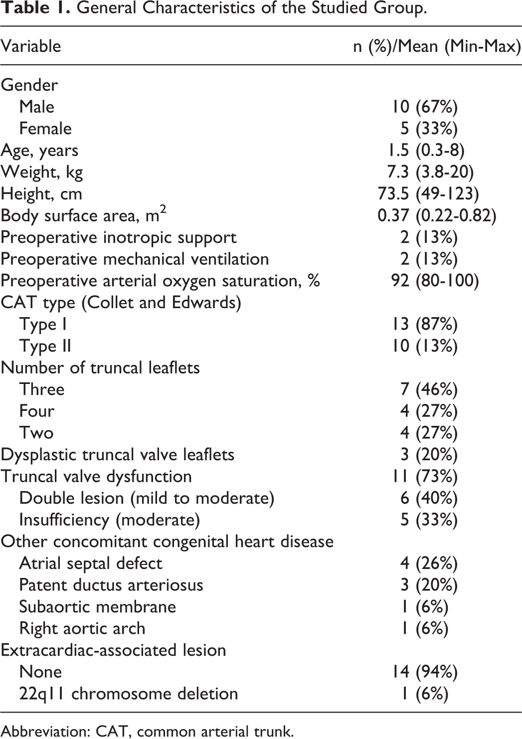
Abbreviation: CAT, common arterial trunk.
Myocardial protection for primary repair of CAT was accomplished using moderate hypothermia of 25.4°C (range 18°C-28°C) and cardioplegia (antegrade in eight cases and combined antegrade and retrograde in seven). Mean time of CPB was 132.4 minutes (range 96-201 minutes) and of aortic cross clamp was 96.7 minutes (range 67-185 minutes). The mean diameter of the handmade bovine pericardial-valved woven Dacron conduit was 15.2 mm (range 12-20 mm).
Early Results
Mortality
Operative mortality was 6.6% (n = 1) and corresponded to an eight-month-old infant who was emergently transferred to our institution with severe cardiac failure associated with pulmonary origin sepsis, requiring antibiotics, inotropics, and mechanical ventilatory support 20 days before the operation. Echocardiography showed a type I CAT with a large ventricular septal defect of 12 mm and a truncal valve with mild regurgitation associated with a stenosis that generated a maximum gradient of 33 mm Hg. Due to the severity of this case, an urgent surgical correction of CAT was performed with a 16-mm handmade graft. This patient was managed by means of a delayed sternal closure protocol due to hemodynamic instability after weaning from CPB. In the intensive care unit (ICU), he developed several hypertensive pulmonary crises that were refractory to the nitric oxide therapy and finally died on the first postoperative day.
Morbidity
Table 2 shows the postoperative surgical and nonsurgical complications. There were four patients who underwent delayed sternal closure as a result of mediastinal bleeding and/or hemodynamic instability. During the postoperative period following the correction of CAT, one patient was reoperated on for immediate mediastinal bleeding, presented cardiac arrest, and required cardiac resuscitation. There were ten cases that required nitric oxide therapy because of postoperative pulmonary hypertensive crisis. There were no sternal wound infections or mediastinitis in this series. Except for one patient, all the others required inotropic support for a mean time of 6 days (range 2-15 days). Excluding the patient who died, the mean duration of mechanical ventilation support was 8 days (range 1-18.7 days), mean ICU postoperative time was 10.5 days (range 1-21 days), and mean total hospitalization time was 24.1 days (range 8-48 days).
Early Postoperative Complications of Woven Dacron Pericardial-Valved Conduit.

Mid-term Results
Results of the bovine pericardial-valved woven Dacron conduit
All of the patients in this series were reoperated at the mid-term postoperative period because of obstruction of the handmade bovine pericardial-valved woven Dacron conduit. The mean duration from implantation to explantation of this graft was 8 ± 3 years (range: 3.5-14.4 years). Only one of these patients that developed stenosis of the graft at the mid-term follow-up received a percutaneous balloon dilation, which increased the survival of the graft for two more years before the peeling operation. In the whole series, only two patients developed major complications unrelated to stenosis of the conduit used for the reconstruction of the right ventricle to the pulmonary artery continuity. The first one was a patient with type I CAT who was repaired with a handmade conduit, and four years later developed a stroke with multiple left temporoparietal ischemic lesions that produced clinical aphasia and right brachial hemiparesis. Echocardiography did not show abnormalities in the handmade conduit or signs of endocarditis but detected a thrombus in the left appendage as the probable cardioembolic focus. This patient received medical treatment and remains with physical rehabilitation until he was reoperated for tube obstruction eight years later from the primary repair. The second mid-term complication was observed in an infant with type I CAT and moderate regurgitation of the truncal valve that was surgically corrected with a handmade graft. This aortic regurgitation remained moderate, but the patient developed progressively severe aortic insufficiency with left ventricular function impairment. For this reason, he received three years later an aortic valve replacement with a mechanical prosthesis. Postoperative evolution was satisfactory and seven years later was reoperated for peeling procedure due to the obstruction of the handmade conduit.
Results of the peeling procedure
Although the patients in this series improved their clinical functional class (NYHA/Ross) after primary repair of CAT by means of a pericardial-valved woven Dacron conduit, all of them developed obstruction of the graft, with subsequent clinical and echocardiographic impairment. Maximum echocardiographic pressure gradient showed a mean value of 77.7 ± 29.8 mm Hg (range: 46-143 mm Hg) at the time of reoperation. Figure 2 shows the survival free from obstruction of the handmade bovine pericardial-valved woven-Dacron conduit for primary repair of CAT.

Survival free from obstruction of the handmade graft Kaplan-Meier curve.
All of the patients were reoperated for graft obstruction by means of a peeling procedure, but only in 26.6% (n = 4) a pulmonary valve bioprosthesis was used. The other 11 cases did not receive a valvular prosthesis. There was no operative mortality after the peeling procedure. Early postoperative period after peeling procedure was uneventful in most of our cases. One patient developed a neurocognitive deficit postoperatively and another one a segmental pulmonary thomboembolism without clinical repercussion.
Long-Term Results
There was a long-term mortality of 13.3% (n = 2) in this series. Excluding the case who died at the early postoperative period of CAT primary repair, the other patient was reoperated 14 years after the primary correction of CAT with a handmade conduit because of obstruction of the graft associated with a residual ventricular septal defect. A peeling procedure was performed without placing a pulmonary valvular prosthesis, and a residual ventricular septal defect closure was also performed. The patient died 14 years after the peeling procedure due to cardiac failure and septic shock. Figure 3 shows the actuarial survival rates as 93.3%, 86.7%, and 86.7% at 5, 10, and 15 years, respectively.

Overall survival Kaplan-Meier curve.
The mean echocardiographic follow-up was 9 ± 2.8 years (range: 5-13 years). At this time, echocardiographic maximum pressure gradient showed a mean value of 27 ± 17 mm Hg (range: 0-55 mm Hg) after the peeling procedure. Mean clinical follow-up was 5 ± 1.3 years (range: 2-6 years). Figure 4 shows the clinical evolution of the 13 survivors of our series, and we can see that at the mid-term follow-up, after the peeling procedure, all of them improved their functional status when compared with the functional class at the preoperative period of the primary CAT correction.

Clinical evolution of the functional class (NYHA/Ross).
Overall follow-up of our series is shown in Figure 5. There were only three deaths: one of them at the early postoperative period of CAT primary repair and two at the mid-term period following the peeling procedure. All of the patients developed stenosis of the handmade pericardial-valved woven Dacron conduit at the mid-term period, but after reoperation (peeling procedure) the survivors remain asymptomatic at the time of closure of this study.

Overall follow-up of primary surgical repair of simple truncus artriosus communis by means of a bovine pericardial-valved woven Dacron conduit.
Comment
First described by Rastelli and Ongley in 1968, 22 successful surgical correction of CAT was performed by means of an aortic homograft to reestablish the continuity between the right ventricle and the pulmonary artery. From that time on, several methods of restitution of the pulmonary circulation continuity have been developed. One of them is the use of xenografts such as bovine jugular-valved conduits (Contegra without support; Medtronic, Inc, Minneapolis) and stentless porcine pulmonary valve with arterial root conduits (Shelhigh; Shelhigh, Inc, New Jersey). However, the ideal right ventricle-to-pulmonary artery conduit remains a matter of controversy, especially in high-pressure systems, as does conduit choice with respect to the age at which they must be implanted. The cryopreserved homograft has features that make it a highly desirable valved conduit for reconstruction of the right ventricular outflow tract. Despite the excellent handling characteristics of these conduits, limited overall supply, availability in pediatric sizes, and increased cost of cryopreservation are among the primary disadvantages recognized for homografts. 23 These are particularly important limitations for hospitals in developing country such as our institution, for they lead us to develop an alternative handmade bovine pericardial-valved woven Dacron conduit for the primary correction of CAT. The advantage of this graft is its availability in different sizes as well as the cheaper cost, compared with those of homografts and xenografts. These aspects acquire special relevance to the population of patients presented at our institution.
Several series have reviewed institutional experiences with cryopreserved homografts in the pulmonary position. 24 –28 These reports have included relatively low numbers of infants and neonates, where the durability of any non-growing conduit will be limited. 29 –31 Kalavrouziotis et al 32 reported a high incidence of reintervention procedures (by means of percutaneous catheterization or surgery) in the groups of patients using aortic homografts (freedom from reintervention survival rate of 14% at ten years). Pulmonary homografts showed a better freedom from intervention survival rate (82% at 8.5 years). Contegra and Shelhigh xenografts had a similar survival rate (70% at 27 months and 67% at 34 months, respectively), but the follow-up time was too short to compare them with homografts and draw definitive conclusions. Sierra et al 33 compared the evolution between homografts and Contegra in right ventricular outflow tract repair in children with an average age of 4.9 years and concluded that freedom from reintervention secondary to stenosis of the conduits is similar (81.3% at seven years for homografts and 90.7% at seven years for Contegra). Forbess et al 23 analyzed the results with pulmonary homografts according to patient age at implantation. They showed that freedom from reintervention at five years is dramatically lower in the patients with age less than one year at the time of implantation (25%) than in patients between one and ten years (61%) or in those more than ten years of age at implantation (81%). Additionally, they conclude that the presence of CAT is a risk factor for restenosis in the mid-term. Lacour-Gayet et al 34 analyzed the evolution of several CAT repair techniques in children with an average age of 41 days and established that the use of homografts at early age is a risk factor for reoperation. Based on the available evidence, the current tendency of our institution is to promote the use of synthetic prostheses for CAT repair in infants, reduce the utilization of xenografts, and definitively avoid the use of homografts.
Independent of the type of conduit used to establish continuity between the right ventricle and the pulmonary arteries, the most frequent causes of surgical repair failure are biological graft stenotic degeneration due to calcification, conduit mismatch because of patient growth, or sternal compression of the graft. These complications need reoperation in the mid-term or long-term to replace the conduit, which is associated with a major risk of morbidity and mortality. The main mode of failure of the bovine pericardial-valved woven Dacron conduit in our series was degenerative calcification of the biologic tissue that leads to stenosis of the tubular graft at the valve level. Survival rate without reoperation due to stenosis of the bovine pericardial-valved woven Dacron conduit at the mid-term period in our series is favorably compared with those of others reported in the literature and shows a clear tendency to offer advantages in comparison with homografts in infants because of its lower incidence of stenosis, greater availability, and reduced cost. It is also important to state that regurgitation is, as well, another long-term problem, but its development is slower than that of stenosis. Therefore, surgical indication for reoperation due to regurgitation of the handmade pericardial-valved woven Dacron conduit is less prevalent, because the need for reoperation due to stenosis develops earlier. Despite the fact that all of the patients in this series required in the mid-term follow-up a new operation because of graft obstruction, the peeling procedure alternative has shown a low-risk operative mortality and its effect on the overall survival is minimized. 35 Mid-term outcomes are good because of lack of obstruction of the new conduit. Long-term follow-up of this procedure is needed in order to establish the freedom from obstruction rate. Innovative interventional techniques such as valve endoprosthesis (valve in tube or valve in ring) are among the potential future alternatives of choice for these patients in an effort to extend the functional durability of their right ventricular outflow tract reconstruction.
In conclusion, complete surgical repair of CAT can be achieved with a relatively low mortality and acceptable early, mid-term, and long-term results, even in cases with late presentation. Surgically created continuity between the right ventricle and the pulmonary artery by means of a woven Dacron prosthetic conduit with a handmade bovine pericardial valve support progresses irremediably toward the stenosis of the graft at the mid-term period but confers a lapse of reasonable time to allow the child’s growth before reoperation. Finally, when obstruction of the graft develops, the peeling procedure is a good alternative because of its low operative mortality risk that does not affect the overall survival, although long-term outcomes warrant further follow-up. Therefore, the handmade bovine pericardial-valved woven Dacron graft may be a good alternative to the xenograft-valved conduit when an allograft conduit is not available.
Footnotes
Declaration of Conflicting Interests
The author(s) declared no potential conflicts of interest with respect to the research, authorship, and/or publication of this article.
Funding
The author(s) received no financial support for the research, authorship, and/or publication of this article.