






Abstract
Anomalous origin of a branch pulmonary artery (PA) from the ascending aorta is rather rare within the spectrum of congenital cardiovascular anomalies. In the scarce subset of patients with anomalous origin of right PA (RPA) from the ascending aorta originating from the lateral aspect of the distal ascending aorta, early neonatal repair by employing native aortic tissue for RPA reconstruction combined with translocation of the aorta posteriorly to the RPA can be an alternative option in the surgical armamentarium.
Keywords
Introduction
Anomalous origin of a pulmonary artery (PA) from the ascending aorta (AAo) represents a rarity within the spectrum of congenital cardiovascular anomalies (CCVAs). In patients with anomalous origin of right PA (RPA) from the AAo (or, much less commonly, left), the PA usually originates from the right posterior wall of the AAo. 1 In a scarce subset of patients, the anomalous origin of branch PA from the AAo arises from the lateral aspect of the AAo.
Surgical correction of anomalous origin of PA from the AAo has evolved over the years since the first successful attempt 2 and is associated with improved outcomes. 3,4 Recommended strategies include interposition synthetic or homograft 1,3,4 and techniques using autologous aortic tissue or pericardium. 5,6
We describe herein a case of anomalous origin of the RPA from the AAo, and its management which consisted of successful early neonatal repair by employing native aortic tissue for RPA reconstruction combined with posterior (to the RPA) translocation of the aorta.
Technique
A one-week-old neonate was transferred to our institution for poor feeding, episodic pallor, and respiratory distress after failing a congenital cardiac screening at an outside facility. Diagnostic workup with transthoracic echocardiogram (TTE) and computed tomography angiography was notable for anomalous origin of RPA from the distal AAo, aberrant right subclavian artery, patent ductus arteriosus on prostaglandin with a right-to-left shunt, and suprasystemic right ventricular (RV) systolic pressure greater than 100 mm Hg (Figure 1). There was a patent foramen ovale (PFO). The pathophysiologically critical nature of the lesion prompted neonatal correction.
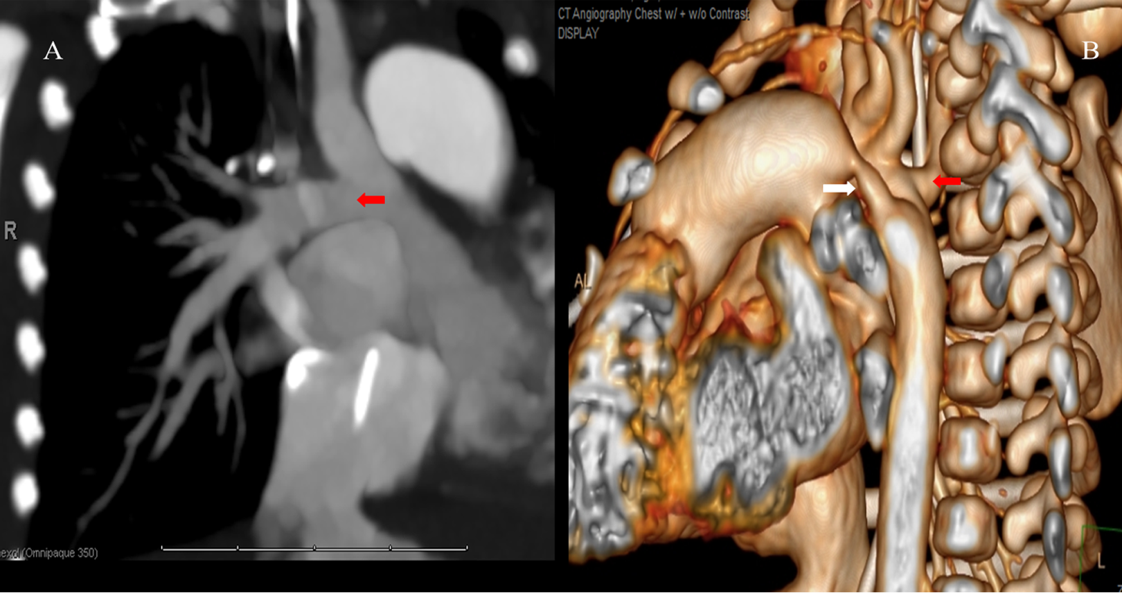
A, Anomalous origin of right pulmonary artery from distal ascending aorta (red arrow highlights takeoff from the ascending aorta). B, Aberrant right subclavian artery posteriorly to the esophagus (red arrow); patent ductus arteriosus (white arrow).
After a standard venous and proximal aortic arch cannulation, mildly hypothermic (32°C) cardiopulmonary bypass (CPB) was initiated. Extensive mobilization from the aortic root to the origin of the aberrant right subclavian artery (off the descending thoracic aorta) as well as the aortic arch was performed. The patent ductus arteriosus was doubly ligated and divided to allow maximum mobilization of PA system. The aberrant right subclavian artery was reimplanted to the proximal right common carotid artery (CCA) to allow maximum mobilization of the aortic arch and prevent future esophageal posterior compression as a result of the anterior and caudal positioning of the aortic arch in order to bridge the circumferential tissue defect of the AAo. The main PA (MPA), left PA (LPA), and RPA were carefully mobilized to the first segmental lobar branching. The anomalous origin of RPA from the AAo was identified and isolated from the systemic circulation using vessel loop. Subsequently, the aortic cross clamp (ACC) was applied and heart arrested using antegrade cold blood cardioplegia. The AAo was transected obliquely, according to the longitudinal axes of the RPA, above and beneath the anomalous origin of PA from the AAo, which provided a symmetric aortic ring at a size slightly larger than the RPA diameter (Figure 2A). The cylindrical aortic flap was unevenly divided providing 60% to 70% of the circumference to the neo-RPA anterior wall and 30% to 40% to the posterior one. The AAo was transposed posteriorly and an end-to-end tension-free anastomosis with 7/0 Prolene suture was performed. The aortic cross clamp was removed and the remainder of the procedure was performed on normothermic CPB with the heart beating. Then, an anteriorly hinged trapdoor incision (with two transverse incisions at a right angle with the longitudinal one) was made at the MPA anterior wall adjacent to the LPA origin extending at least few millimeters beyond the caudal end of the LPA, thus, complementing the aortic flap created for the neopulmonary posterior wall (Figure 2B). First, the new posterior wall of the anteriorly transposed RPA was created by anastomosis between the end of the mobilized MPA trapdoor and the posterior aortic (neopulmonary) flap, using a combination of running and interrupted 7/0 polyglyconate suture (Maxon; Davis-Geck, Inc, Danbury, Connecticut). The anterior aortic flap accommodated the anterior–inferior wall of the neo-RPA, maintaining a native tissue and wide angle anastomosis with the MPA. The suture line at the MPA level was completed with interrupted sutures, in order to allow tissue growth (Figure 3). The final anterior–superior segment of the neo-RPA wall (less than one-fifth of the circumference) was reconstructed with fresh oversized autologous pericardium (Figure 3; inset).
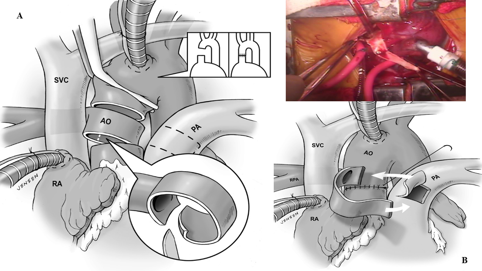
A, Distal AAo is transected obliquely above and below the anomalous origin of the RPA from the aorta, creating a symmetric aortic ring at a size slightly larger than the RPA diameter. B, Anteriorly hinged trapdoor is made at the MPA anterior wall adjacent to the LPA origin extending at least few millimeters beyond the caudal end of LPA. AAo indicates ascending aorta; LPA, left PA; MPA, main PA; PA, pulmonary artery; RPA, right PA.
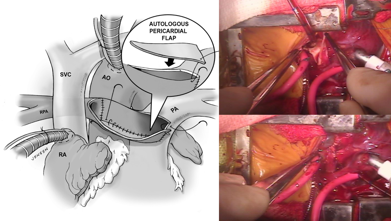
The anterior aortic flap accommodates the anterior–inferior wall of the neo-RPA. The anterior–superior remnant of the neo-RPA is reconstructed with autologous pericardium (inset). PA indicates pulmonary artery; RPA, right PA.
Aortic cross clamp and CPB times were 18 and 79 minutes, respectively. Intraoperative RV pressure was less than 60% of systemic pressure. Echocardiogram demonstrated moderately dilated RV, good biventricular function, and predominantly left-to-right shunt across the PFO. After extubation of the trachea in the operating room, the patient was transitioned to high-flow nasal cannula and inhaled nitric oxide (iNO).
Postoperatively, iNO was weaned off within 48 hours. The mediastinal chest tube was removed on day three. The patient was uneventfully discharged home the following day on diuretics and aspirin. Predischarge TTE demonstrated normal biventricular function, significantly reduced size of RV and RPA peak velocity 1.2 m/s.
At follow-up, three months later, the infant continued to thrive and echocardiography indicated nondilated RV with normalized septal motion, normal biventricular function, and calculated peak gradient <10 mm Hg across the reconstructed RPA (Figure 4).
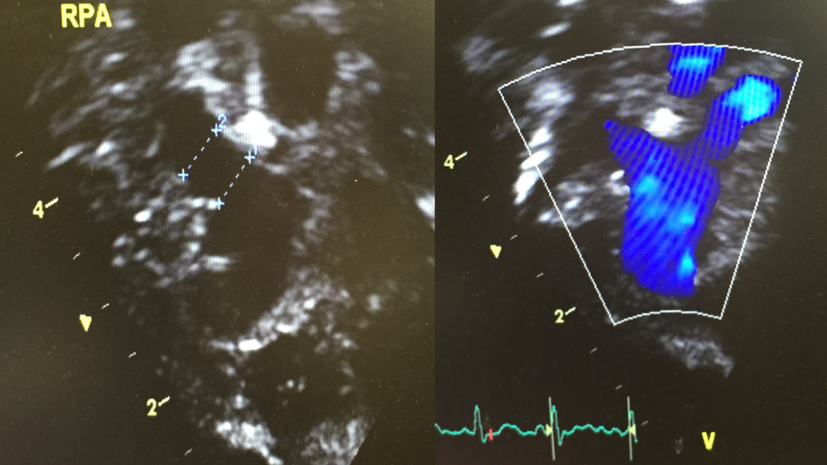
Transthoracic echocardiogram at last follow-up with view of the reconstructed RPA. PA indicates pulmonary artery; RPA, right PA.
Comment
The physiologic sequelae related to anomalous origin of PA from the AAo are based on the predominantly large left-to-right shunt with the contralateral lung subjected to the entire flow from the RV, as well as that contributed by the associated anomalies. 1,6 Without early surgical correction the natural history of this condition is dismal with high early mortality. 1,4
From the reported types of anomalous origin of PA from the AAo 3,4 proximal takeoff from the aorta is the most common. Usually, its origin is from the posterior-lateral aspect of the AAo. 1,5 As in our case, anomalous origin of RPA from the AAo originating from the lateral aspect of the aorta just caudally to the right CCA takeoff is extremely rare, especially when an aberrant subclavian artery is present.
Direct reimplantation or interposing prosthetic graft techniques carry a substantial longitudinal risk of anastomotic stenosis. 1,3,4 Direct retroaortic implantation of the RPA is generally reserved for those patients in whom the RPA originates in close proximity to the MPA. When applied in an RPA origin from the lateral aspect of the AAo, it can be associated with a rather early declining anastomotic integrity. 1,3 To address the stereotactic disadvantage when the anomalous origin of branch PA from the AAo originates from lateral aspect of distal AAo in the face of an aberrant subclavian artery an alternative pulmonary reconstituting approach was entertained using native aortic tissue 6 combined with posterior (to the reconstructed RPA) translocation of aorta and reimplantation of the right aberrant subclavian artery to the right CCA. This strategy exercises (1) reduced tension at the RPA-MPA anastomosis, (2) superior flow kinetics related to the wide angle and kink-free orientation of the reconstructed RPA with the MPA, (3) ameliorated anastomotic configuration and integrity, (4) growth potential inherent to the extensive involvement of native aortopulmonary tissue, and (5) maximum mobilization of the aortic arch and potential prevention of future esophageal posterior compression (by an unrepaired aberrant right subclavian artery) as a result of the anterior and caudal positioning of the aortic arch to facilitate a tension-free aortoaortic anastomosis. The described approach allows for generous in situ native tissue and limited autologous pericardial use which supports a tension-free placement of the newly created RPA anteriorly to the AAo. As a result, the posteriorly transposed AAo may alleviate the hazard of aortic compression on the, now, overlying neo-RPA without any risk for the LPA to be under tension or kinking, considering that the anteriorly hinged trapdoor incision was made at the MPA anterior wall adjacent to the LPA origin, thus, complementing the aortic flap created for the neopulmonary posterior wall without any axial rotation or torsion applied on the LPA. 6
The concept of early neonatal repair safeguards a sustainable reduction of RV volume index and dimensions coupled with drastic resolution of RV pressure load and its deleterious effects. The immediate regression of the septal impingement into the left ventricle has a profound impact on biventricular function. Avoiding prolonged ACC further alleviates the strain to an already deconditioned biventricular myocardium. This complex ventricular interplay can be rapidly reversed on a reproducible fashion and with a remarkable hemodynamic outcome. Long-term follow-up with advanced imaging (functional Magnetic Resonance) or differential lung perfusion scan (nuclear scintigraphy) might help determine the outlook of this strategy with reference to distribution and flow kinetics of pulmonary blood flow.
Native aortic tissue RPA reconstruction combined with posteriorly (to the RPA) transposed aorta can complement the surgical strategies offered in the anomalous origin of branch PA from the AAo. The procedure is safe and reproducible during the early neonatal period and can be tailored to specific anatomic variants encountered in this rare CCVA.
Footnotes
Authors’ Note
Parental consent was obtained. The institutional review board (IRB) of Children’s Hospital of Georgia does not require IRB review and approval for isolated case reports.
Acknowledgment
The authors would like to thank Michael Jensen for his wonderful illustrations.
Declaration of Conflicting Interests
The author(s) declared no potential conflicts of interest with respect to the research, authorship, and/or publication of this article.
Funding
The author(s) received no financial support for the research, authorship, and/or publication of this article.