
Abstract
This case report describes a rare association of tetralogy of Fallot with interatrial communication of the inferior sinus venosus type (inferior sinus venosus defect). The patient underwent intracardiac repair for tetralogy of Fallot at 8 months of age. His postoperative course was complicated by desaturation. On reexploration, interatrial communication of the inferior sinus venosus type was revealed which went unnoticed on transthoracic echocardiogram as well as on computerized tomogram.
Introduction
Tetralogy of Fallot (TOF) is the commonest cyanotic congenital heart disease for which intracardiac repair is being performed. Association of TOF with interatrial communication of the inferior sinus venosus type is extremely rare. This case report describes this rare association in an infant as well as the challenges encountered in the diagnosis and management of this rare combination. A thorough literature search did not reveal any such case report previously.
Case Report
An 8-month-old male infant weighing 8 kg presented with a history of increasing cyanosis since birth. A thorough clinical examination revealed a heart rate of 122 beats per minute, respiratory rate of 34 breaths per minute, and a saturation of 73% on room air. On auscultation, the first heart sound was normal with single second heart sound along with grade 4/6 ejection systolic murmur at left upper sternal border. The rest of the examination was unremarkable.
His chest radiograph and electrocardiogram were suggestive of right ventricular hypertrophy and features suggestive of TOF. Transthoracic echocardiography confirmed the diagnosis of TOF with a small ostium secundum atrial septal defect (OS-ASD) shunting bidirectionally and severe infundibular stenosis with good size pulmonary annulus, main and branch pulmonary arteries. A small left superior vena cava (SVC) draining into coronary sinus (CS) was detected with no patent ductus arteriosus (PDA) or major aortopulmonary collaterals.
The child underwent intracardiac repair of TOF under cardiopulmonary bypass (CPB) with aorto-bicaval cannulation with moderate hypothermia. A trans-right atrial approach was used to perform pericardial patch closure of the ventricular septal defect (VSD) and infundibular resection. The OS-ASD was closed directly. Following repair, he was transferred to the cardiac intensive care with stable hemodynamics. The immediate postoperative period was unremarkable, however, few hours later his saturation on a fractional inspired concentration of oxygen of 60% showed a drop to 80% supported by a PaO2 of 45 mm Hg on arterial blood gas analysis. Ventilatory parameters were confirmed to be as per recommendations for his age and body weight.
Transthoracic echocardiography was done, which confirmed the VSD patch in situ without any residual shunt or infundibular stenosis. Bubble contrast echocardiogram was not conclusive. Saturation continued to be the same; however, the etiology of the hypoxia could not be established. Cardiac catheterization angiography was performed which ruled out the possibility of unroofed CS (Figure 1A). A computerized tomographic (CT) angiography was done, which was also inconclusive for the cause of desaturation. After excluding the usual and possible causes of desaturation, and a thorough discussion with the relatives, the team of cardiologist and cardiac surgeon decided to reexplore the patient to identify the cause of desaturation.
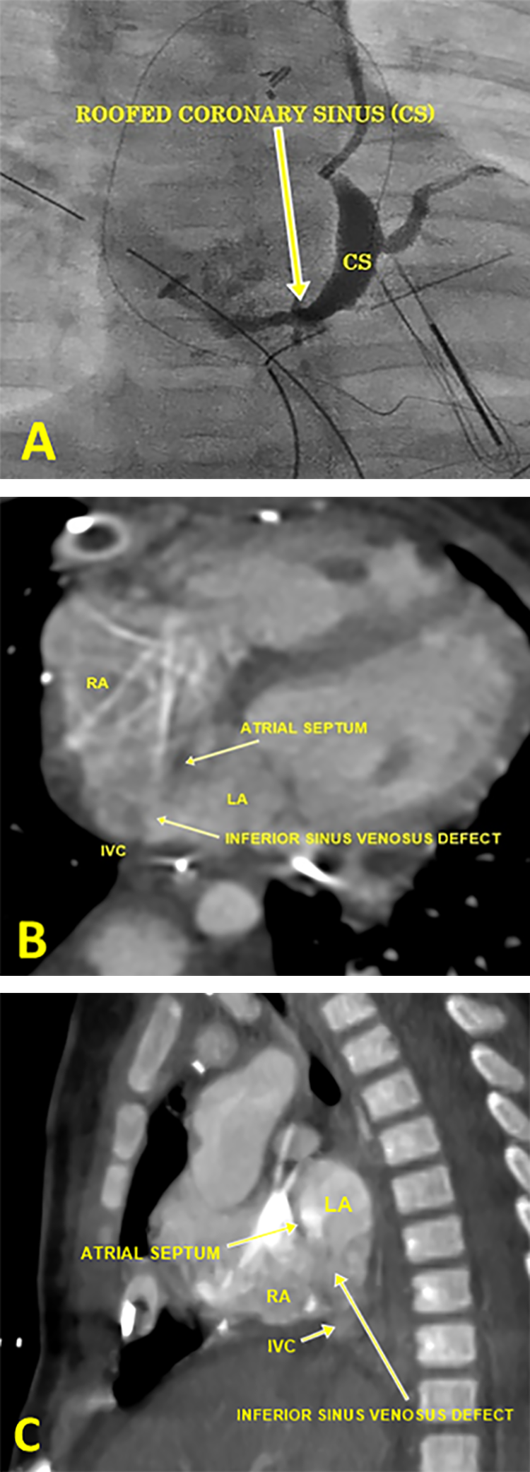
A, Coronary sinus angiogram in antero-posterior (AP) view showing roofed coronary sinus (CS). B and C, Computerized tomographic images showing interatrial communication of the inferior sinus venosus type (inferior sinus venosus defect) along with overriding of inferior vena cava (IVC) on atrial septum.
Under CPB with cardioplegic arrest, the right atrium (RA) was reopened and the anatomy was reassessed. Careful examination showed an interatrial communication of the inferior sinus venosus type (size of 0.5 cm) which was obscured by a redundant Eustachian valve in the RA. The defect was repaired directly.
Retrospectively, the CT images were carefully reviewed two-dimensionally which showed the interatrial communication of the inferior sinus venosus type (inferior sinus venosus defect; Figure 1B and C). Volume rendered three-dimensional CT angiographic images were not of good quality due to motion artifact. The postoperative period was normal and the child was discharged on the seventh day after operation and is doing well on serial follow-up.
Discussion
Tetralogy of Fallot is the commonest cyanotic congenital heart disease, comprising approximately 7% to 10% of all congenital heart disease. 1 Tetralogy of Fallot can be associated with other cardiac lesions as well. Sheikh et al found associated cardiac anomalies such as right aortic arch, additional VSD, PDA, coronary artery abnormalities, bilateral SVC, ASD, and so on. 2
Commonly associated ASD with TOF is OS-ASD; it can be associated with other types of ASDs (mixed atrial septal defect) which accounts for 7% of all ASDs. The interatrial septum is divided into five septal zones and involvement of two or more of these results in mixed ASDs. Caval venous defects are the least common with the SVC type constituting the majority. 3,4 Though well described in the literature, reports of the inferior caval type of defect are relatively uncommon. 5
Al Zaghal et al characterized a caval vein defect as overriding of the intact rim of the fossa ovalis by the caval vein, whether superior or inferior, which may result in shunting at the atrial level. These are, however, not true atrial septal defects, as the interatrial communication does not result from a deficiency of the true atrial septum. A true ASD can coexist with a caval vein defect, the most conclusive diagnosis being made by confirming the intact muscular border of the fossa ovalis. 6
In this case, it appears that while snugging the IVC cannula, the interatrial communication of the inferior sinus venosus type was obscured, which reemphasizes the fact that the IVC cannula needs to be placed neither too low nor too high, so that the entire anatomy of the interatrial septum can be well assessed intraoperatively. Availability of transesophageal echocardiography is therefore an additional advantage in such cases. 7
Although very uncommon, the association of interatrial communication of the inferior sinus venosus type with TOF is not nonexistent. This case report reemphasizes the need for detailed pre- and intraoperative assessment of the cardiac anatomy as well as keeping a high index of suspicion for such unusual associations with TOF to avoid surgical dilemma and rerun of CPB in such patients.
Footnotes
Author’s Note
Consent for publication was granted by patient’s parents.
Declaration of Conflicting Interests
The author(s) declared no potential conflicts of interest with respect to the research, authorship, and/or publication of this article.
Funding
The author(s) received no financial support for the research, authorship, and/or publication of this article.