
Abstract
This article reviews the collaboration between clinician and illustrator throughout the ages while highlighting the era of cardiac surgery. Historical notes are based on Professor Sanjib Kumar Ghosh’s extensive review, literature searches, and the archives of the Johns Hopkins University Department of Art as related to Medicine in Baltimore. Personal communications were explored with medical illustrators and medical practitioners, many of whom are colleagues and trainees, to further chronicle the history of medical illustration and education in the era of cardiac surgery. Medical illustrators use their talents and expressive ideas to demonstrate procedures and give them life. These methods are (1) hovering technique; (2) hidden anatomy, ghosted views, or transparency; (3) centrally focused perspective; (4) action techniques to give life to the procedure; (5) use of insets to highlight one part of the drawing; (6) human proportionality using hands or known objects to show size; and (7) step-by-step educational process to depict the stages of a procedure. Vivid examples showing these techniques are demonstrated. The result of this observational analysis underscores the importance of the collaboration between clinician and illustrator to accurately describe intricate pathoanatomy, three-dimensional interrelated anatomic detail, and complex operations. While there are few data to measure the impact of the atlas on medical education, it is an undeniable assertion that anatomical and surgical illustrations have helped to educate and train the modern-day surgeon, cardiologist, and related health-care professionals.
Introduction
The purpose of this article is to review the collaboration between clinicians and illustrators throughout the ages and concentrate on the contributions that chronicled the era of cardiac surgery. Much of the historical notes from antiquity to the dawn of the 20th century have been referenced in, Evolution of Illustrations in Anatomy: A Study from the Classical Period in Europe to Modern Times, 1 by Sanjib Kumar Ghosh of the Department of Anatomy, Medical College, Joka, Kolkata, India. His literature review of historical sources allowed insight into further investigation. Inquiries with administrators of the Maude Abbott Museum in Montreal and archives of the Johns Hopkins University Department of Art as Applied to Medicine in Baltimore were made and examined. Personal communications were explored with medical illustrators and medical practitioners, many of whom are colleagues and trainees, to further chronicle the history of medical illustration in the era of cardiac surgery
The result of this observational analysis should signal to the reader the importance of the collaboration between clinician and illustrator to accurately describe intricate pathoanatomy, 3-dimensional interrelated anatomic detail, and complex operations. While there are few data to measure the impact of the atlas on medical education, it is an undeniable assertion that anatomical and surgical illustrations have helped to educate and train the modern-day surgeon, cardiologist, and related health-care professionals.
Historical Notes Leading Up To the Era of Cardiac Surgery
Medical illustration has been incorporated into anatomic study for centuries. Hippocrates of Cos (∼460 to ∼377 BCE), long thought to be the Father of Medicine, introduced the concept that “sickness arises from the environment or the patient; not from supernatural influences.” This led practitioners to catalog symptoms and physical findings in order to solve the mysteries of illness. He and his followers were responsible for the Hippocratic Corpus, which was written over a 200-year period. 2,3 The Hippocratics left detailed descriptions of disease entities, but no drawings have survived owing to the difficulty in artistic reproductions.
Galen of Pergamum (130-210 ACE) had an enormous influence on medical research and practice up until the early 19th century. 4 He performed experiments by careful observation and wrote some 22 volumes cataloging his observations and findings. 5,6 His presence and experimentation were so extensive that no one dared contradict him. The anatomic findings of his texts were exclusively descriptive and contained no drawings as noted by scholars. 7 Most of the illustrations ascribed to his work have been chronicled and preserved by medieval artists who interpreted his findings. Figure S1 is a facsimile of his likeness. A drawing of his comprehensive internal anatomy is shown in Figure S2, which attempts to interpret anatomy and physiology. His understanding of the circulation however was never proven nor was it accurate at any level. Figure S3 is an anatomic analysis depicting the physiology of the circulation which, according to Galen, was propelled by the liver. 8 As was proven by Harvey in 1628, Galen’s physiology of the heart and circulation was completely wrong 9 !
The Post-Galenic Era and Introduction of Human Dissection
In subsequent centuries, great advances were made in anatomic and physiologic studies that were chronicled by descriptive passages and artistic examples. Artists associated with anatomists, practitioners, and surgeons to offer a visual understanding of the intricate interrelationship between anatomy and physiology. The study of medicine during the late medieval period (1300-1450), while highly influenced by Galen, showed signs of scientific advances and rational inquiry. There were enclaves of study, later to become universities, which started to emerge as organized institutions of free study. King Frederick II, the Holy Roman Emperor (1194-1250), was influential in advancing the cause of medical education and human dissection. 10 During this period, Mondino de Liuzzi (1275-1326) performed systematic human dissection which resulted in his classic text, Anathomia Mondini. This publication advanced insights and perceptions of human anatomy; however, there were no illustrations within the text. 11,12 It was left to Liuzzi’s student, Guido da Vigevano (1280-1349), to introduce, for the first time, the idea that illustration could enhance the study of anatomy. 10 Vigevano published his manuscript, Anathomia, in 1345 which contained six drawings highlighting trephination and brain structure. 13 This manuscript established the beginning of this new idea of illustration to accompany description and was carried into the late Middle Ages, although the anatomic artistic interpretations were largely unrealistic, elementary, and oftentimes wrong. 1,14 During this period, there was no reliable method to reproduce drawings; they were therefore made manually by the artist. The introduction of wood-cut illustrations in the late 15th century allowed publication of multiple copies of the text and associated drawings. 14
The Renaissance
The change away from Galenic Medicine came with the Renaissance that was highlighted by a return of interest in the human body; a return to Greek art, literature, and music; and the rise of the great universities that were the focus of a new-found inquiry into philosophy, arts, and medicine, to name only a few. 2 A greater understanding of the human body, through human dissection, was necessary to understand anatomy and to postulate physiology based on observed disease patterns in life.
The celebrated University of Padua was among the first universities to make inquiries into these principles, which was supported by Pope Sixtus IV who in 1482 issued a papal bull that allowed human dissection. 2 The influence on artistic expression and medical study was enormous.
Artists as Dissectors, Anatomists as Artists
It was Leonardo da Vinci (1452-1519) who produced anatomical drawings based on human dissection between 1510 and 1511. 15,16 His famous drawing of the heart (Figure S4) included all the characteristics of topical cardiac anatomy including the sinuses of Valsalva. Leonardo, who was an incomparable painter and sculptor, introduced a system of drawings that allowed the observer to appreciate the anatomic details of limbs from different viewpoints (Figure S5) that had the effect of a three-dimensional recreation. 17,18 These principles were to be developed in the ensuing centuries resulting in computed tomographic three-dimensional imaging 19,20 and three-dimensional printed models of congenital heart disease lesions. 21 Other famous artists who performed human dissection to maximize their interpretation and expression of human anatomy included Albrecht Durer (1471-1528), Michelangelo (1475-1564), and Raphael (1483-1521).
The idea that anatomists could create their own illustrations had its origins much later, probably owing to educational emphasis on the sciences rather than the applied arts such as drawing, painting, and sculpture. The same is true nowadays, although that chasm has been bridged by some physician-anatomists with artistic talents. Notable anatomists who have published their own illustrations include Robert Hooke (1635-1703), Antonio Scarpa (1747-1832), John Bell (1763-1820), and Sir Charles Bell (1774-1842).
Anatomists and Artist Collaborators: Renaissance Into the 18th Century
The first recorded collaboration between an artist and an anatomist became manifest in the 16th century. Andrea Vesalius (1514-1564) was the fifth generation of a long line of Flemish doctors. 22 After his education at the Universities of Paris and Padua, he became professor of anatomy. Unlike the established norms, Vesalius (Figure 1) preferred performing dissections himself, oftentimes using live animal vivisection alongside of the human dissection, to prove anatomical details and physiologic principles. 22 Recognizing the power of illustration, he partnered with Jon van Calcar (Figure 1) who was a protégé of the famous Renaissance painter, Titian. They produced six huge anatomical plates that survive. Figure S6 shows van Calcar’s drawings from “De Corporis Fabrica” published in 1538. 23 Using these principles, Vesalius was able to systematically disprove many of Galen’s physiologic interpretations. Other important anatomist-artist teams included Giulio Casseri (ca. 1552-1616) and Odoardo Fialetti; Bernhard Siegfried Albinus (1697-1770) and Jan Wanderlaar (1690-1759); and William Hunter (1718-1783) and Jan van Riemsdyk (ca.1750-1788; Figure S7).
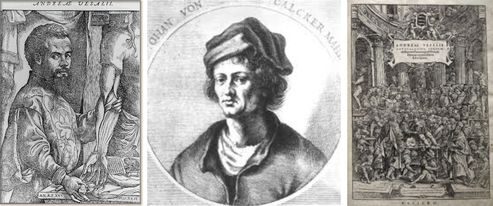
Collage of drawings showing Vesalius, Jon van Calcar, and anatomical plates from their book, “De Corporis Fabrica.” Vesalius “Epistola rationem…,” 1546; portrait of Vesalius. Portrait of Van Calcar, Fair use policy; Courtesy of Wikimedia. Van Calcar Plate Vesalius, De Humani corporis fabrica libri septem; Courtesy of Wellcome Collection. Creative Commons Attribution (CC BY 4.0).
This tradition continued into the 17th and 18th centuries. Govert Bidloo (1649-1713) was a Dutch physician at the Universities of Hague and Leiden who performed dissections on executed criminals. 24 He published an anatomical atlas in 1685, Anatomia Humani Corporis, 25 which was illustrated by the Dutch painter Gerard De Lairesse (1640-1711), a pupil of Rembrandt (1606-1669). The collaboration between anatomist and illustrator however was fraught with controversy, 26,27 since the descriptive narrative was often discordant from the illustration, leading to confusion and disorder.
Another Dutch anatomist, Bernard Siefgired Albinus (1697-1770) professor of anatomy and surgery in the Univesity of Leiden, 28 published Tablua sceleti et musculorum corpris humani in association with the well-known artist Jan Wandelaar (1690-1759). 29 Albinus was a perfectionist whose work was described as “ivory cut” and “attic perfection” by Samuel Thomas Soemmerring (1755-1830) who incorporated this technique into his own illustrations. 30
William Hunter (1718-1783) was a celebrated Scottish anatomist and physician not to be confused with his equally famous brother, the noted surgeon, John Hunter. William Hunter was professor of anatomy to the Royal Academy where he published Anatomia uteri humani gradidi in association with Jan van Rymsdyk (1730-1790), 31 –33 a noted artist. Hunter’s style was noted by the term “grand naturalism,” which depicted the illustrations in a perfected style, often misrepresenting the actual reality of the anatomical details. 27
The French Anatomist and social reformer, Felix Vicq d”Azyr (1748-1794), concentrated his work in neuroanatomy. His comprehensive work on the anatomy of the human brain 34 reproduced life-sized sections of the brain in the axial plane. These were colored by aquatint 35 and were the first comprehensive and accurate coronal sections of the brain. 36 At about the same time in Italy, Paolo Mascagni (1755-1815) published Vasorum lymphaticum which was the first description of the lymphatic system. He partnered with Antonio Serantoni (1780-1837), a well-known artist to create intricate sets of anatomical drawings. 11
At the end of the 18th century in Scotland, John Bell (1763-1820) distinguished himself as an anatomist and surgeon in Edinburgh. He became known as the “father of surgical anatomy” for his enthusiasm for teaching anatomy and perfecting surgical techniques. His drawings were more diagrammatic than ornate and as such ushered in a new era of illustration that reflected a sort of “pleable naturalism,” which became popular among other anatomists. 27
Nineteenth Century
The dawn of the 19th century was highlighted by surgeon-anatomists who further defined the anatomic and technical aspects of surgical procedures. The French anatomist-surgeon, Jules Germain Cloquet (1790-1883), developed illustrations of hernial sac disorders that were instrumental in developing innovative reparative operations. 37 Also in France, Jean-Baptiste Marc Bourgery (1797-1849), a fellow anatomist-surgeon, published his treatise on human anatomy, Traite complet de l’anatomie, which was comprised of some 2,109 pages of folio-sized text and 726 hand-colored lithographs that were completed by the artist, Nichlas Henri Jacob (1782-1871). 38 Bourgery’s student, Ludwik Maurycy Hirschfeld (1814-1876), excelled as a dissector that enabled him to associate with the French artist, Francois Leveille (1769-1829). 39 These two partnerships resulted in excellent illustrative plates and precise descriptions, as they set new standards in illustration. 38
The later half of the 19th century saw further evolution of anatomic illustration. Henry Gray (1827-1861), an English anatomist and surgeon, performed his own intricate human dissections and with his friend, Henry Vandyke Carter (1831-1897), a noted illustrator, published Anatomy: Descriptive and Surgical in 1858. 39 His masterful work has been carried on into the modern age as Gray’s Anatomy. 40 The illustrations used in Gray’s Anatomy were accurate and used sectional anatomy techniques to characterize the localized portions of the body. 41 This concept was later developed further by Christian Wilhelm Braune (1831-1892), a German anatomist who introduced the use of frozen cadavers for precise anatomical inquiry by using the sections to facilitate drawings over transparent paper that added to the detail. 42 Also in Leipzig at the time were Werner Spalteolz 43 and Wilhelm His, Professor der Anatomie an der Universität Leipzig who invented the microtome. The tradition continued in Leipzig. Wilhelm His Jr, a cardiologist and anatomist, discovered the function of the cells that transmit electrical impulses from the AV (Atrioventricular) node in the heart to the muscle cells of the heart wall in 1893 known eponymously as the bundle of His. 44
Twentieth-Century Developments Leading to the Era of Cardiac Surgery
The beginning of the 20th century saw a transformation of medical illustration in North America that established the foundation of the Era of Medical Illustration in Cardiac Surgery. Shortly after the establishment of the Johns Hopkins University School of Medicine (1893), Max Brödel arrived in Baltimore in 1894. 45,46 He was an art student under Dr Carl Ludwig, Director of the Physiological Institute in Leipzig. Drs Franklin Mall, Howard Kelly, and William Welch recruited him to Johns Hopkins. When he arrived, Brödel began illustrating for Howard Kelly, Professor of Gynecology, William Halsted, Professor of Surgery, and Harvey Cushing, Professor of Surgery. Upon Howard Kelly’s retirement, Dr Thomas Cullen created an endowment fund from Mr Henry Walters to establish the first Department of Art as Applied to Medicine with Max Brödel as the Director in 1911. 47 Brödel was responsible for introducing the technique of “carbon dust drawing on Ross stipple board,” which had the effect of making medical, grayscale, tonal illustrations appear like living tissue. Figure 2 shows a collage of Max Brödel and his well-illustrated reproductions of hysterectomy and laryngeal pathology. This was revolutionary, since it reproduced well in an era of mainly black-and-white printing. He incorporated tissue realism with cross-sectional anatomy to emphasize the interrelational anatomy. His teaching style was effective and produced many students who excelled in their own careers. His was a concentrated, instructive, and didactic pattern of medical illustration, and he is considered by many as the father of medical illustration 47 (Figure S8).
Collage of historical notes and drawings showing Max Brödel and his depictions of hysterectomy and otolaryngeal procedures. Brödel established the first Department as Applied to Medicine in the United States at Johns Hopkins University. From left to right: Max Brödel arriving in Baltimore, demonstrations of hysterectomy, and fibroma resection of the head and neck. All illustrations have been reproduced with permission from The Max Brödel Archives of the Department of Art as Applied to Medicine.
Frank H. Netter (1906-1991) was a legendary American illustrator and surgeon who broadened medical illustration by meticulous research, an understanding of pathoanatomy/physiology, and reproduction of inter-relational anatomy. Netter’s Atlas of Human Anatomy was published in 1989 48,49 and rapidly became the most widely used atlas of anatomy in American medical schools. 50
The field of congenital heart study is thought to be started by Maude Abbott, MD, who was a pathologist at McGill University in Montreal. She classified diseases by careful observation and clinical correlation. 51 Maude Abbott published her Atlas of Congenital Cardiac Disease (Figure S9) in 1936. 52 The descriptions were accurate, and the drawings were expertly performed by J. Blankstock and Hortense Douglas Cantile. Hortense studied with Brödel at Johns Hopkins where she mastered the technique of “carbon dust.” Figure S10 shows the exquisite anatomic details of double-inlet left ventricle and pulmonary stenosis. The Osler Library History of Medicine at McGill University has the original carbon dust drawings.
Helen Taussig was the daughter of a well-known Boston physician who urged her to pursue nursing as a career. She chose otherwise. After she was rejected at Harvard Medical School, she was accepted to Johns Hopkins University School of Medicine where she excelled and became interested in congenital heart disease. She correlated history, symptoms, and physical findings of her patients with their autopsy findings upon their death. 53 She published Congenital Malformations of the Heart (Figure S11), which was principally illustrated by Elizabeth Browning, also known as “BJ.” BJ used carbon dusting techniques with coronal section (Figure 3) to show intracardiac pathoanatomy. 54
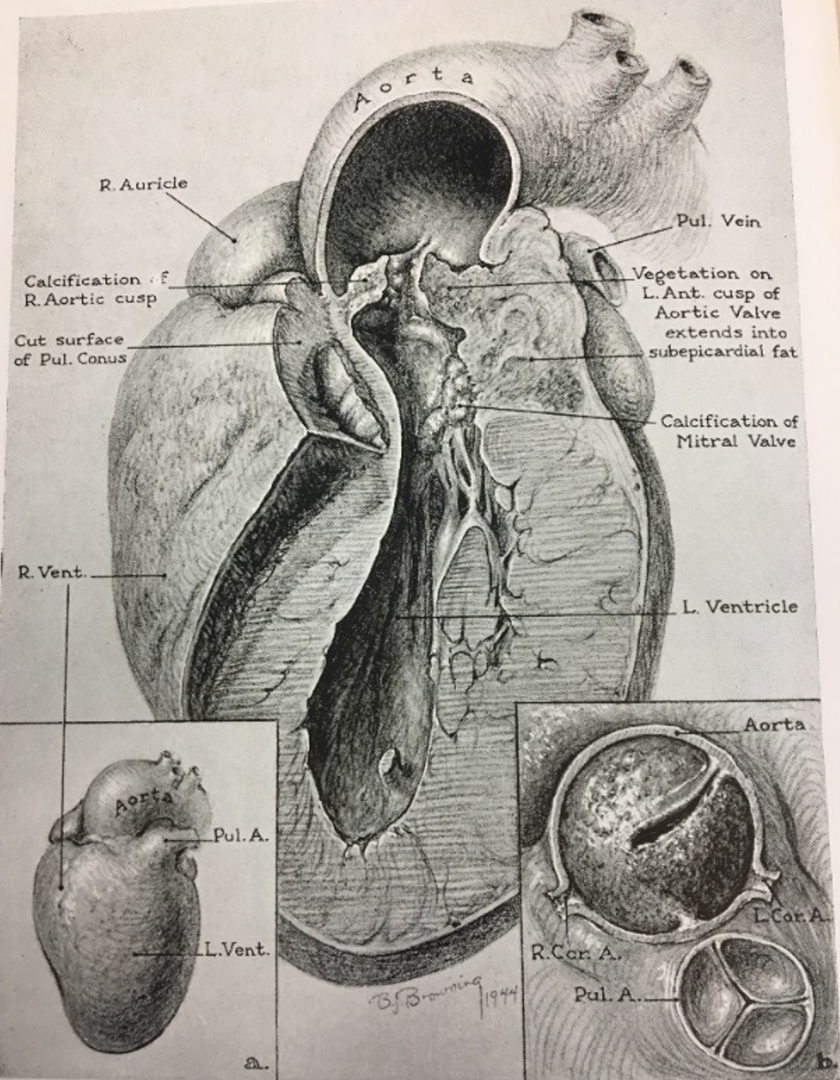
Detailed coronal views of specimens of aortic stenosis by Elizabeth Browning, also known as “BJ.” BJ used carbon dusting techniques with coronal sections. Reproduced with permission from Oxford University Press. Helen B. Taussig. Congenital Malformations of the Heart, Volume II: Specific Malformations. Cambridge, Massachusetts: Harvard University Press, Copyright © 1947, 1960 by The Commonwealth Fund.
The Era of Cardiac Surgery
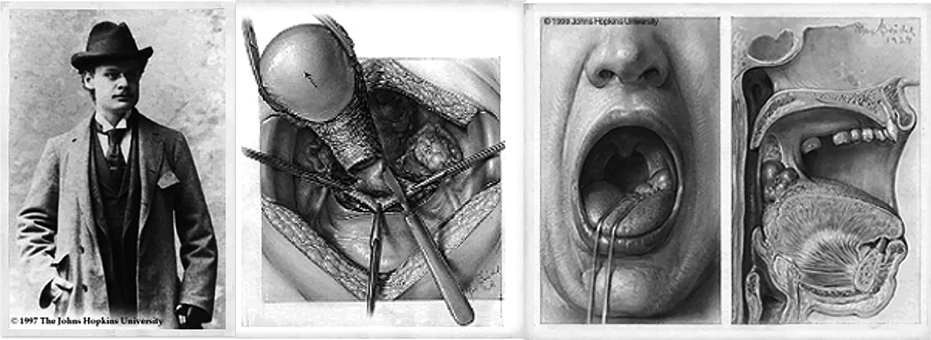
It was Crafoord, 55 Gross, 56 and Blalock 57 whose intrepid efforts with closed heart surgery started the path to open-heart surgery. Some historic photographs are worthy of mention. Figure S12 is a collage of the legendary figures who participated in the first Blalock-Taussig shunt, which included William Longmire (Chief Resident), Alfred Blalock (Surgeon), Hellen Taussig (Pediatric Cardiologist), Vivien Thomas (Laboratory Director), and Denton Cooley (Intern). The operation showing the anatomy, new clamps, and suturing technique was illustrated by Leon Schlossberg (Figure 4), a student of Brödel (1935). 58 He learned the “carbon dust” on Ross board technique and worked closely with Alfred Blalock, Hopkins Professors, and later Paul A. Ebert 59 to accurately assess and illustrate the intricate cardiac surgical methods; the new clamps and tools that were developed to perform the operations; and the ingenious methods to show depth, texture, and anatomic relationships. His career was interrupted by World War II in which he served as a Lieutenant Commander in the Navy as Officer-In-Charge of Medical Illustration. He returned to Hopkins in the late 1940s to be the principle illustrator for Alfred Blalock. He carried on the traditions of carbon dusting until the Ross board material was no longer available. He developed his own material using calcium-coated board applied by a spray gun and gave him the same effect as Ross board. For his efforts, this technique came to be known as “Schlossboard.” His knowledge of the three-dimensional concepts of the heart and vascular system aided surgeons in understanding complex abnormalities and the operative approaches used to repair them. 57 –59 Examples of these talents are shown in Figure S13, which describes surgical treatments of cyanotic congenital cyanotic disease (blue babies) and Blalock-Taussig shunt. The dynamic realism is displayed by tissue reaction to retraction and vessel clamping. New instruments are highlighted and show the technique of vascular mobilization and intracardiac anatomy.
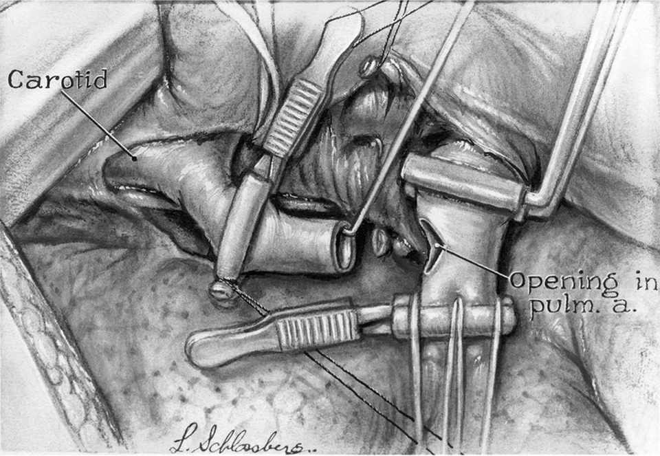
Leon Schlossberg documented the Blalock-Taussig shunt as shown in this drawing. New clamps and tissue texture are highlighted using carbon testing technique. Reproduced with permission from the Journal of the American College of Surgeons, formerly Surgery, Gynecology & Obstetrics.
Ted Bloodheart trained under Max Brödel in 1939 and became one of the first medical illustrators in Los Angeles. He associated with Dr Burt Mayer, John C. Jones, George Lindesmith, and Winfield Wells at the Los Angeles Children’s Hospital, where he utilized new media to create his carbon dust on LetraMax 4000-coated board, similar to the Brödel technique on Ross board. Examples of his work are noted in Figure S14. 60
Gerald P. Hodge graduated from Johns Hopkins under Ranice Crosby in 1949. 61 Ranice Crosby (Figure S15) was the third chairman of the Department of Art as Applied to Medicine at Johns Hopkins 62 and maintained the carbon dust techniques that were established by Brödel. In addition to Hodge’s artistic skills, while at the University of Michigan, he established a training program in 1965 in medical and biological illustration that revealed his alma mater, Johns Hopkins. His award-winning art that was created in association with Drs Herbert Sloan and Otto B. Gago highlighted the techniques of active suturing, hovering perspective, and epicardial texture that was prepared with carbon dusting on video paper (Figure S16). He was a consummate artist and dedicated mentor.
Herb Smith and Barry Baker, both graduates of the Johns Hopkins tradition under Ranice Crosby, accepted medical illustration positions at Baylor College of Medicine in Houston (∼1965) under Dr Michael DeBakey and his celebrated staff which included Drs Crawford, Dietrich, and Denton Cooley until the separation that created The Texas Children’s Hospital and the Institute at St Luke’s Hospital. This was a time of rapid surgical innovation. Both artists chose as their technique line drawings that had the dual effect of portraying accuracy and rapid execution for publication. Herb Smith’s technical highlights included a sequential educational process that was created by using multiple drawings highlighting the specific area of surgical intervention. Figure S17 shows resection of a ventricular aneurysm from incision to closure. Barry Baker portrayed his art using inserts and multiple steps in a single image as noted in Figure S17, which shows aortic valve replacement with coronary artery bypass.
Tim Hengst who also trained at Hopkins under Ranice Crosby in 1974 accepted a position at the Texas Heart Institute under Denton Cooley and was followed by William Andrews who studied at The University of Texas Southwestern Medical School in Dallas. Their illustrations were characterized by full range of values in line with pen and ink. Drawings (Figure S18) by Tim Hengst are shown by highlighted areas of action, sequential illustrations, and human (hand) proportionality. Tim Hengst was eventually recruited back to Hopkins in 1976, where he continued his robust career.
Late 20th Century Leading to the Early 21st Century
The era that spanned the late 20th and early 21st centuries was characterized by a sea change in surgical therapy for patients with transposition of the great arteries, functionally single ventricle, left ventricular outflow tract obstruction, and tetralogy of Fallot, among others. Innovative procedures such as arterial switch operation (ASO), Norwood operation, extracardiac Fontan, Fontan Conversion with arrhythmia surgery, Ross-Konno, and pulmonary valve sparing/restoration for tetralogy of Fallot repair have emerged. 63 Scientific reports were generated with illustrations that demonstrated the fine parts of each operation. While there were many programs and medical illustrators who participated in these advances, concentrated efforts were centered in Chicago at Children’s Memorial Hospital (CMH) starting first with Willis J. Potts whose pioneering efforts with the Potts’ shunt and coarctation of the aorta were illustrated by Virginia Samter, a talented medical illustrator who was graduated from University of Illinois at Chicago Medical Illustration School in 1947. 64 Her drawings showed the step-by-step procedures, highlighting the surgical techniques and new clamps made especially for the operations (Figure S19). Dr Farouk Idriss succeeded Dr Potts at CMH and concentrated his research and clinical efforts on anatomical correction of TGA. The initial drawings were illustrated by Virginia Samter (Figure 5). Subsequently, during the evolving clinical experience with the ASO, it was Zelda Zelinsky 65 who illustrated the progressive steps of the ASO in shades of grey (Figure 6). Interestingly, Zelda Zelinsky and Virginia Samter were both graduated from the University of Illinois at Chicago Medical Illustration School in the same class of 1947. In 1989, Dr Constantine Mavroudis succeeded Dr Farouk Idriss, assumed the research and clinical directions that were established at CMH, and brought his own vision of medical education through publication and illustration. It was at this time that Rachid Idriss, the son of Dr Farouk Idriss, completed his education in medical illustration and associated with Drs Mavroudis and Backer at CMH. Concurrently, Dr Mavroudis assumed the editorship of the textbook, Pediatric Cardiac Surgery from Arciniegas who edited the first edition. 66 The second, third, fourth, 63 and soon to be fifth editions followed and were principally illustrated by Rachid Idriss. In the later stages of this association, the Atlas of Pediatric Cardiac Surgery 67 was published and was followed by the Atlas of Adult Congenital Heart Surgery. 68 Idriss’s style is grounded in his educational experience and his own unique understanding of the anatomy and expression of the details.
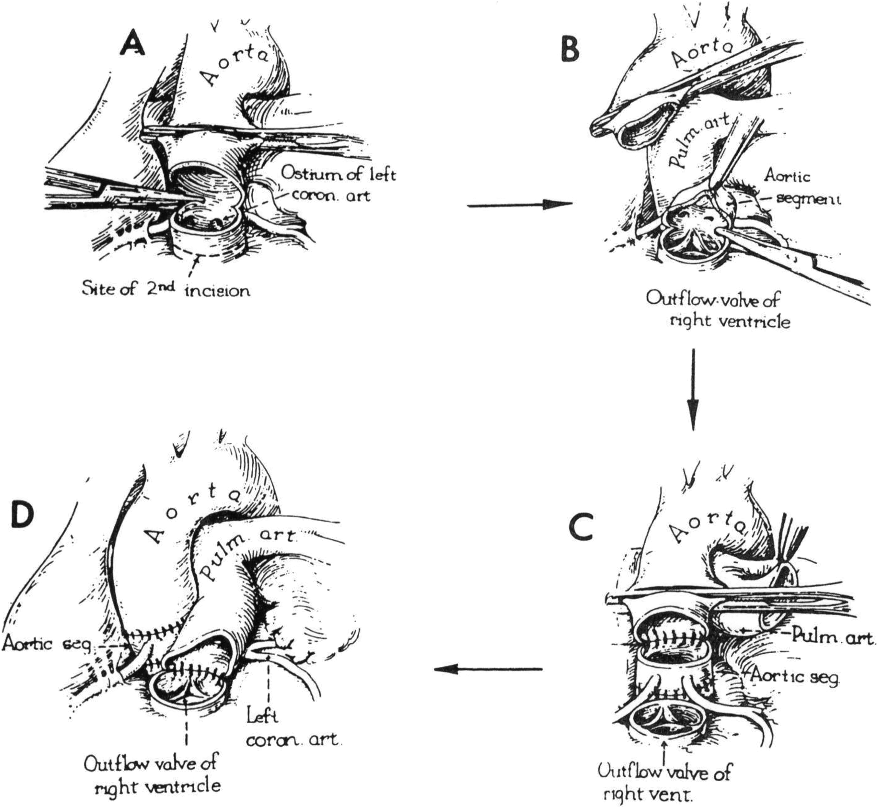
Virginia Sumpter also worked with Dr Farouk Idriss, whose pioneering efforts to develop the arterial switch operation started in 1961. Her rendition of Idriss’s notion of the arterial switch operation is shown in four drawings, depicting the important steps of the procedure. Of some interest is that cardioplegia was not yet introduced and that arterial blood gas determination was not clinically available. Reproduced with permission from Elsevier. Arterial switch published by Hanley and Belfus. Mavroudis C, Backer CL, eds. The Arterial Switch Operation. Philadelphia, PA: Hanley & Belfus, Inc; Cardiac Surgery: State of the Art Review. Vol. 5; 1991.
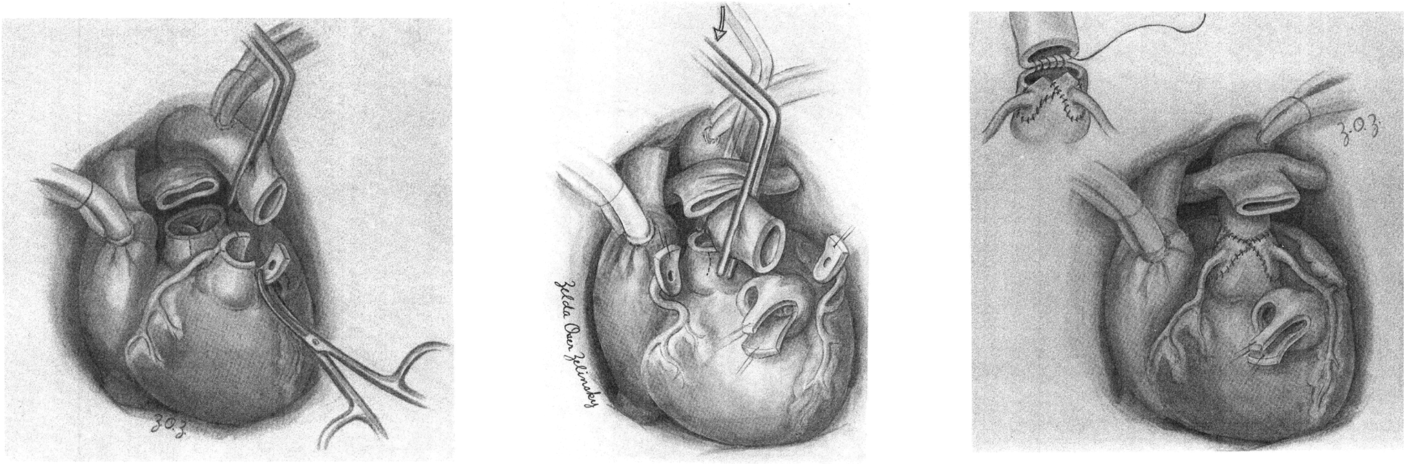
Zelda Zelensky started to work with Farouk Idriss shortly after Virginia Sumpter. Her style was tonal grades of gray and was depicted to give more texture to the illustration. Shown here are Zelensky’s rendition of steps of the arterial switch operation, which were perfected after the introduction of cardioplegia, arterial blood gas determination, and fine suture techniques. Left panel shows the coronary artery dissection after great vessel transection. Middle panel shows the maneuver of Lecompte portrayed by the “Ghost” clamp that was moved from the distal ascending aorta (ghosted clamp) to the proximal ascending aorta (actual clamp). The dissected coronary artery buttons are shown in preparation for the neoaortic reconstruction. Right panel shows the completed neoaortic reconstruction, with the inset demonstrating the suturing technique that was introduced by Dr Idriss. Reproduced with permission from Dr Constantine Mavroudis who retains the copyright.
Annotated Techniques That Highlight Medical Illustration
We have categorized seven different methods how medical illustrators use their talents and expressive ideas to demonstrate procedures and give them life. These methods are (1) hovering technique; (2) hidden anatomy, ghosted views, or transparency; (3) centrally focused perspective; (4) action techniques to give life to the procedure; (5) use of insets to highlight one part of the drawing; (6) human proportionality using hands or known objects to show relative size; and (7) step-by-step educational process to depict the stages of a procedure.
The “hovering technique” may have been introduced by Schlossberg in his illustration of the Blalock-Taussig shunt and Paul Ebert’s description of ventricular septal defect (VSD) closure. These methods (not shown) allow the observer to visualize the safe location of suture placement en route to VSD closure. These techniques can also facilitate the understanding of complex operations such as the Konno procedure. Hovering techniques show the aortic valve prosthesis being implanted, ventricular augmentation patch being sutured, and pulmonary outflow tract patch completing the operation. Rachid Idriss’s hovering technique for VSD closure (Figure S20) follows the same principles. Idriss takes the hovering technique one step further in his demonstration of the Florida Sleeve Operation 68 (Figures S21-S23). This detailed series of step-by-step explanation of a complex operation shows numerous techniques leading to a comprehensive educational experience. In particular, the hovering technique visualizes how the graft can be openly displayed with appropriately placed pledgetted sutures, which when tied will produce the desired effect.
The technique of showing “hidden anatomy” is also known as “ghost views” and “transparency.” This effect is particularly important in congenital heart surgery, since internal anatomy can be so challenging to the conduct of the operation. Figure S24 shows the technique using lighter tones to mimic a ghost appearance. Other methods use dotted lines to indicate a structure that is anatomically behind another. The spatial interrelationships allow the observer to visualize and imagine in three dimensions that aid in the understanding of the anatomy and the operation that is being explained.
The “centrally focused perspective” allows the artist to deemphasize the periphery of the operative field and call attention to the important part of the operation that is directed to the center of the illustration. The camera sees all; the medical illustrator focuses the important elements of the message to the viewer. This is beautifully shown in the greater majority of all the illustrations shown herein.
“Action techniques” animates the drawing that adds life to the procedure. The combination of shades of gray that connote texture along with sutures that are shown in space as serpiginous forms gives the impression that the sutures are in the act of being placed. The result is an active display of the ASO. Figure 7 and Figures S25 to S28 show the serpiginous display of sutures in action.
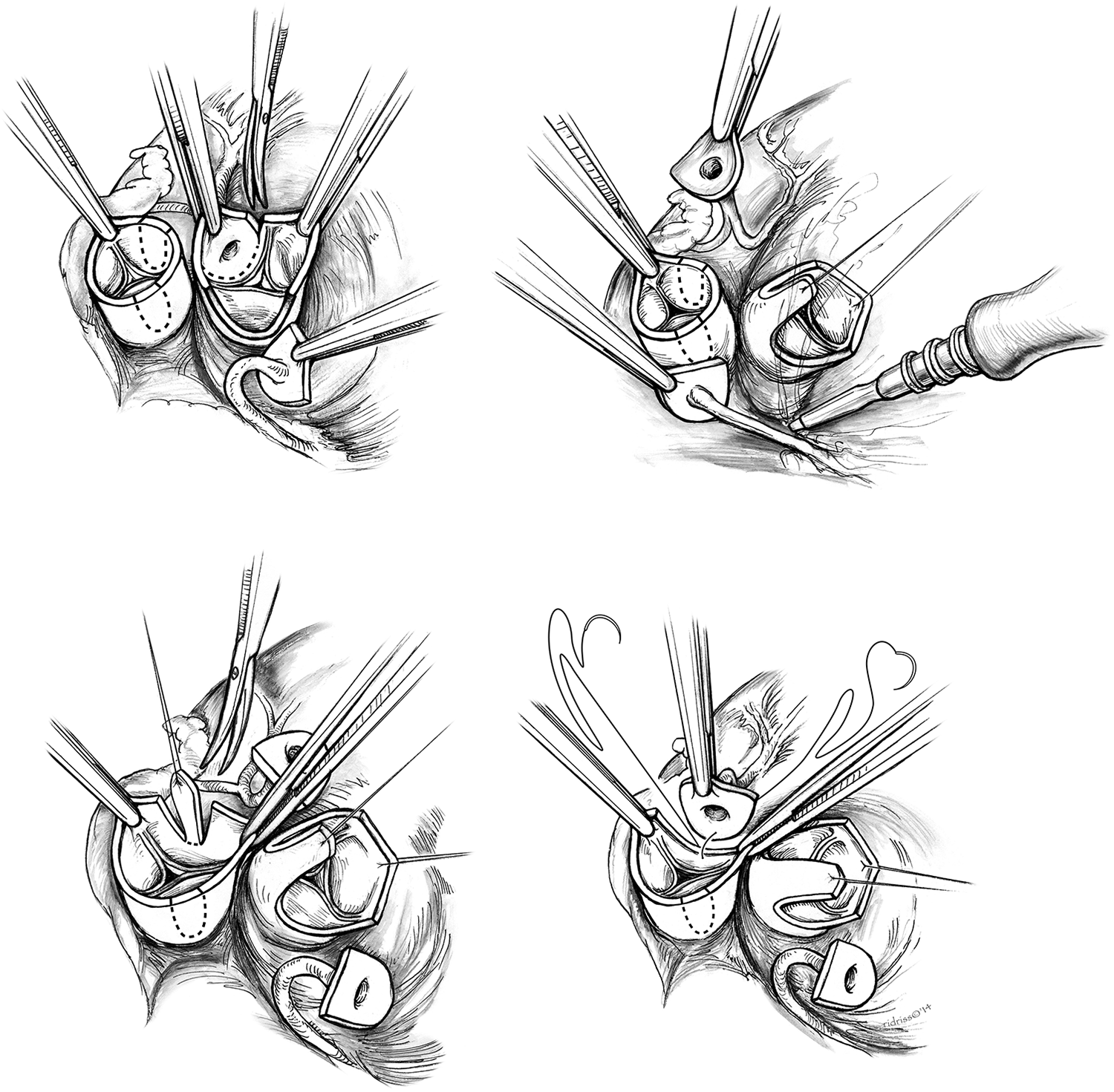
This series of drawings depicts the initial complex steps of the arterial switch operation as illustrated by Rachid Idriss. The important preliminary stages of the operation leading up to coronary artery dissection, which is shown herein (upper left panel), have been omitted. This first drawing (upper left panel) takes up the technique of coronary artery dissection and coronary button creation. The important aspects of this illustration are preparation of the coronary buttons from the ascending aorta with eventual reimplantation to the ascending pulmonary artery, shown with dashed lines, which will become the neoaorta. In the right upper panel, the aorta, soon to be the neopulmonary artery, is shown retracted to confer attention to the neoaortic reconstruction that is shown in progress by aligning the coronary buttons to their proposed reimplantation sites. Low-set electrocautery is shown freeing the right coronary artery attachments for complete mobilization. The smoke is a subtle technique to show action. In the left lower panel, the neoaortic sinuses of Valsalva are being prepared for coronary reimplantation. The indication is that the surgeon removes no more than two-third the size of the coronary button. This principle is shown very nicely in the drawing. In the right lower panel, the left coronary button is being reimplanted into the facing sinus of Valsalva. The idea here is to start the anastomosis as noted to avoid time delays with visualizing the repair. From Atlas of Pediatric Cardiac Surgery, 2015. Reproduced with permission from Springer Nature.
“Insets” in an illustration have the same effects of a maritime map that uses a focused explanation of a complex water passage, hazardous harbor, or mooring site. This method allows the observer the ability to visualize both the Olympian view of the process while making quick references to the focused display, all in one illustration.
“Human proportionality” is a method to indicate the size relationship between the operative subject and the human. The idea is that the observer knows more or less how large her hand, a Kelly clamp, and a scalpel is compared to the anatomic structure.
Perhaps one of the most important techniques of cardiac surgery illustration is the “step-by-step” technique, which depicts, in an organized educational process, the stages of an operation. This illustrative technique is shown for the critical steps of total arch reconstruction and debranching for aortic dissections involving the ascending aorta and transverse arch (Figure S29, S30). Critical to understanding the steps of this operation is minimizing circulatory arrest times and absence of cerebral flow.
Modern Times and the Medical Illustrator
Rachid Idriss is joined by many other medical illustrators 69 –71 who have made sentinel contributions during times of great advances in surgical procedures. Rebekah Dodson 69 is the principle medical illustrator for Comprehensive Surgical Management of Congenital Heart Disease, 2nd edition, by Dr Richard Jonas 72 (Figure S31). She is a talented illustrator who is as comfortable with continuous tone techniques as well as pen and ink watercolor demonstrations. Other medical illustrators include Robert Benassi, 70 who worked at the Mayo Clinic, and Jack Desley, 71 who was the principle illustrator for Dr John Kirklin. 73
There is no intent to exclude any illustrator's work from this analysis. Our purpose was to present a focused historic overview of selected medical illustrators and their creations, highlight the techniques of medical illustration leading up to the era of cardiac surgery, and to extoll the importance of medical illustrators in the educational process of learning complex open-heart procedures. While new technologies of medical imaging are extant 74 and developing rapidly, these methods are the “cameras” of anatomical reproduction. The camera, computed tomography scan, magnetic resonance imaging, reconstructed heart models, and arteriograms are clearly important for diagnosis, operative planning, and therapeutic interventions; however, they do not “illustrate.” Illustration focuses, interprets, emphasizes, directs, cautions, explains, and, in the most fundamental way, teaches. The association between medical illustrator and clinician has ancient roots. There is no substitute for human interaction, spatial interpretation, and medical education in its most robust form, illustration.
Supplemental Material
Supplemental Material, Figures_1-7_presentation_format - Medical Illustration in the Era of Cardiac Surgery
Supplemental Material, Figures_1-7_presentation_format for Medical Illustration in the Era of Cardiac Surgery by Constantine Mavroudis, Gary P. Lees and Rachid Idriss in World Journal for Pediatric and Congenital Heart Surgery
Supplemental Material
Supplemental_Figures - Medical Illustration in the Era of Cardiac Surgery
Supplemental_Figures for Medical Illustration in the Era of Cardiac Surgery by Constantine Mavroudis, Gary P. Lees and Rachid Idriss in World Journal for Pediatric and Congenital Heart Surgery
Footnotes
Declaration of Conflicting Interests
The author(s) declared no potential conflicts of interest with respect to the research, authorship, and/or publication of this article.
Funding
The author(s) received no financial support for the research, authorship, and/or publication of this article.
Supplemental Material
Supplemental material for this article is available online.
References
Supplementary Material
Please find the following supplemental material available below.
For Open Access articles published under a Creative Commons License, all supplemental material carries the same license as the article it is associated with.
For non-Open Access articles published, all supplemental material carries a non-exclusive license, and permission requests for re-use of supplemental material or any part of supplemental material shall be sent directly to the copyright owner as specified in the copyright notice associated with the article.