
Abstract
Ulvan from green algae (Ulva lactuca L.) has been reported to possess potential anti-inflammatory activity, and nanotechnology-based topical formulations have been widely explored to enhance its efficacy. This study aimed to develop an ulvan nanoemulgel and evaluate its in vivo anti-inflammatory activity in mice. Ulvan was extracted by ultrasonication at 70 °C using distilled water, followed by ethanol precipitation. Ulvan extraction yielded 65.73% with a melting point of 196 °C–198 °C, and Fourier Transform Infrared Spectroscopy (FTIR) analysis confirmed characteristic functional groups of ulvan. The optimized nanoemulsion has a good physical appearance, droplet size of 35.20 ± 2.77 nm, polydispersity index (PDI) of 0.428 ± 0.053, and zeta potential of −12.54 ± 0.41 mV. The optimized nanoemulsion formula was incorporated into a 2% hydroxypropyl methylcellulose (HPMC) gel base containing 8% ulvan. The nanoemulgel has a droplet size of 197.23 ± 15.32 nm, PDI 0.437 ± 0.016, pH 6.04 ± 0.02, viscosity 1,421.47 ± 59.31 centipoise (cps), spreadability of 6.82 ± 0.03 cm, and adhesiveness 12.54 ± 0.41 seconds. Anti-inflammatory activity was evaluated in mice using four mice per group (determined according to Federer’s formula). Statistical analysis was performed using analysis of variance with a 95% confidence interval. The anti-inflammatory activity increased from 7.66% (ulvan) to 24.96% (ulvan gel) and 38.41% (ulvan nanoemulgel), indicating enhanced efficacy through formulation. These results demonstrate the potential of ulvan, particularly in nanoemulgel form, as a promising topical anti-inflammatory agent.
Introduction
Ulvan is one of the main components of Ulva lactuca seaweed, which has been reported to possess biological activity both in vitro and in vivo. 1 Ulvan content in seaweed is derived from the cell wall of the Ulva genus. 2 Previous studies have reported the potential of the compound as anti-inflammatory 3 and analgesic. 4 In addition, ulvan can be extracted using distilled water, acid, and enzymes. 5 Extraction with an autoclave at 121 ℃ for 15 minutes produced ulvan sediment with a yield of 1.98%, a total sugar content of 46.1%, and a sulfate content of 21.53%. 6
According to previous studies, ulvan, which consists of rhamnose, iduronic acid, xylose, glucuronic acid, and galactose, is a polymer with a large molecular weight (1,068.2 kDa). 7 Consequently, it has a low solubility, absorption, and bioavailability, particularly when used orally.8–10 This study formulated ulvan into nanometer-sized droplets to increase the pharmacological effect. Despite the potential, to our knowledge, ulvan-loaded nanoemulgel for topical anti-inflammatory evaluation in a carrageenan-induced mouse back-skin model has not been previously reported, but has been more developed in oral preparations.11–13 This topical preparation is a good alternative as an anti-inflammatory agent because it is delivered directly to the inflamed area. Topical use can also improve patient compliance and reduce side effects or unwanted effects. 14
Nanoemulgel in this current study consists of an oil-in-water emulsion incorporated in a gel base. The formation of nanoemulgel makes nanoemulsion more stable, increasing viscosity and adhesiveness. 15 The formation of nanoemulgel with the incorporation of ulvan in nanometer-sized globules has the potential to improve the pharmacological effect of the compound. In addition, the high water concentration in the gel system hydrates the stratum corneum, making the drug easily penetrate the skin. 16 Nanometer-sized nanoemulsion globules as the dispersed phase can carry the drug through the largest barrier in the skin, moving easily across the stratum corneum.14,17 The active substance in the nanoemulsion system often moves from the internal phase (nanoemulsion) to the external phase (gel), thereby creating a drug reservoir. The oil globules are then released from the gel base and delivered to the skin, where the globules penetrate the stratum corneum. In a formula consisting of oil, surfactants, and a combination with cosurfactants, the gel base can act as a penetration enhancer. The addition of a gelling agent can increase adhesion to the skin, helping to increase effectiveness. Therefore, nanoemulgel effectively retains the drug, enabling controlled release while also increasing permeation into the skin. 18 Previous studies have shown that curcumin nanoemulgel preparations can increase the permeation flux of compounds through the skin up to two times compared to regular gels. 14 In a previous study, 19 ropinirole penetration increased 7.5 times with nanoemulgel compared to regular gels. Compared to oral tablet preparations, the formation of carvedilol nanoemulgel increased the area under the curve (AUC) by 1.72 times. 20 These results indicate that the formation of nanoemulgel has the potential to improve the anti-inflammatory effect of active substances.
Carrageenan is a compound widely used as an inflammatory agent in animal models to evaluate the activity of anti-inflammatory drugs. Several in vitro analyses have shown that carrageenan can induce inflammation by triggering the innate immune pathway, involving the activation of nuclear factor kappa B (NF-kB), which plays a role in the activation of tumor necrosis factor alpha (TNF-α) and Interleukin-6 (IL-6).21,22 In silico studies have also shown the potential of ulvan polysaccharides as anti-inflammatories through inhibition of cyclooxygenase (COX)-2 enzyme equivalent to diclofenac sodium. 23 The potential of green algae ethanol extract as an inhibitor of COX-2 expression was also demonstrated in ulcer-induced mice. 24 Green algae water extract has been shown to possess analgesic and anti-inflammatory effects in vitro using the membrane stabilization method and in vivo using carrageenan induction with diclofenac sodium as a comparator. 4 In the present study, in vivo anti-inflammatory activity was evaluated using the carrageenan induction method on the backs of mice. Therefore, this study aims to extract and formulate ulvan from Ulva lactuca L. into gel and nanoemulgel, as well as to test its topical anti-inflammatory activity in mice.
Materials and Methods
Materials
The material used in this study was Ulva lactuca L., obtained from Drini beach, Gunung Kidul Regency, collected in May 2025, and had been identified by the Department of Pharmaceutical Biology, Faculty of Pharmacy, Gadjah Mada University, under number 2439/UN1/FA.2/BF/PT.01.06/2025. Material for ulvan extraction comprised distilled water and 96% ethanol (Merck, USA). Gel and nanoemulgel making materials consisted of hydroxypropyl methylcellulose (HPMC) (SARDA Manufacturing Substance Pharmaceutical, Taiwan), methyl paraben (Bratachem, Indonesia), propylene glycol (Bratachem, Indonesia), olive oil (Sasso, Italy), Tween 80 (Bratachem, Indonesia), and PEG 400 (Bratachem, Indonesia). Furthermore, the materials for the anti-inflammatory test consisted of type I carrageenan (Merck, Germany), diclofenac sodium gel (Voltadex, Dexa Medica, Indonesia), and 0.9% physiological NaCl (Brataco, Indonesia).
The experimental animals used were two-month-old male mice (BALB/c) weighing around 25–30 g, obtained from the Pharmacology Laboratory of Universitas Ahmad Dahlan. The animals were given pre-treatment in the form of a new habitat adjustment in a 45 × 30 cm cage with a room temperature of 24 °C–25 °C, with a humidity of 40%–70% in the Pharmacology Laboratory of Universitas Ahmad Dahlan. The experimental animals were given standard feed and drinking water with a light cycle setting of 12 hours of light and 12 hours of darkness. The test procedure received ethical approval from the Study Ethics Committee of Universitas Ahmad Dahlan, number REC-UAD/02/01/07-2025/086.
Methods
Extraction of Ulvan from Ulva lactuca L.
Extraction process was conducted using sonication at a frequency of 40 kHz at a temperature of 70 °C for 90 minutes. A total of 100 grams of fine green algae simplicia was dissolved in distilled water with a sample and solvent ratio of 1:20. 25 After the extraction process produced seaweed slurry, the extract was filtered using a vacuum pump (Krisbow, Indonesia). The filtrate obtained was then evaporated with a water bath at a temperature of 50 °C to remove water content. 26 Subsequently, the filtrate was precipitated with 96% ethanol to obtain pure ulvan with a ratio of 1:2 and then allowed to solidify for 12 hours at a chilling temperature. 27 The solid phase of the filtrate (pure ulvan) was dried in an oven (Memmert, Germany) at 40 °C for 6 hours. 28
Characterization of Ulvan from Ulva lactuca L.
Characterization of ulvan-based on melting point measured by Stuart Scientific melting point apparatus-SMP3 (United Kingdom) 29 and infrared spectrophotometer FTIR/UATR spectrophotometer (Spectrum Two by Perkin Elmer and Nicolet Avatar 360 IR, USA) 2 has been equipped with the attenuated total reflectance technique. The sample, which was about 5 mg, was placed on a plate containing a single germanium crystal. By reducing atmospheric background interference, the spectrum was obtained using 128 scans with a resolution of 4.0 cm⁻ 1 and in the range of 4,000–400 cm⁻ 1 .
Formulation of Ulvan Nanoemulsion
The development of ulvan nanoemulsion was done using a spontaneous emulsification method consisting of 10% olive oil, 67.5% Tween 80, and 22.5% PEG 400. In the first stage, Tween and PEG 400 were mixed using a vortex (KA Eurostar, Staufen im Breisgau, Germany) for ±1 minute until homogeneous. Subsequently, a mixture of olive oil that had been mixed with ulvan was added to the mixture. The mixing process was continued using a vortex for ±2 minutes until homogeneous. 30 To produce nanoemulsion, distilled water was added in a ratio of 1:10 and continuously agitated with a magnetic stirrer until a transparent system was formed. 31 Furthermore, the amount of ulvan incorporated into nanoemulsion was gradually increased from 90.0, 100.0, 110.0, and 120.0 mg per gram of nanoemulsion to determine the maximum ulvan concentration that produced a clear formulation.
Characterization of Ulvan Nanoemulsion
Characterization of the nanoemulsion included determination of particle size, polydispersity index (PDI), and zeta potential. A total of 1,000 µL of sample was placed into a cuvette to be analyzed 30 for globule size and zeta potential using a particle size analyzer (Malvern, Germany).
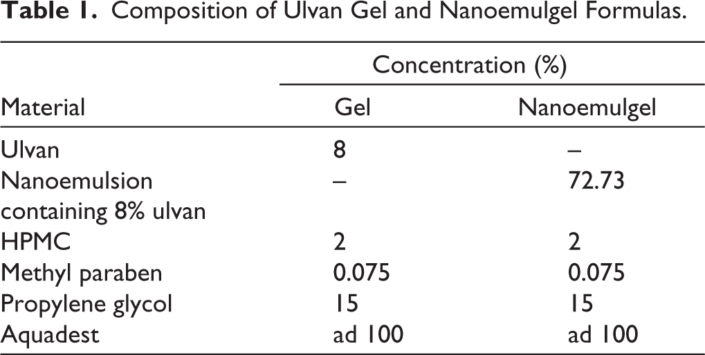
Formulation of Ulvan Gel and Nanoemulgel
Gel and nanoemulgel were formulated from ulvan, as shown in Table 1. Gel preparation was started by dissolving HPMC in 30 mL of heated Aquadest at 80 °C–90 ºC until a homogeneous dispersion was formed. Subsequently, methyl paraben was added to the mixture using a stirrer at 700 rpm for 15 minutes. Propylene glycol and ulvan were added little by little while stirring with a stirrer, and Aquadest was added up to 100 mL into the formula until homogeneous using a stirrer at 700 rpm for 25 minutes, and stored in a tightly closed container. 32 The same method was used to develop a nanoemulgel containing ulvan nanoemulsion.
Composition of Ulvan Gel and Nanoemulgel Formulas.
Physical Evaluation of Ulvan Gel and Nanoemulgel
Physical properties of ulvan gel and nanoemulgel included a viscosity test using a Brookfield viscometer at 1–10 rpm. In this study, viscosity results were expressed in centipoise (cps) as the average of three repetitions. 33 The spread diameter test was carried out with a glass object given a load of 150 g for one minute, and the diameter of the nanoemulgel that spread was measured by taking the average length of the four sides. 34 Furthermore, the adhesion test was conducted on the surface of the object glass, given a load of 1 kg for five minutes, mounted on an adhesion tester with a load of 80 grams, and the time for the glass to release the adhesive was recorded, 34 while the pH was measured using a calibrated pH meter (Mettler, Swiss).
Topical In Vivo Anti-inflammatory Test with Carrageenan Induction
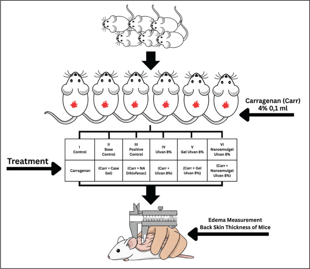
Mice were divided into six groups, with four mice per group, namely the control group (carrageenan), base control group (carrageenan + gel base), positive control group (carrageenan + diclofenac sodium gel), 8% ulvan treatment group (carrageenan + 8% ulvan), 8% ulvan gel treatment group (carrageenan + 8% ulvan gel), and 8% nanoemulgel treatment group (carrageenan + 8% nanoemulgel). Gel base, gel, and nanoemulgel treatments were given at 0.02 g. The number of replications for each group (four mice per group) was determined using Federer’s formula, (t − 1) (r − 1) ≥ 15, where t represents the number of treatment groups and r represents the number of replications. 35 Based on this calculation, a minimum of replications per group was required to ensure sufficient degrees of freedom for the experimental error. Since the study involved six treatment groups (t = 6), the formula (t − 1) (r − 1) ≥ 15 yields a minimum requirement of four replications (r = 4). Accordingly, four mice were used per group in this study to satisfy this criterion.
In vivo anti-inflammatory activity testing was conducted using the carrageenan induction method on the backs of mice. 36 Mice were shaved on the back area of 2 × 2 cm, put back into the cage, and left for 1 × 24 hours. Inflammation in mice was induced by injecting 0.1 mL of 4% carrageenan in physiological NaCl solution subcutaneously into the backs of mice. Before being given 4% carrageenan, the initial skin thickness (V0) was measured using a caliper. Edema measurements were carried out on the back folds of mice with a caliper every 30 minutes for four hours, and an anti-inflammatory testing scheme is shown in Figure 1.
Schematic of Anti-inflammatory Test Using Carrageenan Induction Method on the Back of Mice.
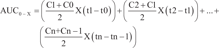
In this study, the fold thickness data were expressed as edema volume, and a time versus edema volume relationship curve was created to calculate AUC using the formula Equation 1. AUC data were statistically analyzed using Statistical Product and Service Solution (SPSS) with the analysis of variance test at a 95% confidence level, and continued with the Bonferroni test for differences between groups. The percentage of anti-inflammatory power was calculated from the comparison of AUC values of the treatment group with the control group, which was presented in Equation 1.
Description:
AUC0-x:
AUC of back-skin edema thickness in mice from 0 to 4 hours
Cn-Cn-1:
Amount of edema thickness from 0 minutes to 4 hours
tn-tn-1:
Measurement time from 0 minutes to 4 hours
AUC(0-x)0:
Average AUC value of the negative control group (mm/hour)
AUC (0-x)n:
Average AUC value of the treatment group given the test substance at dose n
Results and Discussion
Extraction of Ulvan from Ulva lactuca L.
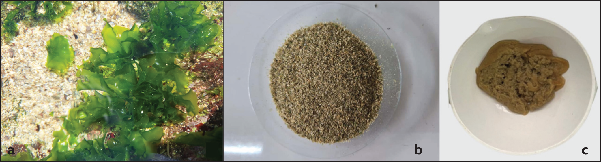
Ulvan was extracted from Ulva lactuca L. using an ultrasonicator (Elmasonic P, Germany) (ultrasonication method), resulting in a yield of 65.73%, greater than the previous study using the hot water extraction (HWE) method. 37 Fresh Ulva lactuca L., dried simplicia, and ulvan were presented in Figure 2. The superiority of the sonication method was also supported by previous results that reported a yield of around 30% in just 30 minutes. 5 However, previous studies showed that the HWE method combined with ethanol precipitation produced a yield of 36.5% after 20 hours of extraction. 38 This comparison showed that although conventional methods still had the potential to provide competitive results, ultrasonic technology offered more optimal time efficiency and yield.
(a) Fresh Ulva lactuca L., (b) Ulva lactuca L. Simplicia Powder, (c) Ulvan.
Characterization of Ulvan from Ulva lactuca L.
Ulvan was characterized by measuring the melting point compared to a previous study. The test results showed that the extracted ulvan had a melting point of 196 °C and began to change color to brown at 198 °C. This value was consistent with reports29,39 stating that ulvan had a high melting point (190 °C–200 °C) due to its complex polysaccharide structure with many hydrogen bonds and sulfate groups. A high melting point indicated a high degree of polymerization, because the longer the polysaccharide chain, the stronger the intermolecular forces that must be overcome during the melting process. 40 Therefore, a value of 196 °C showed that the isolated ulvan had a relatively high degree of polymerization. The color change at 198 °C indicated the beginning of thermal degradation and caramelization.
Ulvan was characterized using an infrared spectrophotometer based on the functional groups C=O carbonyl, carboxylic acid, hydroxy O–H, and sulfate. FTIR analysis in Figure 3 showed a strong absorption at a wave number of 3,284 cm⁻ 1 , which indicated the presence of hydroxyl groups (–OH). This result was consistent with a previous study that identified an absorption band around 3,400 cm⁻ 1 , showing stretching of the hydroxyl group. 25 Another study also revealed that O–H groups usually appeared at wavelengths around 3,300 cm⁻ 1 . 33 Meanwhile, it was reported that the absorption of ulvan appeared at 3,395 cm⁻ 1 , with O–H stretching vibration in the range of 3,000–3,600 cm⁻ 1 . FTIR spectrum at wavenumber 2,928 cm⁻ 1 indicated the presence of alkane (C–H) functional groups in the sample. 6 This result was in accordance with a previous study reporting that the absorption at 2,927 cm⁻ 1 was related to alkane groups. 6 Furthermore, the absorption band at 2,937 cm⁻ 1 also reflected C–H vibration, which was one of the main characteristics in the structure of polysaccharides. 2 This result was also supported by previous results, which showed that the weak peak in the range of 2,925–2,928 cm⁻ 1 originated from the aliphatic C–H stretching vibration of the methyl group. 41
Infrared Spectrum of Extracted Ulvan.
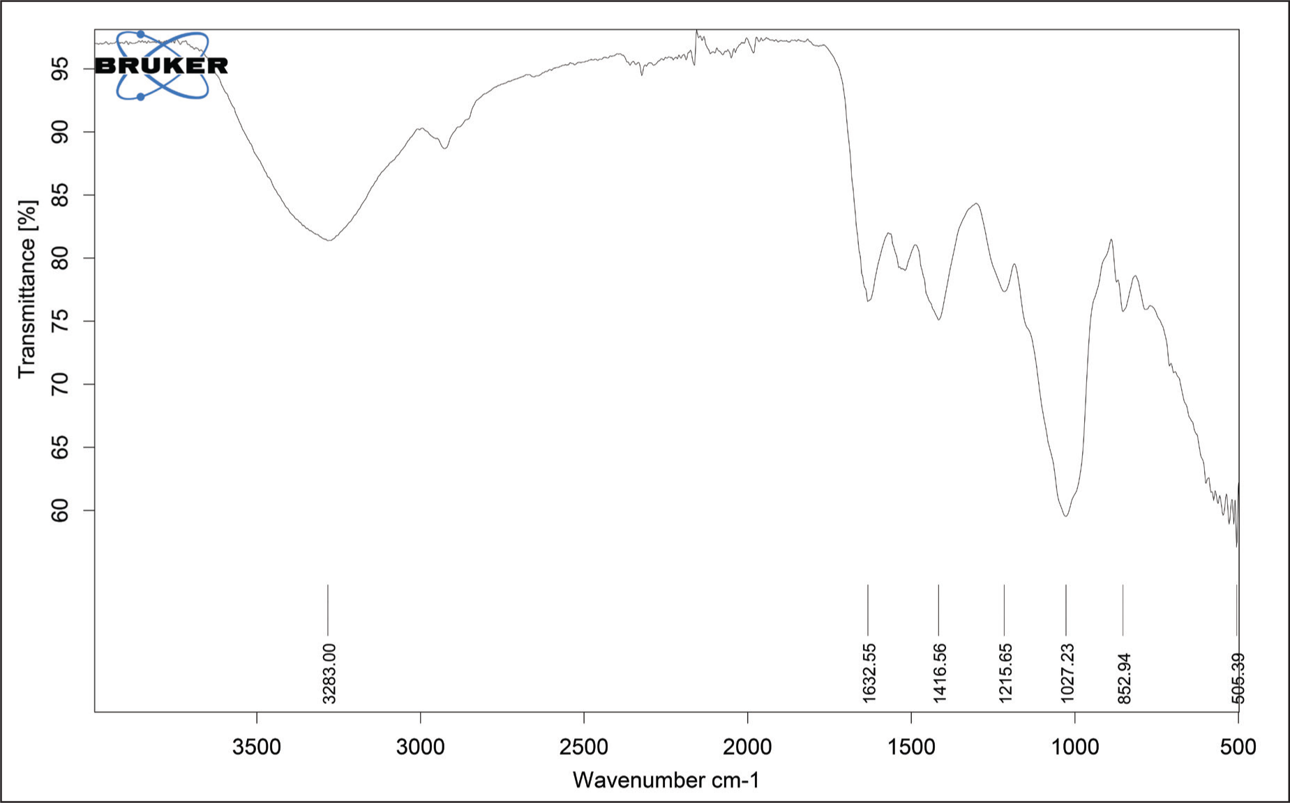
The spectrum at wave number 1,632.55 cm⁻ 1 indicated the presence of a carboxylic group (C=O) in the green algae sample. Furthermore, it was reported that absorption bands with similar intensity related to the carboxylic group of uronic acid appeared around 1,651 and 1,435 cm⁻ 1 . 2 Previous research identified the carboxylic group at an absorption of 1,637 cm⁻ 1 . 6 In general, this group was detected in the wave number range of 1,632–1,597 cm⁻ 1 for the asymmetric vibration of carboxylic acid, and between 1,448–1,400 cm⁻ 1 for its symmetric vibration. 6 Furthermore, absorption peaks were also obtained at 1,416.56, 1,215.65, and 1,027.32 cm⁻ 1 . The absorption indicated the presence of C–O functional groups (single bonds between carbon and oxygen) and of ether-containing compounds. The wave number for the C–O group ranged from 1,200 to 1,000 cm−1, which described C–OH and C–O–C groups of the sugar ring and glycosidic bonds. 6 While in the FTIR spectrum, approximately 1,027 cm⁻ 1 referred to C–O stretching of the main rhamnose sugar. 41 Based on a previous study, the wave number 1,150–750 was a typical region for ulvan. 38 In the range of 1,400–1,600 cm⁻ 1 wave absorption bands, stretching of the O–C–O group was also found as a marker for carboxyl groups and uronic acids in ulvan. 38 Meanwhile, according to previous references, the C–O functional group could also be marked in the 983 cm−1 wave absorption band. The results of the determination of the extracted ulvan functional group based on infrared spectra are shown in Table 2.
Determination of Ulvan Functional Groups from Extraction Results Based on Infrared Spectra.
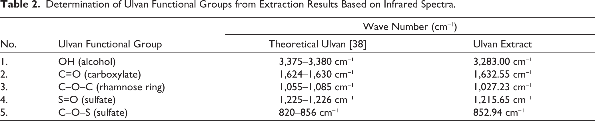
The peak of 1,215.65 cm−1 obtained in this study corresponded to the S=O stretching of the sulfate group. 42 This was consistent with previous results, stating that the S=O functional group was found in the absorption bands of 1,215 cm−1 and 1,257 cm−1.6,38 According to previous research, the S=O group was indicated by a wave region of 1,260–1,240 cm−1. 6 The data above also showed the presence of FTIR spectrum absorption at a wave of 852.94 cm−1. This spectrum indicated that there was a glycoside group in the sample. Based on a previous study, the peak of 845 cm−1 was the C–O–S stretching region of the sulfate group. 42 The presence of absorption in the form of a small band in the 852.94 cm−1 wave region showed the presence of the C–O–S functional group (usually found in ulvan, as a marker of the polysaccharide form and the presence of β-glycosides). 25 The C–O–S functional group was often found as the main marker of ulvan. Based on the analysis of the characteristics of the melting range and identification of functional groups with infrared spectra, it was concluded that the extraction result was an ulvan-rich extract.
Formulation and Characterization of Ulvan Nanoemulsion
Several studies revealed that ulvan is a polymer with a large molecular weight (1,068.2 kDa), 7 consequently, it has a low solubility and absorption.8–10 Therefore, incorporation into nanoemulsion is a suitable option to increase the pharmacological effect. The surfactant and cosurfactant in this formula follow the proven ratios established in our earlier research, which produce a stable and clear emulsified form. 30 Nanoemulsion components in this study, including olive oil, Tween 80, and PEG 400, were also selected based on low cost and availability. To develop the oil-in-water nanoemulsion, Tween 80 (HLB 15) and PEG 400 were combined to emulsify olive oil, a natural source of long-chain triglycerides (C18). The low water solubility of such vegetable oils requires elevated surfactant levels to achieve proper dispersion. While Tween 80 provides the necessary HLB for the o/w system, the addition of PEG 400 as a cosolvent is critical for minimizing interfacial tension. This combination enhances the fluidity of the surfactant at the oil interface and boosts drug solubility, which collectively improves the homogeneity of the system. 43 An ideal nanoemulsion often has a clear, homogeneous system with no apparent precipitation or phase separation either in the emulsified or original form, and this indicates the solubility of the incorporated drug. 31 Figures 4a–c showed the clear appearance of ulvan nanoemulsion with no visible sedimentation.
Visual Appearance of Nanoemulsion Containing Ulvan at Concentrations of 90.0 mg (a), 100.0 mg (b), and 110.0 mg (c) per g Nanoemulsion; A Cloudy Visual Appearance was Found After the Addition of 120 mg Ulvan/g Nanoemulsion (d).
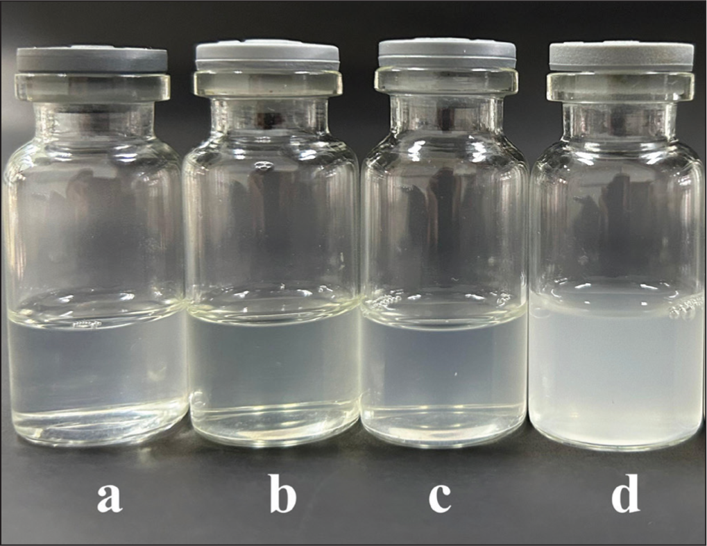
Globule size, PDI, and zeta potential were then used to determine the influence of drug concentration in the formula. Furthermore, to determine the drug loading capacity, ulvan was added to the nanoemulsion in stages, and the highest amount of ulvan that produced a clear nanoemulsion without precipitation was determined as the drug loading capacity. Table 3 and Figure 4 showed that nanoemulsion can solubilize up to 110 mg of drug per 1 g nanoemulsion, and a cloudy visual appearance (Figure 4d) was found after the amount of drug was increased to 120 mg and had a globule size above 700 nm (Table 3). This high drug loading capacity was influenced by the high surfactant concentration in the formula. The formula with the highest amount of drug (110 mg/g nanoemulsion) produced a clear, nanometer-sized, and highly monodispersed emulsion. This showed that the active ingredient was well incorporated. 44 This particle size met the target globule size of <200 nm.44,45
Characteristics of Ulvan Nanoemulsion with Various Amounts of Ulvan (Mean ± SD, n = 3).
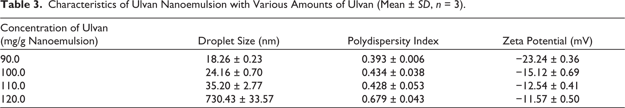
The addition of the drug to the formula led to an increase in droplet size of nanoemulsion (droplet size ranged from 18.26 to 35.20 nm) (Table 3). When a drug is incorporated into the oil phase, it occupies space within the internal phase. As the concentration of the drug molecules increases, the total volume of the oil droplets expands. Since the number of droplets is often limited by the surfactant concentration, each droplet must accommodate more material, leading to a physical increase in diameter.30,46 Furthermore, adding high amounts of drug often increases the viscosity of the oil phase. According to the principles of emulsification, a more viscous internal phase is harder to break down into smaller droplets during homogenization or sonication. This resistance to shearing results in larger mean droplet diameters. 47 Moreover, at high concentrations, drug molecules may migrate to the oil-water interface, competing with surfactant molecules. This can disrupt the stability of the surfactant film or reduce its efficiency in lowering interfacial tension, which prevents the formation of ultra-fine droplets.46,47 Furthermore, the packing parameter of the surfactants at the interface can be altered by the presence of drug molecules. If the drug is bulky, it can interfere with the tight packing of the surfactant tails, leading to a less curved interface and, consequently, larger droplets. 48
Aside from mean droplet size measurements, the size distribution of oil droplets, which was expressed as PDI, is also an important factor that must be considered. PDI is the ratio between the standard deviation and the mean droplet size, and it indicates the degree of non-uniformity during the production process. Low PDI indicated the uniformity and narrow size distribution of the polydispersed phase. 49 The results showed that nanoemulsion containing 90.0, 100.0, and 110.0 mg ulvan per g nanoemulsion had values of 0.393 ± 0.006, 0.434 ± 0.038, and 0.428 ± 0.053, respectively, as shown in Table 3. The higher amounts of the ulvan (120.0 mg per g nanoemulsion) have large PDI values (>0.5), showing that the emulsion produced has a large droplet size distribution. For a lipid-based carrier, a PDI of 0.5 and below was considered to be acceptable because a highly polydisperse droplet size can lead to aggregation and phase separation upon storage. 49
According to the data presented in Table 3, higher drug concentrations led to a reduction in the zeta potential of the nanoemulsion. This was probably due to the molecular interactions between the functional groups of ulvan and other formulation components. A similar phenomenon was observed by Zhang, 50 who identified a differentiation in zeta potential between loaded and unloaded nanoemulsions. It was attributed to the formation of intermolecular hydrogen bonds between the drug’s hydroxyl groups and the oxygen or nitrogen-containing moieties in the surfactants and oil phase. Such interactions modify the surface charge density of the droplets. Zeta potential is the electrostatic charge at the nanoparticle surface. 51 Based on previous findings, the nanoemulsion could be said to be stable when it had a zeta potential value of above ±20 mV due to the presence of a sufficiently large repulsive force between globules. As a result, it could prevent coalescence and stabilize the system. 52 The zeta potential value of nanoemulsion containing 90.0 mg/g nanoemulsion was higher than −20 mV (−23.24 ± 0.36 mV), and the other formula was lower than −20 mV (−15.12 to −12.54 mV), but it was still not appropriate to conclude that nanoemulsion was unstable. In the system, there was Tween 80, a nonionic surfactant, which did not provide an electrostatic barrier between the oil droplets, resulting in a lower zeta potential and limiting the system’s ability to prevent aggregation through electrical repulsion. 53 Negative values in the zeta potential were also caused by anionic impurities in the surfactant, such as fatty acids, and anionic species from water, including hydroxyl ions on the droplet surface. This could also be caused by the charge on the encapsulated drug, ulvan, which is an anionic sulfated polysaccharide 54 and has negatively charged oxygen groups. 54
Based on globule size, PDI, and zeta potential, a nanoemulsion with a maximum loading of 110 mg was selected as the best formula and subjected to preparation of the nanoemulgel and anti-inflammatory activity evaluation. The summarized physical characteristic of the nanoemulsion was presented in Table 3.
Formulation and Physical Properties Evaluation of Ulvan Gel and Nanoemulgel
Gel was formulated using 8% ulvan as the active ingredient, and the selection of the 8% concentration was based on the results of previous research that reported that 5% ulvan had shown anti-inflammatory effects, 55 as well as the results of preliminary analysis that 8% gel provided the best physical properties. Furthermore, gel form was selected because it matched the properties of ulvan, which was a hydrophilic polysaccharide, making it more stable and easily dispersed in water-based systems. 56 Gel had the advantages of a light texture, spread easily on the skin surface, provided a cooling sensation, and allowed for a more even release of the active ingredient. 57 HPMC was used as a gel base, which functioned as a gelling agent to form a stable gel consistency. Propylene glycol was also added as a humectant and penetration enhancer, methyl paraben as a preservative to prevent microbial growth, and distilled water as the main solvent. This combination of ingredients was expected to produce ulvan gel with good physical properties and optimal stability for topical application. The selection of nanoemulsion form was based on the advantages of the nanoemulsion system, which produced particle sizes on the nanometer scale. Therefore, the pharmacological effect was more optimal compared to conventional gel preparations. 58
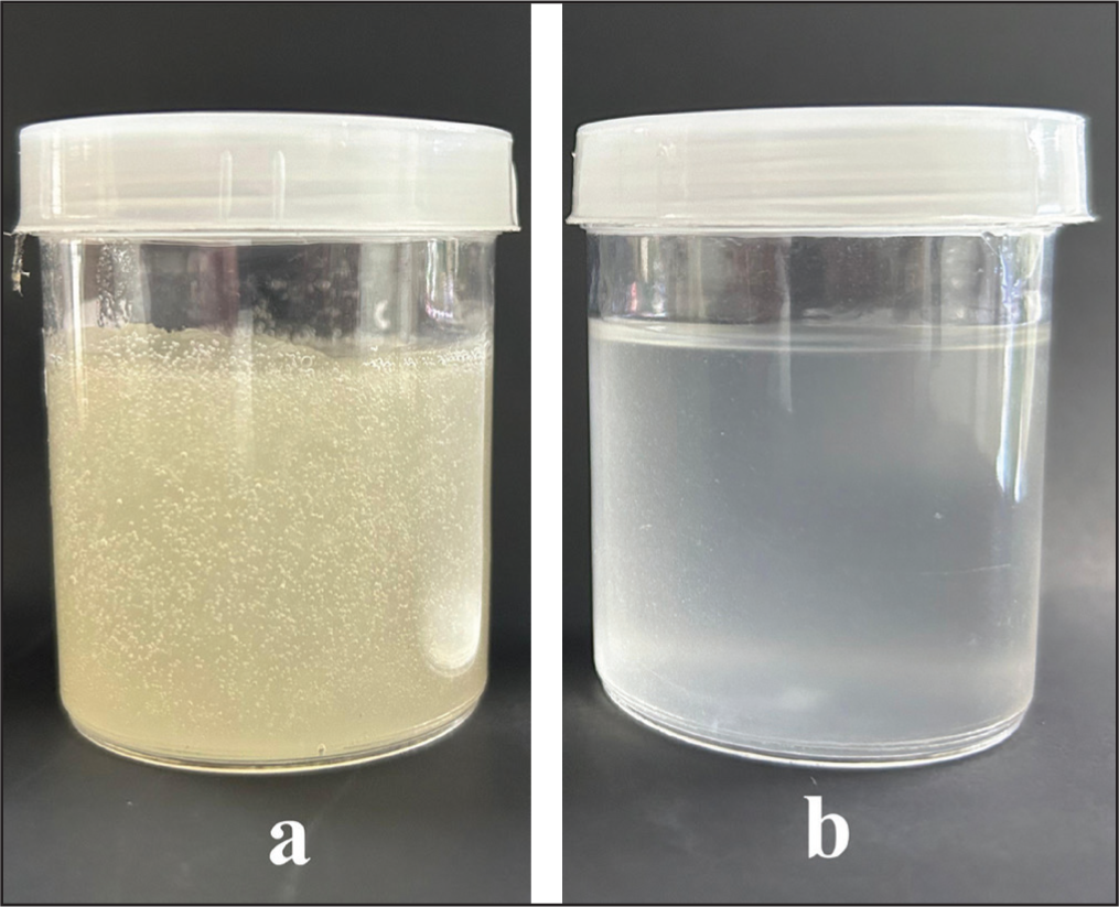
Based on Figure 5, the nanoemulgel formula produced a transparent and homogeneous appearance. Meanwhile, the gel formulation produced a slightly yellowish color and also a homogeneous appearance (Figure 5). The physical properties of the ulvan gel and nanoemulgel are presented in Table 4. The nanoemulgel exhibited a droplet size within the nanometer range (197.23 nm). The nanoemulgel exhibited a PDI of less than 0.5 (Table 4), signifying a uniform and narrow size distribution of the internal nanoemulsion droplets. 31
(a) Ulvan Gel, (b) Ulvan Nanoemulgel.
Characteristics of Ulvan Gel and Nanoemulgel (Mean ± SD, n = 3).
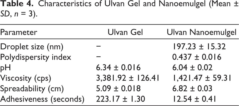
The viscosity test results showed that gel had a value of 3,381.92 ± 126.41 cps, while nanoemulgel exhibited a smaller value, namely 1,421.47 ± 59.31 cps. Moreover, gel had a higher polymer content; as a result, it formed a dense three-dimensional network and increased the viscosity of the preparation, while nanoemulgel contained an oil phase, surfactants, and nanoparticles that could disrupt or loosen gel network structure, making the viscosity lower. 59
Regarding its rheological properties, the nanoemulgel exhibited a gel-to-sol transition and demonstrated shear-thinning behavior upon the application of mechanical stress. The system subsequently regained its gel-like consistency once the stress was withdrawn, a phenomenon indicative of pseudoplastic flow combined with thixotropic characteristics. 60 Formulations possessing these pseudoplastic traits maintain high viscosity during storage but become easily pourable and spreadable during application. Such rheological profiles are advantageous for topical delivery. 61
The pH measurement aimed to determine whether the resulting preparation could be well accepted by the skin and cause no irritation when it was in accordance with the skin pH. In this study, the pH value of gel and nanoemulgel preparations must be maintained, which is not too acidic, causing skin irritation, and when it is too alkaline, leading to dry or scaly skin. 62 The results of the pH measurement showed that gel preparation had a pH of 6.34 ± 0.016, while nanoemulgel had a pH of 6.04 ± 0.02. This difference occurred due to differences in the composition of the ingredients used. In nanoemulgel, olive oil contained free fatty acids, and there was an interaction between PEG-400 and Tween, which reduced the pH value. 52 Both gel and nanoemulgel preparations were in the skin pH range of 4.5–6, 60 which was expected not to irritate.
The results of the spreadability test showed that gel had a value of 5.09 ± 0.018 cm, while nanoemulgel had a larger value of 6.82 ± 0.03 cm. This difference was caused by the presence of an oil phase in the form of olive oil, as well as additional ingredients, PEG-400 and Tween surfactant, in the nanoemulgel. The combination of these ingredients reduced the surface tension of the preparation, making it easier to spread when applied. 52 Furthermore, the very small oil droplets in nanoemulgel made the system more fluid and not as dense as gel, making the spreadability wider. 60 In this study, nanoemulgel was easier to apply and could cover a wider area of skin than gel. The ideal expected adhesion parameter was not less than four seconds, 63 and the adhesion value of gel was 223.17 ± 1.30 seconds, while nanoemulgel was only 12.54 ± 0.41 seconds. The higher adhesion value of the gel was due to the hydrophilic polymer structure, which formed a dense network, making it adhere more strongly to the skin surface. However, the presence of olive oil and PEG-400 in nanoemulgel made the gel structure looser, plus the Tween surfactant reduced surface tension; therefore, the bond with the skin was weaker. 52 This caused the nanoemulgel to be less sticky than the gel. The greater the adhesiveness of the preparation, the better its ability to adhere to the skin. As a result, the process of absorption of the active substance through the skin was also more optimal. 60 Based on these results, the gel and nanoemulgel formula of 8% ulvan had physical properties that meet the requirements to continue in vivo topical anti-inflammatory activity testing.
Topical In Vivo Anti-inflammatory Evaluation with Carrageenan Induction
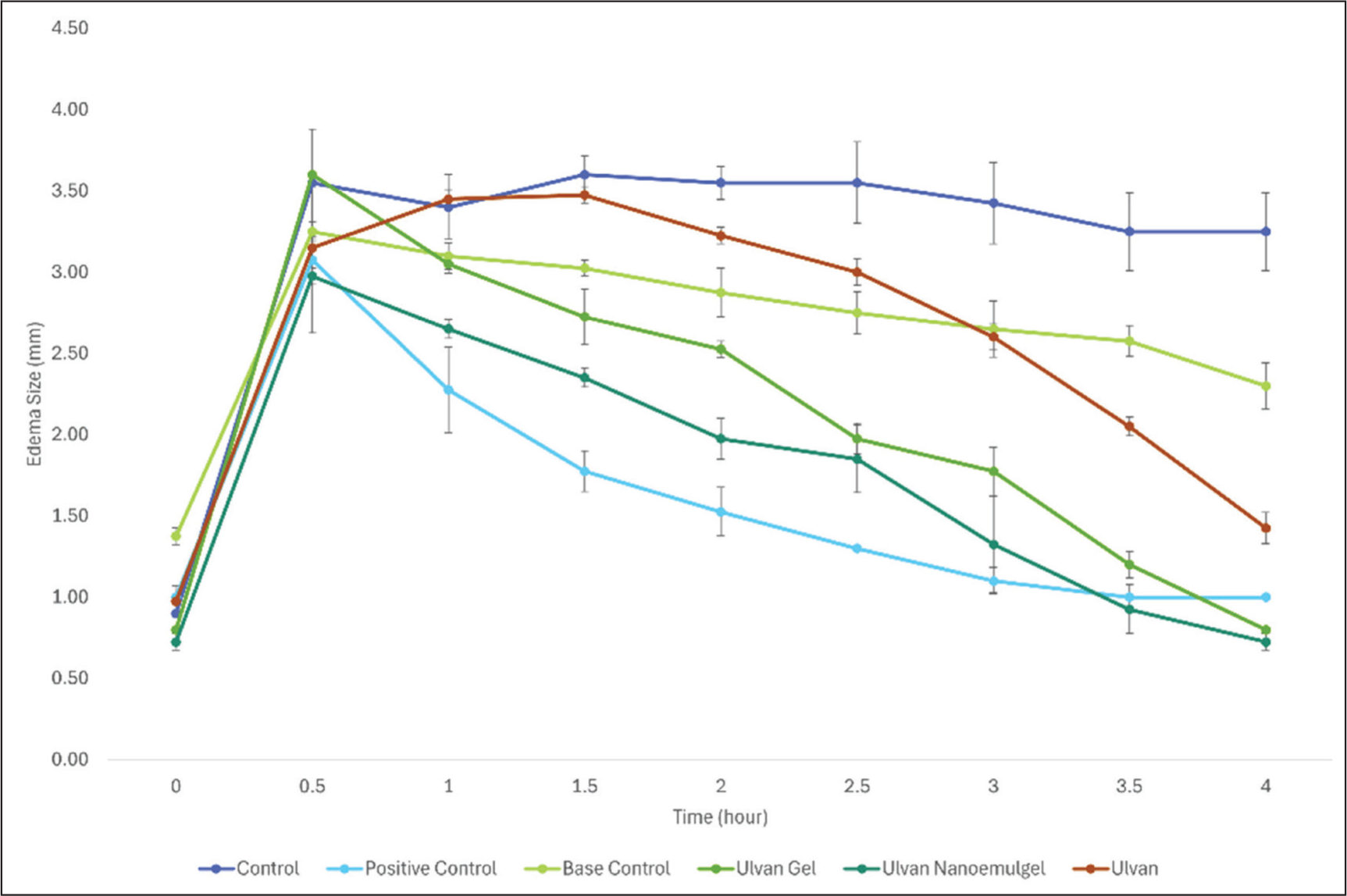
Anti-inflammatory activity was measured using the thickness of the back-skin of mice induced by carrageenan, which was then presented as edema volume. The results of edema volume every 30 minutes over a period of four hours are presented in Figure 6. Furthermore, the AUC value showed the ability to reduce edema volume over four hours, as observed from the decrease in the thickness of the back-skin edema of mice induced by 4% carrageenan, presented in Table 5. Figure 6 showed that 4% carrageenan provided the highest edema, as indicated by the highest AUC value. These results were in accordance with a previous study that 4% carrageenan could be used as an inflammation model. 22
Curve of Relationship Between Time and Edema Size Every 30 Minutes.
AUC0–4h Values of Back-skin Edema Thickness in Mice for Each Treatment Group.
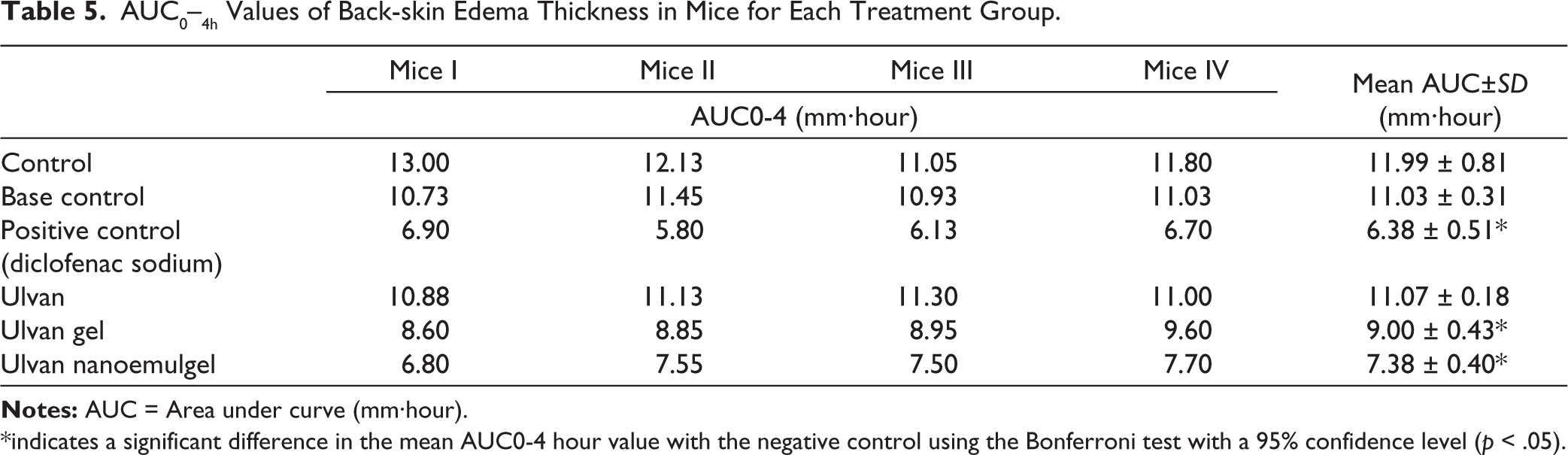
*indicates a significant difference in the mean AUC0-4 hour value with the negative control using the Bonferroni test with a 95% confidence level (p < .05).
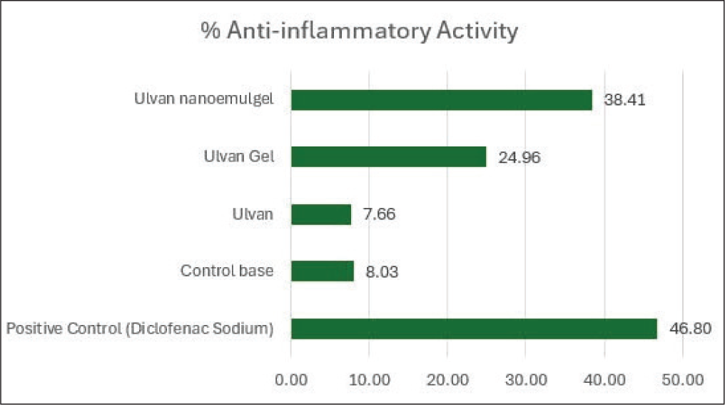
The results of the anti-inflammatory activity test for several treatment groups were presented in Figure 7, with the positive control group (diclofenac sodium) showing the highest inhibitory effect of 46.8%. This result was in accordance with its pharmacological profile as a non-steroidal anti-inflammatory drug, which functioned by inhibiting the COX enzyme, thereby suppressing the synthesis of proinflammatory prostaglandins. 64 The 8% ulvan extract nanoemulgel preparation showed anti-inflammatory activity of 38.41%, higher than the 8% ulvan gel (24.96%) and 8% ulvan (7.66%). These results showed that the 8% gel formulation could increase anti-inflammatory activity by 3.26 times, while the 8% nanoemulgel preparation increased anti-inflammatory activity by 5.01 times compared to 8% ulvan. Furthermore, gel- and nanoemulgel-based topical delivery systems were capable of enhancing the anti-inflammatory effect of active compounds. 65 Nanoemulsion was known to have small particle sizes and physicochemical stability that supported the bioavailability of active ingredients, including ulvan polysaccharides. 60 The 8% ulvan without formulation only produced an anti-inflammatory effect of 7.66%, which was relatively low compared to gel and nanoemulsion forms. This was explained by the limited absorption of hydrophilic compounds such as ulvan through the stratum corneum when administered without a proper delivery system. 60
Anti-inflammatory Activity of Ulvan, Ulvan Gel, and Ulvan Nanoemulgel.
These results showed that gel and nanoemulsion formulations based on ulvan extract had quite potent anti-inflammatory activity, although lower than the positive control/marketed product (diclofenac sodium gel). Ulvan, a sulfated polysaccharide from Ulva lactuca, was known to have anti-inflammatory activity through inhibition of inflammatory mediators and modulation of immune cells such as macrophages.66,67 A previous study showed that ulvan or ulvan oligosaccharides reduced lipopolysaccharide (LPS)-induced phosphorylation of Mitogen-Activated Protein Kinase (MAPK) (p38, Extracellular signal-Regulated Kinase [ERK], and c-Jun N-terminal kinase [JNK]), as well as IBα phosphorylation and nuclear translocation of NF-κB p65. The effects of these pathways led to decreased expression of iNOS, COX-2, and proinflammatory cytokines such as TNF-α, IL-6, and IL-1β.68,69 Wang et al. (2021) reported that selenized Ulva pertusa polysaccharides suppressed COX-2 mRNA expression in a DSS-induced colitis model. 70 These results were associated with inhibition of the NF-κB pathway, decreased proinflammatory cytokines such as TNF-α, IL-1β, and IL-6, and improved gut barrier integrity. Based on the result of this study, ulvan could affect inflammatory pathways leading to COX-2 regulation, although no test for COX-2 enzyme inhibitory activity was carried out. These pathway effects also explained the in vitro reduction in NO, PGE2, and cytokine release seen in a macrophage model. Several sulfated seaweed polysaccharides inhibited NF-κB/MAPK activation. Previous analysis also showed that green algae aqueous extracts had anti-inflammatory activity in vitro. 4
In addition to demonstrating enhanced pharmacological activity, ulvan extracted in this study showed a high yield (65.73%) and characteristic functional groups, including carboxylic acid (C=O), hydroxyl (OH), and sulfate moieties, confirming its chemical identity. The successful formulation of ulvan into gel and nanoemulgel systems with acceptable physical characteristics—such as viscosity, spreadability, adhesiveness, and pH—further supports the feasibility of these delivery systems for topical application. Overall, the findings indicate that incorporating ulvan into gel and especially nanoemulgel formulations significantly enhances its topical anti-inflammatory efficacy, highlighting the potential of ulvan-based nanoemulgels as promising natural topical anti-inflammatory agents. While the current results provide valuable insights, the relatively small sample size (n = 4 per group) remains a limitation. Therefore, we recommend that larger confirmatory studies be conducted in the future to validate these preliminary findings and ensure their generalizability to a broader population.
Conclusion
In conclusion, ulvan can be extracted from Ulva lactuca L. with a yield of 65.73% and a melting point of 196 °C–198 °C. Identification of functional groups using an infrared spectrophotometer shows the presence of carboxylic acid groups C=O, hydroxy sugar OH, and sulfate groups that correspond to the chemical structure of ulvan. Ulvan is formulated in the form of gel and nanoemulgel with an ulvan content of 8%, and has met the physical properties of gel and nanoemulgel preparations. Nanoemulsion has a good physical appearance, droplet size of 35.20 ± 2.77 nm, PDI of 0.428 ± 0.053, and zeta potential of −12.54 ± 0.41 mV. The nanoemulgel has a droplet size of 197.23 ± 15.32 nm and PDI 0.437 ± 0.016. All results of physical characterization of viscosity, spreadability, adhesiveness, and pH of nanoemulgel and gel meet the specified requirements. Anti-inflammatory properties of ulvan, 8% ulvan gel, and 8% ulvan nanoemulgel are 7.66%, 24.96%, and 38.41%, respectively. Therefore, gel and nanoemulgel formulations of ulvan increase anti-inflammatory properties significantly (p < .05) by 3.26 times and 5.01 times, respectively, compared to ulvan. The novelty of this study lies in the development of gel and nanoemulgel formulations incorporating ulvan, which have not been previously reported. Furthermore, both formulations were demonstrated to significantly enhance the anti-inflammatory activity of ulvan.
Footnotes
Acknowledgements
The authors gratefully acknowledge financial support from the Directorate of Research and Community Service, Ministry of Higher Education, Science, and Technology, Fiscal Year 2025, under Grant No. 125/PFR/LPPM.UAD/V/2025.
Authors’ Contribution
All authors made substantial contributions to conception and design, acquisition of data, or analysis, and interpretation of data; took part in drafting the article or revising it critically for important intellectual content; agreed to submit to the current journal; gave final approval of the version to be published; and agreed to be accountable for all aspects of the work. All the authors are eligible to be authors as per the International Committee of Medical Journal Editors’ requirements/guidelines.
Consent to Participate
Not applicable.
Consent for Publication
Not applicable.
Data Availability Statement
All the data is available to the authors and shall be provided upon request.
Declaration of Conflicting Interests
The authors declared no potential conflicts of interest with respect to the research, authorship, and/or publication of this article.
Ethical Approval
Ethical approval details are given in the “Materials and Methods” section.
Funding
We would like to thank to KEMENRISTEK DIKTI Indonesia for supporting this research through the PFR.
Informed Consent
Not applicable.
Use of Artificial Intelligence-assisted Tools
The authors declare that they have not used artificial intelligence (AI) tools for writing and editing of the manuscript, and no images were manipulated using AI.