
Abstract
The development of suitable bioinks for bioprinting is still a key issue in the field of tissue engineering. Most of the biomaterials used are derived from animal sources. A novel bioink for corneal stroma bioprinting based on chemically modified keratin derived from human hair waste was developed and evaluated. To confirm the successful functionalization of methacrylated keratin (KerMA), various characterization methods such as 1H-NMR, IR as well as a cytotoxity assay were performed. The newly developed biomaterial KerMA was mixed with methacrylated hyaluronic acid and bioprinted with cells as a corneal stroma substitute. Two different cell types were encapsulated in the bioink, immortalized corneal keratocytes and human corneal fibroblasts. The printed constructs were crosslinked under UV light and cultured for up to four weeks. The bioprinted constructs were monitored for four weeks for optical transparency, cell viability (live/dead staining), biomechanical properties and protein expression by indirect immunofluorescence. Furthermore, human corneal epithelial cells were seeded on top of the bioprinted constructs and demonstrated high cell viability. The bioprinted constructs showed good optical transparency and decent biomechanical properties. The addition of KerMA to methacrylated hyaluronic acid led to an improvement in cell viability and protein expression. This bioink approach offers a sustainable, ethically sourced alternative to animal-derived materials. It forms the basis for future in vitro and in vivo studies on the development of corneal tissue.
Introduction
The cornea is the outermost barrier of the eye and plays a crucial role in protecting the eye from environmental damage, serving as a barrier with the tear film, and refracting light, thus enabling vision. 1 The corneal stroma has a unique, anisotropic ultrastructure consisting of parallel collagen fibril lamellae that are essential for optical transparency. 2 Vision is a critical role in everyday life as it affects almost everything from reading to simply walking. 3 However, there are many diseases such as infection, degenerative diseases or scarring affecting the cornea and thus leading to vision impairment or in worst cases blindness. 4 While a high level of hygiene and medicine can be an important tool to prevent corneal blindness, corneal transplantation is often still the primary treatment to restore vision. To date, there is a discrepancy between donors and recipients, so that only one in 70 people can receive a cornea. 5 These obstacles have sparked interest in corneal tissue engineering and regenerative medicine. 6
One emerging technology in the past has been bioprinting. Bioprinting uses a computer-aided design and the additive manufacturing of biomaterials and bioink to recreate the structure and function of a native cornea. 7 Over the past years corneal bioprinting has risen and produced a promising future. One key interest of bioprinting is developing a bioink that demonstrates sufficient transparency, biocompatibility, mechanical stability and is capable of supporting cell viability and differentiation. 7 Most corneal bioprinting uses natural biomaterials from animal sources such as gelatin or collagen derivates. Although, the possibility of using human derived gelatin or collagen exists, it is very scarce and expensive. The option of using recombined collagen or gelatin still lacks wide usage and has been utilized, to our knowledge, only once in corneal bioprinting. 8 The use of animal derived biomaterials is not sustainable and for many animal products, animal ethics play a crucial role in the search for other materials. This field of bioprinting without animal derived biomaterials has been proposed by Berg et al. as “clean bioprinting”. 9
One alternative material that is abundant and can be gained from humans is keratin, which can be obtained from hair and nails. Hair and nails are often waste products and not further used. Thus, keratins are cheap and easily obtainable. Nevertheless, keratin is utilized in many ways in the tissue engineering field due to demonstrating good biodegradability, excellent biocompatibility and having cellular attachment sites such as arginine-glycine-aspartic acid (RGD) and leucine-aspartic acid-valine (LDV). 10 For example, keratins have been applied in skin tissue engineering and wound healing. 11 Beyond skin tissue engineering, keratins have also been implemented in ocular tissue engineering.12–14 Keratin scaffolds have supported epithelial wound healing comparable to that of an amniotic membrane in an in vivo rabbit model for ocular surface reconstruction.15,16 Furthermore, keratin films have showed good transparency and biocompatibility in another ocular surface application. 17 These promising results promote the use of keratin in corneal bioprinting.
Previous mentioned keratin film preparations are based on thermal crosslinking, which makes it incompatible with cell-laden hydrogels for bioprinting. Thus, to overcome this, we propose modifying keratin with methacrylate to yield methacrylated keratin (KerMA). Keratin is naturally insoluble and is often isolated from hair by using the Shindai method, which is applied here. After aqueous dialysation, a suspension of keratin nanoparticles is gained. 12 This nanosuspension is used and modified with methacrylate to gain KerMA. For confirmation of the methacrylation process, 1H-NMR, IR and a 2,3,6-trinitrobenzene sulfonic acid (TNBSA) assay were conducted. KerMA being a nanosuspension, has a low viscosity, thus to make it suitable for extrusion bioprinting, we used methacrylated hyaluronic acid (HAMA) as a high viscosity bioink. Hyaluronic acid (HA) is a natural polysaccharide found abundant in humans and to a certain extent also in the human cornea. 18 HA can be used as a viscosity enhancer in bioprinting but does not provide stability after printing. Thus, it is often methacrylated to HAMA and has been extensively used in the bioprinting field, but rarely used in corneal bioprinting.7,19,20 After printing, the methacrylated groups can undergo rapid photocrosslinking with UV-light to obtain a stable scaffold. Furthermore, HAMA is a hydrogel with high transmission and decent biomechanical properties making it attractive for corneal bioprinting. 21
In this study, we combined high molecular weight HAMA and KerMA to print a disc that mimics corneal stromal tissue. Two different cell types were used, an immortalized human corneal keratocyte cell line as well as primary human fibroblasts isolated from human corneas. Fibroblasts are active in corneal wound healing, whereas keratocytes maintain the intact, healthy state of the cornea. 22 The two cell types were integrated to understand the interaction with the bioprinted 3D matrix. After bioprinting, light transmission, biomechanical properties, cell viability and protein expression of collagen type I and α-smooth muscle actin (αSMA) over four weeks was investigated. Furthermore, corneal epithelial cells were seeded on the printed discs and analyzed. This study serves to introduce the new biomaterial KerMA in corneal bioprinting.
Materials and methods
Materials
Hyaluronic acid was purchased from Acros Organics, Geel, Belgium. Dimethylformamide was obtained from VWR Chemicals, Radnor, USA. Methacrylic anhydride was purchased from Alfa Aesar, Haverhill, USA. Sodium hydroxide, sodium chloride, sodium dodecyl sulfate (SDS), urea, thiourea, mercaptoethanol, sodium bicarbonate, cysteine, calcium chloride, ascorbic acid, paraformaldehyde and Triton X-100 were obtained from Carl Roth, Karlsruhe, Germany. A SpectraPor® dialysis membrane (MWCO 6–8 kDa) was purchased from Repligen, Waltham, USA. Vivspin® 20 (MWCO 5 kDa) were bought from Sartorius, Goettingen, Germany. TRIS was obtained from Caesar & Loretz, Hilden, Germany. Deuterium hydroxide and 2,3,6-trinitrobenzene sulfonic acid (TNBSA) were obtained from Sigma-Aldrich, St. Louis, USA. Hydrochloric acid, 3-(4,5-dimethyl-2-thiazolyl)-2,5-diphenyl-2H-tetrazolium bromide (MTT), sucrose, fetal bovine serum (FBS), antibiotic/antimycotic solution, dispase, insulin, polysorbate 20 (Tween® 20) and bovine serum albumin (BSA) were purchased from Sigma-Aldrich, München, Germany. Keratinocyte growth medium (KGM) was obtained from Lonza, Basel, Switzerland. DMEM and L-glutamine were purchased from Biochrom, Berlin, Germany. Phosphate buffered solution (PBS) was obtained from MP Biomedicals, Irvin, USA. Collagenase was purchased from Merck Millipore, Darmstadt, Germany. TGF-ß1 was obtained from PeproTech, Hamburg, Germany. Isopropanol was purchased from Fisher Scientific UK, Loughborough, UK. Lithium phenyl (2,4,6-trimethylbenzoylphosphinat) (LAP) was obtained from BLDPharm, Shanghai, China. CalceinAM was purchased from AAT Bioquest, Pleasanton, USA. Ethidium homodimer I was obtained from ION Biosciences, San Marcos, USA. Normal goat serum was purchased from Vector Lab, Peterborough, UK. Rabbit anti-collagen antibody, mouse anti-αSMA, FITC-conjugated anti-rabbit antibody and AlexaFluor568-conjugated anti-mouse antibody were obtained from Abcam, Cambridge, UK. Hoechst33342 was purchased from Thermo Fisher Scientific, Waltham, USA. An internal standard procedure was used to extract collagen type I from rat tails.
Synthesis of HAMA
Methacrylated hyaluronic acid (HAMA) has been synthesized as previously described. 23 Briefly, high molecular HA (2000 kDa) was dissolved in double destilled water and stirred at room temperature overnight. On the following day, dimethylformamide was added in a 1:2 DMF/water ratio. Followed by the dropwise addition of methacrylic anhydride (MAA), while the pH was maintained with NaOH between 8 and 9. After 20 h of stirring under light exclusion, the pH was adjusted to 7 and further diluted with water. Sodium chloride was added to obtain a final concentration of 0.6 M. Finally, the solution was transferred to the dialysis membrane (SpectraPor® MWCO 6–8 kDa) and dialyzed against distilled water for 10 days at 4°C, followed by lyophilization. HAMA was stored at −20°C for future use.
Keratin extraction
Human hair was attained from multiple donors from a local hairdresser. First, the hair was pooled and washed with 0.5% SDS in water and followed by rinsing with water before air-drying. For keratin extraction the Shindai Method was used. 24 For the preparation of the Shindai solution 25 mM Tris, 2.6 mM thiourea, 5 M urea and 5% 2-mercaptoethanol were dissolved in water and the pH was adjusted to 8.5. Next, for the extraction, the hair (20 g) was stirred gently for 72 h at 50°C in the Shindai solution (400 mL). Subsequently, the mixture was centrifuged at 5000 rpm for 30 min before the supernatant (Shindai extract) was filtered and stored at −20°C until further use.
For the removal of cytotoxic agents (e.g. thiourea and 2-mercaptoethanol) from the extract, dialysis against water was done.12,14,25 For aqueous dialysis, 100 mL of extract was dialyzed (SpectraPor® MWCO 6–8 kDa) at room temperature with a daily change of water for 6 days in 5 L of water. Before further use for synthesis, the aqueous keratin dialysate was centrifuged for 30 min at 10,000 rpm and the supernatant was used. The same batch was used in further experiments.
To monitor the protein amount a Bradford assay as described previously was used. 26 UV absorption was measured with an Infinite M Plex multiplate reader (Tecan, Männedorf, Switzerland).
Synthesis of KerMA
The aqueous keratin dialysate was diluted with PBS in a 1:5 ratio. Methacrylic anhydride was added dropwise, while maintaining the pH at 7.4. Two different syntheses were conducted to achieve different rates of methacrylation. The first synthesis, KerMA low, used a MAA to keratin dialysate ratio of 1:133.33 (V/V) and reaction time of 2.5 h. The second synthesis, KerMA high, used a MAA to keratin dialysate ratio of 1:66.66 (V/V) and reaction time of 4 h. After stirring under light exclusion and maintaining the pH at 7.4, the solution was transferred to the dialysis membrane (SpectraPor® MWCO 6–8 kDa) and dialyzed against PBS for 6 days followed by dialysis against water for 6 days at room temperature. After dialysation the KerMA was lyophilized and stored at −20°C until further use. KerMA was suspended at a concentration of 50 mg/mL in PBS. Before use, the KerMA nanosuspension was centrifuged for 4 min at 4000 rpm and the supernatant was used.
1H-NMR and FT-IR
1H-NMR spectra were obtained using an AV III HD 500 Spectrometer by Bruker (Billerica, MA, USA). Samples were dissolved in D2O and measurement was conducted at room temperature. Tetramethylsilane was used as an internal standard. Data evaluation was carried out using MestReNova-Software Version 15.1.0-38027 by Mestrelab (Santiago de Compostela, Spain).
FTIR spectroscopy was carried out using a Nicolet iS50 FT-IR spectrometer equipped with an ATR Unit (Thermo Scientific, Osterode am Harz, Germany). The IR spectra were recorded in the range of 4000 to 400 cm−1, baseline corrected and normalized. Spectragryph software was used for further analysis.
TNBSA assay
A TNBSA assay can be used to quantify the remaining free amino groups and thus the degree of methacrylation (DoM) can be determined. 27 This is achieved by comparing the number of free amino groups of keratin before the reaction and KerMA after the reaction. Briefly, KerMA und keratin samples were separately suspended at concentrations of 0.2 mg/mL in sodium bicarbonate buffer. A calibration was done with cysteine. Each solution was mixed with 0.01% TNBSA (prepared in 0.1 M bicarbonate buffer) and incubated for 2 h. To terminate the reaction, hydrochloride acid and 10% sodium dodecyl sulfate were added. The absorbance of each sample was measured at 335 nm with an Infinite M Plex multiplate reader (Tecan, Männedorf, Switzerland). The degree of methacrylation was calculated as followed:

Cell culture
Human corneal keratocytes (HCK)
In this study, SV40 immortalized human corneal keratocytes (HCK), which were gifted by Dr. M. Zorn-Kruppa, were used. 28 The HCKs were cultured in serum-free KGM supplemented with CaCl2 (0.5 M) in a humidified atmosphere at 37°C and 5% CO2.
Human corneal epithelial cells (HCET)
An SV40-immortalized human corneal epithelial cell line (HCET) was further used. The cells were obtained from RIKEN Cell Bank (Tsukuba, Japan). HCET were cultivated with KGM supplemented with CaCl2 (0.5 M) under standardized conditions (37°C, 5% CO2) in a humidified atmosphere.
Human corneal fibroblasts (HuFib)
Primary corneal stromal cells were isolated from human corneas that were not suitable for transplantation provided by Gesellschaft für Transplantationsmedizin Mecklenburg-Vorpommern gGmbH. The isolated cells were cultured in DMEM containing 10% FBS, 4 mM L-glutamine and 1% antibiotic/antimycotic solution. For the enzymatic isolation, first the corneal stroma was stripped from the sclera and epithelium with a scalpel. The stroma was then washed with PBS and digested with dispase solution (1.5 U/mL) for 1 h at 37°C followed by another PBS washing. Next, the stroma was further digested in collagenase solution (100 U/mL) for 12 h on a shaker. The suspension was then centrifuged for ten minutes at 1500 rpm. The remaining cell pellet was resuspended in cell culture medium and cultivated under standard conditions (37°C, 5% CO2). To minimize effects based on donor variability, cells from 5 donors were pooled. Cells of passage 3–5 were used for bioprinting. After bioprinting the cells were cultured in a differentiation medium (DMEM, 10% FBS, 4 mM L-glutamine, 1% antibiotic/antimycotic, 10 µg/mL insulin, 50 µg/mL vitamin C, 1 ng/mL TGF-ß1), which stimulates the cells to proliferate and produce more extracellular matrix such as collagen. 29
MTT assay
HCK and HCET cells were used for the MTT test. Cells were seeded into a 96 well plate and incubated until 80% confluency. The KGM medium was aspirated and replaced with 100 μL test solutions. The incubation time was set to 1 h. KerMA high concentrations of 20, 10, 5, and 2.5 mg/mL in KGM were used as test solutions. After the incubation time, the test solution was removed and replaced with 100 μL diluted MTT solution (1:10 of 0.5% MTT with KGM) per well. After 2 h incubation time at 37°C, the diluted MTT solution was removed and replaced with 100 μL lysis solution per well. The cells were lysed for 1 h under shaking and light exclusion. The lysis solution contained 90.55% isopropanol, 8.82% purified water, 0.36% hydrochloric acid and 0.27% SDS. Absorbance was measured using an Infinite M Plex multiplate reader (Tecan, Männedorf, Switzerland) at 570 nm.
Bioprinting
For bioprinting a commercial printer (BioScaffolder 3.3, GeSiM mbH) was used. To demonstrate the feasibility of the hydrogel, a simple disc was chosen as a model, with dimensions similar to the native cornea, diameter of 11 mm and three layers corresponding to a height of 0.5 to 0.6 mm. The discs were printed via pneumatic extrusion using a 250 µm nozzle, a printing speed of 5 mm/s and an extrusion pressure ranging from 50 to 60 kPa. Following extrusion, UV crosslinking with a UV solo pen (Opsytec Dr. Gröbel GmbH) of 405 nm was applied. For the curing process, the UV pen was placed at a distance of 50 mm for a duration of 6 s. According to the manufacturer’s specification, this setup provides an estimated irradiance of 100 mW/cm2.
Based on a previous study, HAMA was used in a final concentration of 30 mg/mL. In this previous study, printability and shape fidelity were demonstrated. 30 For crosslinking, the photoinitiator LAP was added to achieve a final concentration of 1.32 mmol/L. KerMA was added to achieve a final concentration of 20 mg/mL. Bioink formulations were prepared using either HCKs or HuFibs. In both groups the cell suspension was mixed with HAMA before printing to obtain a final cell concentration of 1.5 × 10⁶/mL. Therefore, two different bioinks were created, HAMA and HAMA with KerMA (HAMAKerMA).
HCET seeding
HCET cells were seeded on top of the bioprinted discs at a concentration of 400,000 cells/disc after one week of printing. Microscopic pictures were taken over the course of four weeks with an inverted light microscope (Olympus IX50 with Olympus DLP28). Live/dead staining was conducted after 28 days.
Cell viability (live/dead staining)
To observe the viability of cells directly, the discs were stained with calceinAM (5 µM) and ethidium homodimer I (2 µM). After incubation of 30 min the discs were observed using a fluorescence microscope (Olympus IX50 with Olympus DLP28). Live/dead staining was performed after 2 h of printing and on day 7, 14, and 28.
Light transmission
Extinction was measured in the wavelength range of 300–800 nm with a resolution of 1 nm. From the extinction the light transmission was calculated. Measurements were performed on three discs with a spectrophotometer (Infinite M Plex, Tecan, Männedorf, Switzerland). Before measurement, the discs were rinsed with PBS and left with a small amount of PBS to keep them moist and positioned in the center of a 6-well plate.
Furthermore, each disc was placed on a printed “B” to evaluate the macroscopic appearance and transparency.
The refractive index was measured with an Abbemat-WR refractometer (Anton Paar, Ostfildern-Scharnhausen, Germany) at 25°C.
Mechanical testing
To assess biomechanical properties a compressive test and tensile test were conducted using a Zwicki-Line Z 0.5 machine (Zwick, Ulm, Germany) with a 10 N load cell and testXpert® II software. Moist discs were tested at 1 mm/min, starting after reaching a preload of 0.03 N.
A modified version of ASTM D412c was printed. A 5 mN preload at 10 mm/min was followed by Youngs’s modulus testing at 5 mm/min, then continued at 25 mm/min until breakage.
Swelling
The discs were measured from images captured using a stereomicroscope and camera (Stemi 508 and Axiocam208, both Zeiss, Jena, Germany) and analyzed with Zeiss Labscope software. Immediately after printing and after 2 h, 7, 14, and 28 days the diameter was measured. The increases in size were calculated as percentages.
Scanning electron microscopy (SEM)
The printed discs were frozen with liquid nitrogen and lyophilized (Epsilon 2–4 LSCplus; Martin Christ Gefriertrocknungsanlagen GmbH, Osterode am Harz, Germany). The samples were then sputtered with gold in a sputtering device 07 120 (Balzers Union Limited, Balzers, Principality of Liechtenstein) at a current of 5 mA, an argon pressure of 0.7 mbar and a working distance of 40 mm.
The images were taken using a Helios G4 CS (FEI Deutschland GmbH, Frankfurt am Main, Germany) with an Everhart-Thornley detector in secondary electron mode, an acceleration voltage of 3.0 kV, a beam current of 0.2 nA and a working distance between 3 and 4 mm. Pore size was calculated with ImageJ.
Immunofluorescence (IF)
3D printed discs were fixed for IF after day 7, 14, and 28 of printing with 4% paraformaldehyde and 10% sucrose in PBS for 1 h. The discs were then washed thoroughly with PBS. For permeabilization a 0.1% Triton-X-100 was used for 30 min. This was followed by another wash with PBS, 3 times in total. Next, the non-specific bindings were saturated with 10% normal goat serum and 0.1% Tween® 20 in PBS for 1 h. The solution was then aspirated and replaced with the antibody solution. This consisted of 1:175 rabbit anti-collagen I and 1:200 mouse anti-αSMA in a tris buffer with Triton-X-100 and BSA. After 48 h incubation, the primary antibody solution was removed and washed 3 times with PBS. This was followed by incubation with the secondary antibodies for 24 h. After the incubation period, the discs were washed and the cell nuclei were stained with Hoechst 33342. This was followed by microscopy under a fluorescence microscope (Olympus IX50 with Olympus DLP28, Hamburg, Germany). Images were analyzed using CellSense (Olympus).
Statistical analysis
Unless otherwise stated, all measurements were carried out at least three times. Data are presented as mean ± standard deviation. Data were analyzed with one-way analysis of variance (ANOVA) with a Tukey post hoc test using OriginLab 2024b software. Prior to ANOVA, data were tested for outliers using the Grubbs’ test and for normal distribution using the Shapiro-Wilk test. Homoscedasticity was assessed using the Levene’s test. If homoscedasticity was significant with Levene’s test a Welch ANOVA was conducted. A significance level of *p < 0.05 was used and was considered statistically significant.
Results and discussion
KerMA characterization
Keratin methacryloyl (KerMA) had been successful synthesized. In the 1H-NMR spectroscopy distinct peaks at 5.7 and 5.4 ppm corresponding to the olefinic hydrogens of methacrylate could clearly be identified in KerMA low and KerMA high (Figure 1(a)). Furthermore, a new peak at around 1.9 ppm further confirmed the methacrylation. The peaks between 6.5 and 6.0 could be attributed to methacrylation of other groups such as hydroxyl groups, which is a common observation in GelMA samples when a high molar excess amount of MAA is used. 31
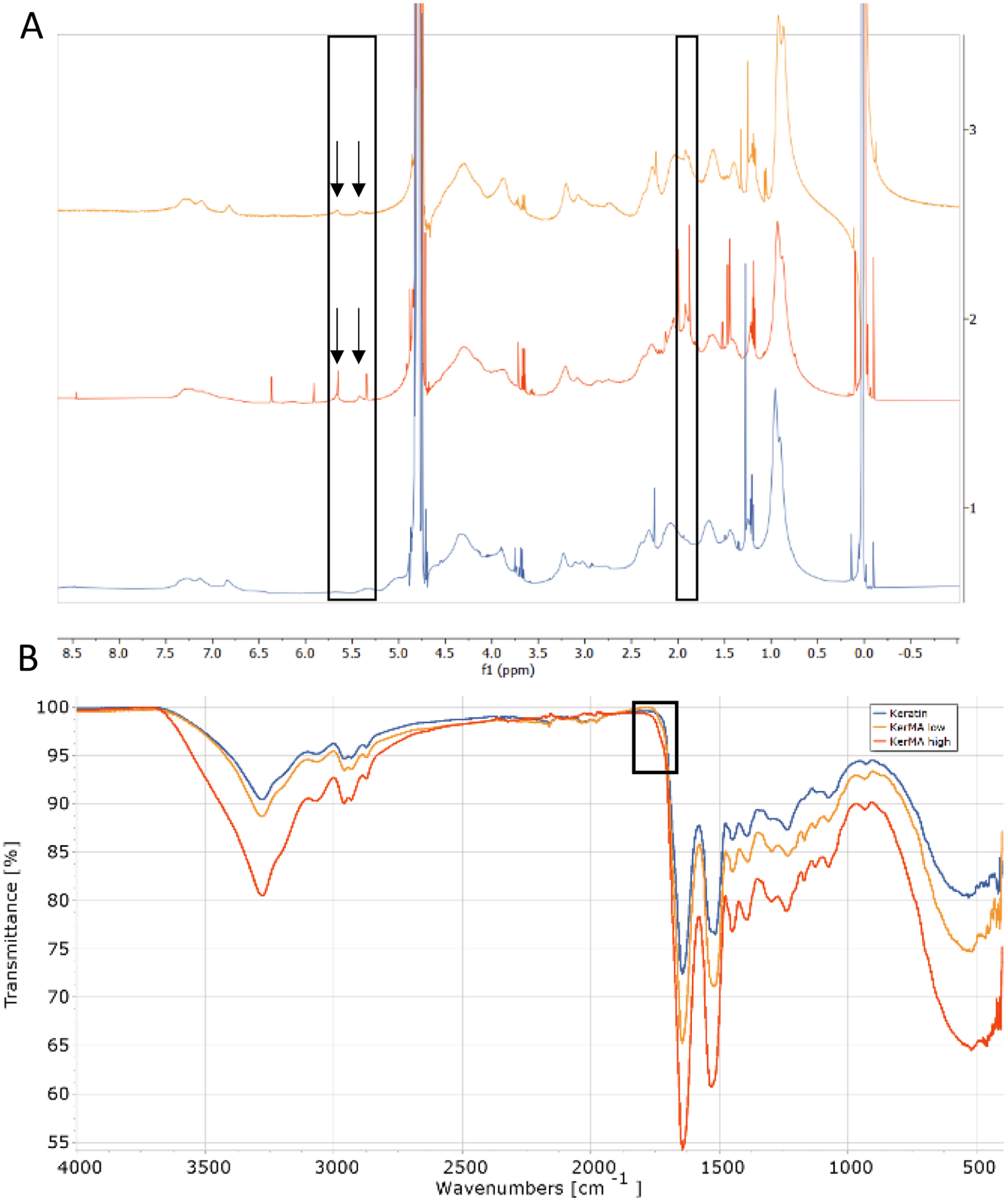
(a) 1H-NMR of KerMA low (yellow, top), KerMA high (red, middle) and pure keratin (blue, bottom) and (b) FT-IR of KerMA low (yellow), KerMA high (red) and pure keratin (blue).
FT-IR spectral analysis was also used to compare keratin with the two KerMA samples (Figure 1(b)). A new ester peak could be seen around 1720 cm−1, which was slightly more distinct in the KerMA high spectrum than in the KerMA low spectrum and was absent in the keratin spectrum, indicating a progressive ester formation and thus a higher degree of methacrylation. Furthermore, KerMA high exhibited the most noticeable spectral changes, hinting at a higher degree of modification. The IR results confirm a possible stepwise chemical modification across the samples, which is more pronounced in KerMA high. A similar trend was observed in the 1H-NMR spectra, as the area under the curve of KerMA high appeared to be higher than KerMA low, supporting a higher methacrylation degree for KerMA high. To further prove this and due to the currently unknown nature of amino acid sequences in aqueous keratin, the degree of methacrylation was calculated by a TNBSA assay. The DoM for KerMA low was 60.07 ± 8.02% and for KerMA high 63.07% ± 6.60%. Although this supports the slightly higher degree of methacrylation for KerMA high, the difference is not statistically significant. While with an increase of methacrylic anhydride typically a higher methacrylation could be achieved, this was not the case here despite attempts with different reaction conditions. 31 This hints that a possible saturation point could be found for the methacrylation. This could be attributed to steric hindrance and thus limited accessibility of the amino acids for methacrylation. Keratin is a filament-forming protein that assembles into a dense network of intermediate filaments. 32 Even when keratin is extracted and processed into a nanosuspension, it is likely to retain its densely packed state, resulting in the maximum methacrylation observed here. 25 To further prove this more future experiments are needed to fully understand the methacrylation process of keratin. Furthermore, a DoM of 60% appeared to be relatively high compared to the relatively small peaks seen in the IR and NMR spectra. Therefore, this value should be interpreted with caution. Although quantitative 1H-NMR integration would provide further clarity, it was beyond the scope of this study due to the heterogeneous nature of keratin. The exact ratio of methacrylatable amino acids remains unknown, making a definitive integration currently unfeasible. Nevertheless, KerMA high has been chosen for further experiments and will now be referred to as KerMA throughout the following manuscript.
MTT
The high cell viability observed in HCK and HCET cells confirms the biocompatibility and non-cytotoxic properties of KerMA within this concentration range (Figure 2). The MTT assay is a cellular metabolic activity test that revealed no major adverse effects on the cells. The tetrazolium dye, MTT, is reduced by mitochondrial enzymes to insoluble formazan, which amount is directly proportional to the number of viable cells. It could therefore hint as a possible positive proliferative effect, as a higher cell viability could be seen with HCK and increasing KerMA concentration, suggesting it provides a more favorable microenvironment. However, this finding should be interpreted with caution as the incubation time was only 1 h, which is sufficient to assess initial cytotoxicity but not sufficient to make definite claims about proliferative effects. To study the long-term effect of KerMA on the cell viability, a live/dead assay of the bioprinted discs is conducted over the time of four weeks. Nevertheless, this initial MTT assay was performed as a preliminary step to clarify non-toxic properties of KerMA before starting bioprinting. The primary goal of the further experiments was to investigate the effect of KerMA on corneal bioprinting, thus testing lower concentrations was not considered to be necessary.
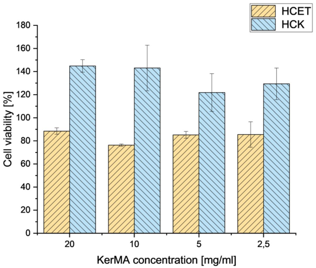
MTT assay of KerMA high on HCK and HCET cells.
Bioprinting
Light transmission
A previous study demonstrated the printability and printing settings of the HAMA bioink. 30 The printability of the HAMAKerMA bioink was demonstrated by the high morphological similarity between the HAMA and HAMAKerMA printed constructs (Figure 3(a)–(d)). Furthermore, a comparative analysis of disc diameters was conducted (n = 24–26). The average diameter of the HAMA disc was 11.17 ± 0.20 mm, and the average diameter of the HAMAKerMA disc was 11.09 ± 0.35 mm. There was no statistically significant difference between the two formulations (p > 0.001), and both discs matched the theoretical target of 11 mm. This corresponds to a high shape fidelity of 0.98 and 0.99, respectively. These results suggest that the addition of KerMA does not disrupt the structural integrity provided by HAMA. However, the effect of KerMA nanosuspensions on shear-thinning behavior and viscosity requires further investigation. The macroscopic clarity of the bioprinted discs, as the letter “B” was clearly visible, demonstrated the transparency essential for a corneal stroma substitute. (Figure 3(a)–(d)). Any major opacities would hinder its application. The light transmission of the hydrogel discs with HCKs was determined at the visible light wavelength ranging from 300 to 800 nm. The light transmission of the HAMA bioink without KerMA was very high and stayed high over the course of four weeks with over 80% transmittance. With the addition of KerMA to the HAMA gel, the transmission decreased to around 70% at day 0, this slight decrease could also be seen in the macroscopic view, as it appeared very slightly cloudy, although not enough that the letter was not visible anymore. This initial decrease of transmission was very likely attributed to the nature of KerMA being a nanosuspension. The keratin nanosuspension used as the starting material for the KerMA synthesis had an average particle size of 113 nm. 25 However, a detailed investigation of the properties of the KerMA nanosuspension still needs to be carried out. This information could help us understand how the nanosuspension affects light transmission. The light transmission then increased over the course of 14 days until it decreased again after four weeks (Figure 3(e)). The increase from day 7 to day 14 might suggest that a reorganization or integration of KerMA took place, although to further prove this, other methods such as small-angle x-ray scattering could be useful. The decrease from day 14 to day 28 might be due to the increase of cell clusters, as seen in Figure 5(b), as those cell clusters may have formed structures that reduced transmission. It has also been reported that cells scatter light and thus lead to a decrease of transmission. 33 Furthermore, the refractive index of both materials was also tested. Due to the nature of hydrogels, the refractive index was very close to water. However, with the addition of KerMA the refractive index increased from 1.3353 ± 0.0002 to 1.3360 ± 0.0001. In the literature the refractive index of a cornea varies from 1.335 to 1.4391. 34 Although the HAMA KerMA hydrogel was at the lower end of the literature values, the addition of KerMA improved the refractive index. This highlights the importance of not only assessing transmission but also considering other crucial optical properties such as the refractive index in the field of corneal bioprinting. It should be noted that the native cornea has a highly organized anisotropic ultrastructure that remains a challenge to be replicated using extrusion bioprinting. However, the hydrogel provides an environment in which the cells could actively remodel and deposit the extracellular matrix, resulting in a more organized ultrastructure similar to the native cornea.
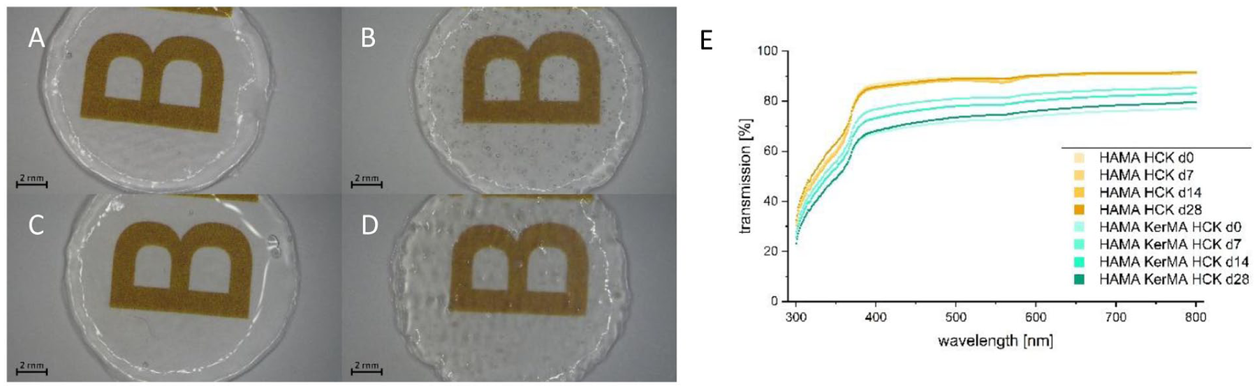
(a) HAMA day 0, (b) HAMAKerMA day 0, (c) HAMA day 28, (d) HAMAKerMA day 28, and (e) transmission of discs over 28 days.
Swelling and mechanical testing
An important feature for the cornea is the swelling behavior of hydrogels, as it indicates the degree of hydrophilicity. 35 Therefore, the swelling behavior of the discs was observed over 28 days. For the HAMAKerMA gel an equilibrium was determined on the first day as no significant change to day 7 could be seen. For the HAMA gel an equilibrium was determined after 7 days (Figure 4(a)). Significant change was not observed over the following three weeks. Although swelling in general is not desired in corneal grafts as it can signal corneal graft failure and often compromise transparency, a stable and controlled swelling behavior may be desired for tissue engineered corneal transplants.36,37 A certain swelling might mimic physiological hydration and promoting cell viability. It is also important that with swelling the hydrogel does not lead to a loss of optical clarity. 38 Faster and controlled swelling is therefore desired. This data could indicate that HAMAKerMA achieved equilibrium faster, although more data points are needed to determine a more precise time point of the swelling equilibrium to further support this.
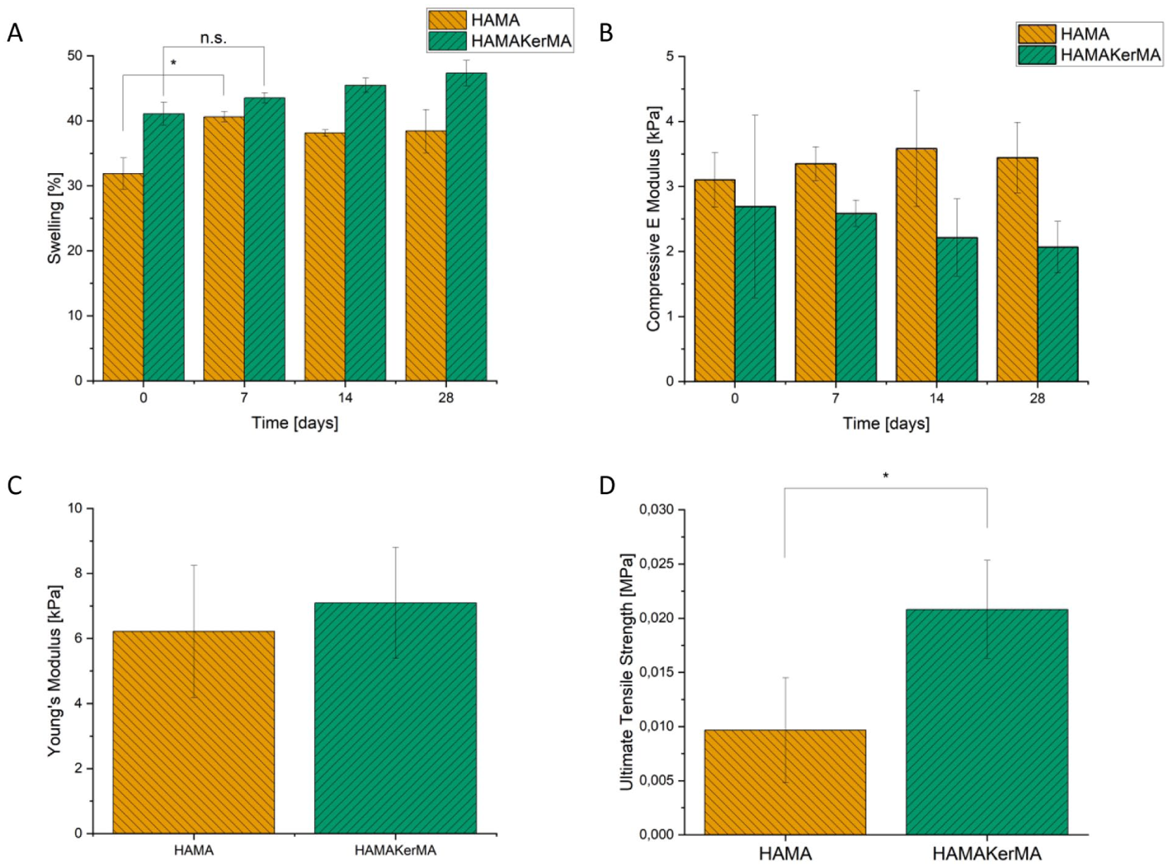
(a) Swelling of the HAMA and HAMAKerMA discs over the course of 28 days, (b) compressive E modulus of the HAMA and HAMAKerMA discs over the course of 28 days, no significant difference, (c) young’s modulus of the HAMA and HAMAKerMA by tensile testing, and (d) ultimate tensile strength by tensile testing (KerMA n = 2).
Two different methods were conducted for the mechanical testing, first a compression test on the bioprinted discs over a period of 28 days and second a tensile test on printed dumbbells according to a modified version of ASTM D412c. The compressive E modulus of the discs did not differ significantly over the course of 28 days (Figure 4(b)). The values were between 2 and 4 kPa, which indicated that the material was quite elastic. A slightly higher Young’s modulus was measured in the tensile test, where the value was around 6 kPa (Figure 4(c)). In terms of the ultimate tensile strength (UTS) there was a significant difference between HAMA and HAMAKerMA (Figure 4(d)). The addition of KerMA almost doubled the UTS. This could be related to the photocrosslinking between KerMA and HAMA. It is likely that the methacrylate groups of KerMA react with the methacrylate groups of HAMA, forming a more stable network, leading to a higher UTS. This was also demonstrated in another study using silk fibroin methacrylate and HAMA. 39 While the mechanical properties appeared to be at the lower end compared to reported values of the cornea, 40 a softer environment could be beneficial because it may support the quiescent state of keratocytes. 41 Higher stiffness is associated with myofibroblast differentiation. 42 Despite these lower values, the discs were easy to handle with tweezers. Further biomechanical studies, such as ex vivo suture tests or in vivo tests could provide clarity on the suitability of the bioprinted material.
Cell viability
Cell viability is a crucial indicator of biocompatibility. A high viability is essential for tissue engineering and especially bioprinting, where not only the material itself plays a role but also the crosslinking and printing process. In this case, extrusion printing has a higher impact on cell viability than other printing methods. 43
The viability of the HCK cell line and HuFibs, encapsulated within the hydrogels HAMA and HAMAKerMA, was monitored over 28 days to assess biocompatibility and the short cellular response to the hydrogel environment. For this purpose, a live/dead staining was performed on day 0, 7, 14, and 28. On the day of printing, the majority of both cells, HCK and HuFibs, showed very high viability with only a few dead cells in both bioinks, confirming that the cells survived the initial bioprinting and crosslinking process (Figure 5).
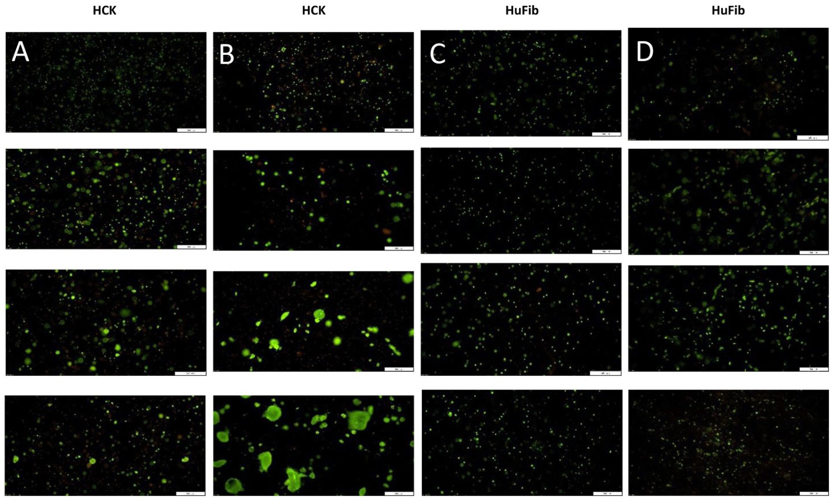
Live/dead staining of: (a) HAMA with HCK, (b) HAMAKerMA with HCK; (c) HAMA with HuFibs; (d) HAMAKerMA with Hufibs. First row is on day 0, second row is on day 7, third row is on day 14, fourth row is on day 28.
HCKs exhibited high viability across all time points, indicating that the hydrogel was non-toxic. Although, the cell viability of the HCKs in the HAMAKerMA bioink decreased slightly on day 7 and 14, it increased again from day 14 to day 28 (Figure 5(b)). However, on day 28, cell clusters of HCKs could be seen in the HAMAKerMA and HAMA hydrogel, which was more pronounced in the HAMAKerMA gel. The increased cell number in the HAMAKerMA bioink may be attributed to its rapid attainment of swelling equilibrium (Figure 4(a)). The swelling kinetics are related to the porosity of the polymer network, which could suggest a highly permeable architecture with superior diffusion kinetics for nutrients and waste products. Furthermore, keratin and KerMA possesses bioactive peptide sequences that can bind integrin receptors on the cell surface. 10 This interaction could trigger intracellular signaling pathways, which could promote proliferation. 44 However, further quantitative analysis using metabolic assays, such as Alamar Blue or DNA-based quantification, is required to confirm this trend.
Throughout the 28 days, the cell viability of the HuFibs remained high in both bioinks (Figure 5(c) and (d)). No morphological changes in cellular distribution were observed, indicating that the HuFibs were alive, but possible in a more quiescent state. Although a high proliferation medium was used, this did not lead to any proliferation of the HuFibs in either bioink. Furthermore, this data suggests that even with the addition of KerMA to the HAMA hydrogel, the proliferation of the HuFibs was not increased, as it was the case for the HCKs. It might be because of the new 3D bioprinted matrix of HAMA.
Immunofluorescence
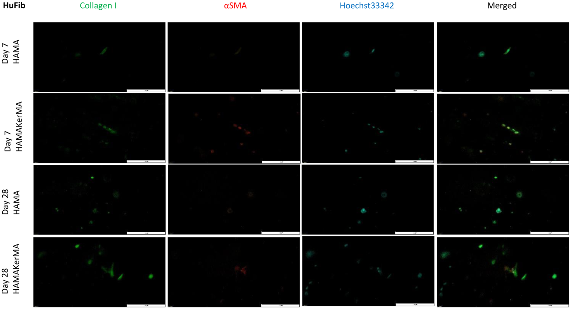
Indirect immunofluorescence staining was conducted at day 7, 14, and 28 to evaluate expression of collagen type I and αSMA in both cell types, HCKs and HuFibs. A negative control can be found in the supplementary information (Supplemental Figure S1). Collagen type I is the most common protein in the cornea and highly expressed by HCKs and HuFibs.29,45 Highly ordered collagen fibrils are abundant in the corneal stroma. 46 αSMA on the other hand is a protein that is expressed by myofibroblasts and after in vivo injury. Myofibroblasts are responsible, among other things, for the structure and organization of extracellular matrix.47–49 When cultured in a serum-free medium, as in this case, HCKs should not express αSMA. 45 In contrast, HuFibs express αSMA, when they are cultivated with TGF-ß1. In the differentiation medium used here, TGF-ß1 was added to increase the collagen I production, but also αSMA was expected to increase. 29
On day 7 little collagen type I and no αSMA was expressed in the HAMA hydrogel for both cell types (Figures 6 and 7). Due to the formation of cell cluster on day 7 in HAMAKerMA gel, more collagen type I was expressed in HCK cell line. However, αSMA was also expressed. Till day 28 the expression of both markers increased in both hydrogels. Nevertheless, the expression of collagen type I was higher in HAMAKerMA bioink. This could correlate to the live/dead staining seen in Figure 5, as more cells proliferated in the HAMAKerMA hydrogel. The addition of KerMA to the HAMA bioinks lead to an increased expression of proteins, further highlighting a possible advantage of adding KerMA to the bioink in terms of cell viability and protein expression for the HCKs.
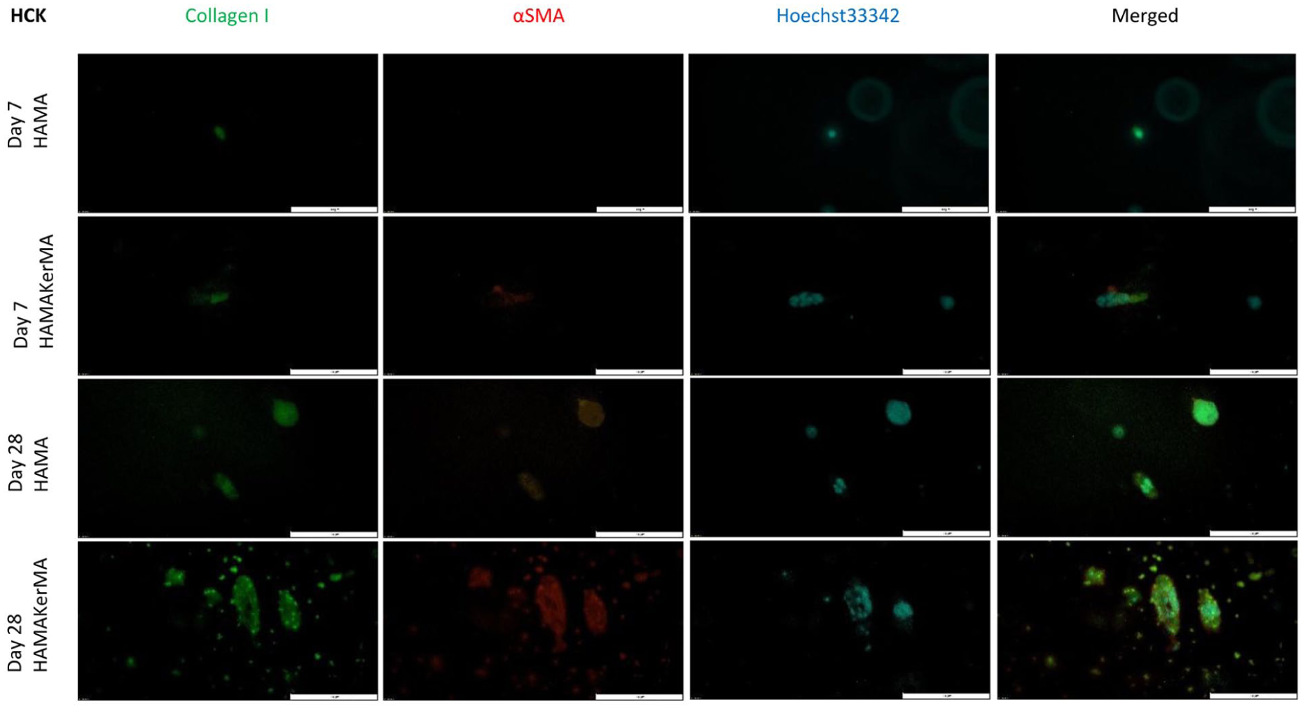
Immunofluorescence of HCKs.
Immunofluorescence of HuFibs.
These preliminary observations, however, provide a first qualitative assessment of collagen type I and αSMA expression. Due to the low cell density and spatial overlap of the immunofluorescence signals, a definitive quantitative comparison between the bioinks is not possible based on these images. Further quantitative studies, such as RT-qPCR, are needed to confirm the claim that adding KerMA increases the protein expression.
In both bioinks, only small amounts of collagen type I or αSMA were expressed by the HuFibs. Over time, a slight increase in collagen type I was observed. In addition, a slightly higher expression of αSMA could be seen in the HAMAKerMA bioink. Nevertheless, the expression of collagen type I and αSMA in the 3D printed construct appeared lower than the typically observed very high expression of collagen type I and low expression of αSMA in 2D cultures under the identical differentiation medium. 29 This could suggest that the newly created environment for the HuFibs had a greater influence on their behavior than the culture conditions. However, it could also suggest that the addition of KerMA promoted a marginally higher expression of collagen type I and αSMA. As stated in our previous study, it was likely that HAMA itself had the greatest influence on this behavior, overshadowing any signals from the medium or KerMA. 21 This could be due to the mechanotransduction of the fairly soft environment, as shown by the biomechanical measurements. Although collagen type I and αSMA expression provides initial insights into the cellular state after printing, these markers are not cornea-specific. To definitively confirm the keratocyte phenotype and the assumed quiescent state of the HuFibs, corneal proteoglycans such as keratocan or lumican must be included. Therefore, further studies on stroma-specific markers are needed to confirm that the bioink environment maintains native cell behavior. Additionally, the UV crosslinking conditions must be considered. Free radicals generated during crosslinking could lead to oxidative stress, impacting both cell viability and protein expression. These microenvironmental cues could alter the cell phenotype. Although a low energy irradiance was chosen here, assessing reactive oxygen species markers could help understand the cells’ behavior. Furthermore, using primary HuFibs in this study provides a preliminary insight into the initial compatibility of HAMA and HAMA-KerMA. Primary cells have been used in corneal tissue engineering to produce cell sheets for regenerative ocular surface therapies. 29 However, it should be acknowledged that human stem cells, such as limbal stem cells or induced pluripotent stem cells (iPSCs), may offer superior clinical scalability. Future research using the aforementioned stem cells is therefore recommended to further evaluate the suitability of these biomaterials and the bioprinting process.
HCET seeding
The corneal epithelium is important for maintaining corneal transparency and for the barrier function of the cornea. 1 Successful epithelialization is crucial for a stromal corneal substitute. Therefore, to investigate that epithelial cells can also attach and proliferate on the bioprinted material, HCETs were seeded on top of the bioprinted construct and cultivated for 28 more days. Cell adhesion was only seen in HAMAKerMA printed discs (Figure 8). This was likely due to the incorporation of KerMA, which provides additional cell attachment motifs. While cell attachment and high viability were observed, the morphology altered and confluence could not be achieved after 28 days. Earlier time points only showed sparsely distributed cells. The lack of confluency prevented meaningful phenotypic characterization, such as indirect immunofluorescence, beyond viability assays. These results suggest that the bioprinted discs could not fully support HCET attachment and require further optimization. Previous studies demonstrated successful attachment, high viability and proper cell morphology of HCETs cultured on keratin-coated substrates.16,25 The modification of keratin to KerMA might have altered these beneficial properties. A more plausible explanation is that the KerMA concentration was too low compared to HAMA to have an impact. To further investigate whether KerMA is suitable for HCETs, a study focusing exclusively on KerMA could support this claim. Furthermore, to understand whether HAMAs intrinsic properties are a limiting factor, the bioprinted constructs could be coated with cell-adhesion proteins such as fibronectin, for example. This would help isolate the problem to understand the mechanism of the lack of cell confluency and change of morphology. However, it is also important to note that the immortalized cell line might not accurately reflect natural behavior. HCETs were used here to gain an initial understanding of their behavior on bioprinted discs with this new material. Limbal epithelial stem cells are commonly used as they play a major role in reepithelization of the cornea. 50 Future work should aim to use limbal stem cells as well, as these are likely to reflect the natural behavior.
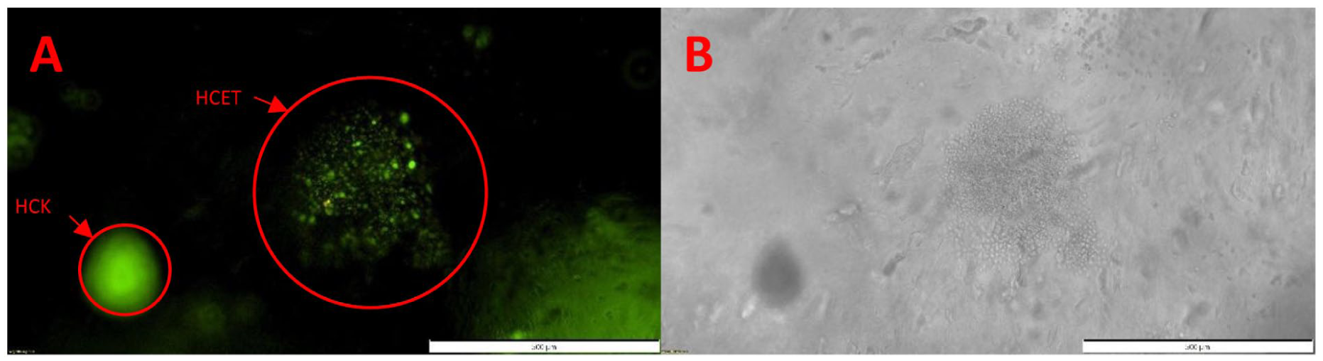
(a) Live/dead staining of HCET on HAMAKerMA bioprint on day 28 and (b) corresponding brightfield image to A.
SEM
Hydrogel porosity plays a pivotal role for cell migration and nutrient diffusion. 51 A higher porosity and pore size supports cell viability and cell morphology. 52 Since both cell types did not express their normal morphology, one explanation was that the pore size and porosity of the HAMA hydrogel is too low due to high molecular weight of the HA, preventing the cells from expressing their dendritic morphology. To assess the pore size of a hydrogel, SEM was conducted here. SEM images revealed pore sizes of 75.80 ± 36.84 µm and 115.46 ± 73.36 µm for the HAMA and the HAMAKerMA hydrogel, respectively (Figure 9). The addition of KerMA resulted in slightly higher pores (Figure 9(b)). This could be one possible explanation for why the cell cluster building was more pronounced in the hydrogel with the addition of KerMA. However, no morphological differences were observed with regard to the porous network. This indicates that the addition of KerMA has only a minor influence on the porous architecture. Nevertheless, it is important to acknowledge the limitation of SEM in assessing pore size and porosity. The evaluation is highly dependent on the sample preparation. 53 Due to the nature of hyaluronic acid hydrogels consisting of high amounts of water, a normal dehydration process was not practical. Therefore, the samples were prepared with liquid nitrogen freezing followed by lyophylisation to obtain a realistic image of the native porous structure. In summary, it is likely that the pore size is sufficient for the cells and therefore does not explain the change of cell morphology.
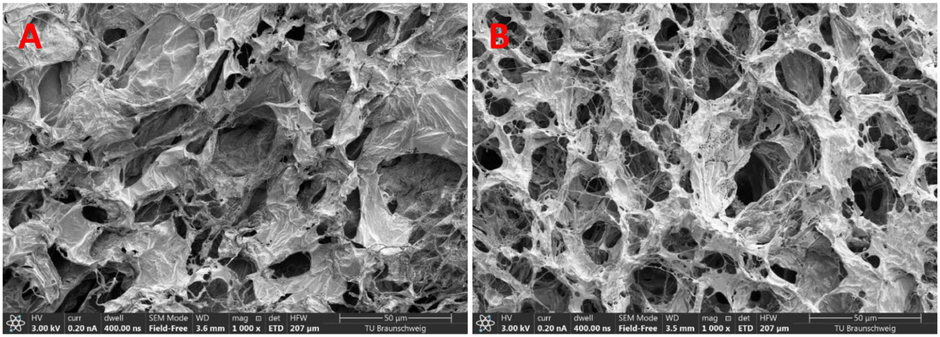
SEM micrographs of: (a) HAMA and (b) HAMAKerMA.
Conclusion
In this study, we were able to successful synthesize a novel biomaterial KerMA based on human waste products and implement it in corneal bioprinting in combination with a HAMA hydrogel. The addition of KerMA to the HAMA Gel led to improved mechanical stability, appropriate swelling and decent light transmission. Both cell types, corneal keratocytes and fibroblasts, were encapsulated in the bioink and demonstrated high cell viability over the course of four weeks. In the combined bioink of HAMAKerMA, the corneal keratocytes showed an increased cell number and expressed a higher amount of collagen. Although a proliferation medium was used, the primary corneal fibroblasts stayed in a quiescent state in both hydrogels and expressed minor amounts of collagen and αSMA. These findings support the incorporation of KerMA as a sustainable biomaterial to improve HAMA hydrogels. We acknowledge, that this study is a preliminary step for corneal bioprinting, as the geometry has not been reproduced. Nevertheless, we believe that KerMA, as a novel biomaterial holds potential in broader use beyond corneal application in other soft tissue applications as a promising alternative to animal-derived biomaterials.
Supplemental Material
sj-docx-1-jbf-10.1177_22808000261453778 – Supplemental material for Development of a human-derived bioink based on keratin methacrylate for corneal bioprinting
Supplemental material, sj-docx-1-jbf-10.1177_22808000261453778 for Development of a human-derived bioink based on keratin methacrylate for corneal bioprinting by Leon Balters and Stephan Reichl in Journal of Applied Biomaterials & Functional Materials
Footnotes
Acknowledgements
Open access funding was provided by the Technische Universität Braunschweig. We would also like to thank Martina Knežević for her dedicated assistance with isolating and cultivating the human corneal fibroblasts.
Author contributions
L.B. and S.R. did conceptualization and design of study. L.B. did visualization, methodology, investigation, data curation, and writing of original draft. S.R. did supervision, resources, project administration, and writing – review and editing.
Funding
The authors received no financial support for the research, authorship, and/or publication of this article.
Declaration of conflicting interests
The authors declared no potential conflicts of interest with respect to the research, authorship, and/or publication of this article.
Data availability statement
All data or related information supporting the conclusions of this study is included in the article or can be obtained separately.
Supplemental material
Supplemental material for this article is available online.
References
Supplementary Material
Please find the following supplemental material available below.
For Open Access articles published under a Creative Commons License, all supplemental material carries the same license as the article it is associated with.
For non-Open Access articles published, all supplemental material carries a non-exclusive license, and permission requests for re-use of supplemental material or any part of supplemental material shall be sent directly to the copyright owner as specified in the copyright notice associated with the article.