






Abstract
This study aimed to develop a novel hemostatic dressing capable of exerting pressure-assisted hemostasis, promoting platelet aggregation, and enhancing the absorption of blood and tissue exudates. The Alginate/calcium chloride (Alg/CaCl2) dressing was fabricated by crosslinking sodium alginate with calcium chloride, resulting in the formation of a stable gel network, which was subsequently freeze-dried to obtain a porous structure. In vitro evaluations were performed using L929 mouse fibroblasts to assess cytotoxicity, platelet aggregation, and platelet activation. The biosafety of the dressing was further examined using a rabbit skin irritation model following both topical application and subcutaneous injection. In addition, the pressure-assisted hemostatic effect of the dressing was evaluated using a customized compression device incorporating the Alg/CaCl2 dressing.
The results demonstrated that the Alg/CaCl2 dressing promoted platelet aggregation and activation while exhibiting minimal cytotoxicity at most tested concentrations, indicating favorable biocompatibility. The dressing possessed a highly porous structure and excellent fluid absorption capacity. Furthermore, the sustained release of physiologically active calcium ions may facilitate platelet activation and fibrin formation, thereby accelerating the hemostatic process. In vivo assessments revealed no significant skin irritation or adverse tissue reactions following either topical application or subcutaneous injection, supporting its safety for biomedical applications. Moreover, compression testing in the animal model demonstrated that the Alg/CaCl2 dressing generated greater compressive force than commercially available hemostatic products, contributing to enhanced pressure-assisted hemostasis.
Highlights
1. A novel Alg/CaCl2 hemostatic dressing was developed by crosslinking sodium alginate with calcium chloride and freeze-drying to achieve both passive fluid absorption and active pressure-assisted hemostasis. 2. CaCl2 crosslinking dramatically increased dressing porosity from 9.3% to 92.3%, enabling rapid absorption of blood and wound exudates to concentrate platelets and coagulation factors at the injury site. 3. The dressing extract induced platelet activation and fibrin network formation in vitro, demonstrating that sustained calcium ion release effectively triggers both intrinsic and extrinsic coagulation pathways. 4. In a rabbit ear artery model, the Alg/CaCl2 dressing achieved hemostasis within 60 seconds and generated a compressive force of 109.3 mmHg, outperforming commercial hemostatic products. 5. The dressing demonstrated favorable biosafety with no significant skin irritation or allergic reactions, and histological analysis confirmed superior wound healing with minimal abnormal tissue reactions compared to commercial dressings.
1. Introduction
When the integrity of the skin is disrupted, the body initiates a highly coordinated wound healing process consisting of four overlapping phases: hemostasis, inflammation, proliferation, and remodeling. 1 Immediately following tissue injury, the hemostatic phase is activated, during which platelets rapidly adhere to the injured site and trigger the coagulation cascade, resulting in clot formation and cessation of bleeding. 2 Subsequently, the inflammatory phase regulates the local immune response, removes cellular debris and pathogens, and prepares the wound microenvironment for tissue repair. 3 During the proliferative phase, fibroblasts migrate into the wound bed and synthesize extracellular matrix components, particularly collagen, thereby promoting granulation tissue formation and tissue regeneration. 4 The final remodeling phase may persist for weeks to months and is characterized by extracellular matrix reorganization and maturation, during which type III collagen is gradually replaced by the mechanically stronger type I collagen. 5
To facilitate and accelerate wound healing, a variety of wound dressings have been developed and are widely used in clinical practice. Current dressing materials include films, foams, composites, and hydrogels, each possessing distinct advantages and limitations. Among these materials, alginate-based dressings have received considerable attention owing to their excellent biocompatibility, gel-forming ability, and intrinsic hemostatic properties. In the presence of calcium ions, alginate undergoes rapid ionic crosslinking to form a stable hydrogel network capable of absorbing several times its own weight in wound exudate. This characteristic enables alginate dressings to maintain a moist wound environment while effectively managing exudate, making them particularly suitable for highly exudative and deep wounds. 6
Despite these advantages, conventional alginate dressings primarily function by providing passive hemostasis and promoting a favorable wound-healing environment. Although they have been shown to shorten healing time and reduce wound-associated pain, their ability to generate mechanical compression is limited.7,8 Since external compression plays a critical role in controlling acute bleeding, particularly in traumatic injuries and arterial hemorrhage, the development of a compression-type alginate dressing with enhanced hemostatic performance may provide additional clinical benefits.
In the present study, we developed a compression-type hemostatic dressing based on sodium alginate crosslinked with calcium ions (Ca2+). The interaction between the carboxyl groups of alginate and calcium ions forms a stable three-dimensional network with high absorbency, adequate gel strength, and excellent conformity to moist wound surfaces. The hemostatic potential of the dressing was comprehensively evaluated through in vitro platelet aggregation and activation assays, in vivo biocompatibility testing, and pressure-induced hemostatic assessments in an animal model. In brief, our dressing could provide extra compression capability up to approximately 100 mmHg and enhance hemostatic performance. Especially no prominent scar tissue formation was observed as compared to the commercial product group.
2. Materials and methods
2.1. Preparation of wound dressings
Sodium alginate (1.2 g) was dissolved in 60 mL deionized water at 95°C with continuous stirring. After cooling, the solution was poured into culture dishes, frozen at −80°C, and freeze-dried for 1–2 days.
The freeze-dried alginate dressings were crosslinked by immersion in CaCl2 ethanol solutions (0.15%, 0.3%, or 0.6%) for 30 s, followed by drying at 65°C.
2.2. Analysis of the dressing surface and porosity
For scanning electron microscopy (SEM) analysis, dried Alg/CaCl2 dressings were cut to 0.5×0.5×0.5 cm3 and mounted onto conductive copper tape. We attached the copper tape with the sample onto a specimen holder and coated the sample with platinum in a vacuum sputter coater (sputtering time: 60 s). The coated sample was then observed by SEM under an accelerating voltage of 10 kV. We used ImageJ to read and estimate the porosity.
2.3. In vitro experiments: MTT assay, platelet aggregation, and platelet activation
In this study, we conducted cell experiments using L929 mouse fibroblast cells to evaluate the cytocompatibility of the extract obtained from Alg/CaCl2 dressings. L929 is widely used in in vitro cytotoxicity testing of biomaterials.9,10
2.3.1. Cytotoxicity test (MTT assay)
Cytotoxicity was evaluated using the Thiazolyl Blue Tetrazolium Bromide (MTT) assay, a standard method for assessing cell viability based on mitochondrial activity. Viable cells reduce MTT to insoluble formazan crystals, which can be quantified spectrophotometrically.
An indirect extraction method was applied according to ISO 10993–12. 11 The dressing materials were incubated in serum-free medium at 37°C for 24 hours to obtain extract solutions. After centrifugation to remove debris, the supernatant was collected and serially diluted (undiluted, 1/2, 1/4, 1/8) with SFM. SFM alone served as the control.
L929 mouse fibroblasts were seeded in 24-well plates at a density of 5 × 10⁴ cells/mL (500 µL/well) and incubated at 37 °C in a humidified atmosphere with 5% CO2. After 24 hours, the culture medium was removed, cells were washed with PBS and treated with either extract solutions or control medium for an additional 24 hours.
Subsequently, cells were washed and incubated with MTT solution (5 mg/mL, 250 µL/well) for 2 hours in the dark. The resulting formazan crystals were dissolved in DMSO (500 µL/well), and the absorbance was measured at 570 nm using a microplate reader. Cell viability was calculated relative to the control group.
All experiments were performed in triplicate. Results are presented as mean ± SD. Statistical differences between groups were analyzed using Student t-test using Excel software. Differences were considered statistically significant when P < 0.05.
2.3.2. In vitro platelet aggregation test
Rabbit blood was centrifuged at 160 g for 10 min at 25°C to obtain platelet-rich plasma (PRP), which was diluted with deionized water. 12 Fifty milligrams of dressing (0.3% Alg/CaCl2 or a commercial product) were incubated with 1 mL of Tyrode’s solution for 6 min, followed by centrifugation to collect the supernatant.
For aggregation analysis, 0.5 mL of PRP was mixed with 1 mL of Tyrode’s solution, and optical density was continuously recorded for 6 min. The dressing-derived supernatant was then added, and measurements were continued for an additional 8 min. PRP treated with thrombin served as a positive control. Absorbance was measured using a plate reader, and platelet aggregation was quantified by calculating pre- and post-reaction transmittance as follows:


2.3.3. In vitro platelet activation test
Rabbit blood was centrifuged at 160 g for 10 min at 25°C to obtain platelet-rich plasma (PRP), which was diluted with deionized water. Fifty milligrams of the 0.3% Alg/CaCl2 dressing were incubated with 1 mL of Tyrode’s solution for 6 min, followed by centrifugation to collect the supernatant.
The supernatant was mixed with 1 mL of diluted PRP and 0.5 mL of Tyrode’s solution, then added to 24-well plates containing 12-mm coverslips and incubated at 37°C with 5% CO2 for 2 h. After incubation, samples were washed with PBS, fixed with 4% formaldehyde for 30 min, washed again, freeze-dried, and examined by SEM. The diameters of fibrin fibers at ten randomly selected locations in the SEM image were measured using ImageJ. The porosity was also calculated.
2.4. In vivo experiments: Allergic reactions
2.4.1. In vivo allergic reaction of New Zealand white rabbits on skin application
All procedures were approved by the Institutional Animal Care and Use Committee of the National Defense Medical Center [IACUC approval No. IACUC-24-087]. To assess for any allergic effects of the Alg/CaCl2 dressing in vivo, we prepared Alg/CaCl2 dressing extract with 0.6% CaCl2 by immersing the dressing in 1 PBS and shaking it in a 37°C water bath for 24 h, followed by centrifugation to collect the supernatant.
We shaved the backs of New Zealand White rabbits and monitored for any signs of skin irritation within 24 h. If no skin irritation was observed at 24 h, we marked and divided the shaved area into multiple zones, each measuring 2 × 3 cm. We then applied 500 µL of the following solutions: 1-chloro-2,4-dinitrobenzene for the positive control (PC) group, 1X PBS for the negative control (NC) group, and the Alg/CaCl2 extract solution for the experimental group. Observations were recorded before shaving and at 0, 1, 4, 24, 48, and 72 h postshaving. We assessed skin reactions according to the ISO 10993 irritation grading scale. 12
2.4.2. In vivo allergic reaction of New Zealand white rabbits on subcutaneous injection
To evaluate the effects of subcutaneous injection of dressing extracts, the fur on the rabbits’ backs was shaved, and the skin was monitored for signs of irritation. If no irritation was observed after 24 hours, the dorsal area was divided into multiple labeled sections. A total of 200 µL of the following solutions was subcutaneously injected into separate sites: 1-chloro-2,4-dinitrobenzene for the positive control (PC) group, 1× phosphate-buffered saline (PBS) for the negative control group, and Alg/CaCl2 extract solutions (crosslinked with 0.15%, 0.3%, or 0.6% CaCl2) for the experimental groups.
Local reactions were recorded at 0, 24, 48, and 72 hours, as well as 7 days post-injection, according to the ISO 10993-10 irritation scoring criteria. 13
2.5. Compression device for in vitro and in vivo pressure hemostasis testing
A hemostatic compression device incorporating the Alg/CaCl2 dressing was developed. As shown in supplemental figure S1, circular base dressings (1 cm in diameter, 8 mm thick) composed of alginate, 0.3% Alg/CaCl2, or a commercial product were fabricated and compressed to approximately 3 mm upon application to the wound surface. A cylindrical pad (1 cm × 7.5 mm) and a second larger pad (3 cm × 4.5 mm) were sequentially affixed using 16% polyvinyl alcohol (PVA) as an adhesive. The assembled device was secured with an elastic bandage to provide sustained compression. A piece of pressure sensor was inserted and placed between alginate dressing and pressure pad to detect the pressure by FlexiForce ELF Single-Point Force Measurement System. The type of the membrane sensor was B201(FlexiForce, Tekscan, MA, USA). To calibrate, apply a known force to the sensor (conventional balance weight was used), and equate the sensor resistance output to this force. Repeat this step with a number of known forces that approximate the load range to be used in testing. Plot Force versus Conductance (1/R). A linear interpolation can then be done between zero load and the known calibration loads, to determine the actual force range that matches the sensor output range.
Hemostatic efficacy was evaluated in a rabbit ear artery injury model. Three rabbits were randomly assigned to three groups (0.3% Alg/CaCl2, pure alginate, or commercial dressing; n = 3 per group, six wounds per group). Hemostasis time was recorded at 60 seconds and extended to 90 or 120 seconds if necessary. Wound healing was assessed 10 days post-hemostasis. Repeated measures one-way ANOVA with Dunnett’s multiple comparisons test were applied for statistical analysis.
2.6. Pathology of the wounds
Tissue sections were obtained from the ear wounds of three rabbits in each of the three groups and subjected to histopathological examination with hematoxylin and eosin staining. Scar formation area of commercial product was marked and its scale relative to entire image was calculated.
3. Results
3.1. Effect of CaCl2 concentration
We compared sodium alginate dressings crosslinked with 0.15%, 0.3%, and 0.6% calcium chloride in alcoholic solutions, as shown in Figure 1(a). All three sample groups were similar in appearance and shape, with no significant differences in size. All three dressings displayed a circular structure with a similar degree of structural integrity. (a) Effect of CaCl2 concentration on the crosslinked dressing: (a) 10 mL Alg/0.6% CaCl2, (b) 10 mL Alg/0.3% CaCl2, and (c) 10 mL Alg/0.15% CaCl2. (b) SEM images before and after crosslinking: (a) alginate and (b) Alg/CaCl2.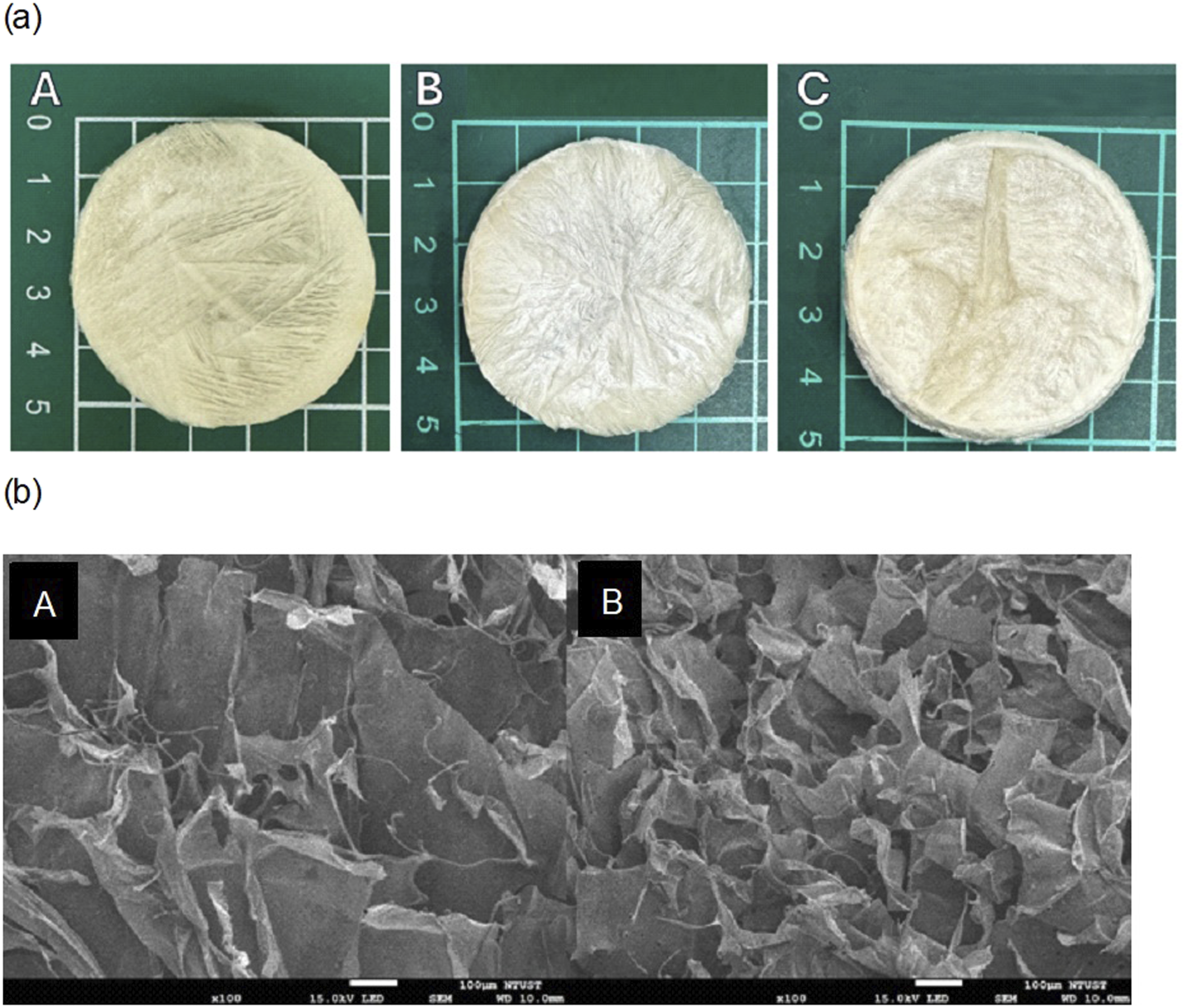
3.2. Porosity analysis
We examined the surface morphology of the dressings using SEM, as shown in Figure 1(b). The uncrosslinked sodium alginate dressing exhibited a compact and relatively smooth sheet-like structure with a porosity of only 9.3%. In contrast, the dressing crosslinked with 0.3% CaCl2 displayed a three-dimensional structure characterized by numerous folds and pores, reaching a porosity of 92.3%, significantly higher than that of the uncrosslinked sample.
3.3. Results of in vitro experiments
3.3.1. Cytotoxicity test (MTT assay)
Cell viability of L929 fibroblasts was evaluated using extracts obtained from sodium alginate dressings crosslinked with different CaCl2 concentrations to assess biocompatibility.
As shown in Supplemental Figure S2(a), extracts from the 0.6% CaCl2–crosslinked dressing exhibited marked cytotoxicity. Cell viability was 49% in the undiluted extract and slightly increased to 54% and 61% following 1/2 and 1/4 dilutions, respectively, indicating concentration-dependent toxicity.
In the 0.3% CaCl2 group shown in Figure 2, the undiluted extract showed 41% cell viability, which remained below the cytotoxicity threshold after 1/2 dilution (51%). Cell viability increased to 75% and 82% at 1/4 and 1/8 dilutions, respectively, suggesting reduced cytotoxicity upon dilution and acceptable biocompatibility at appropriate concentrations. Cell viability of sodium alginate dressing crosslinked with 0.3% CaCl2. *** p<0.001; ** p<0.01; * p<0.05; ns: not significant.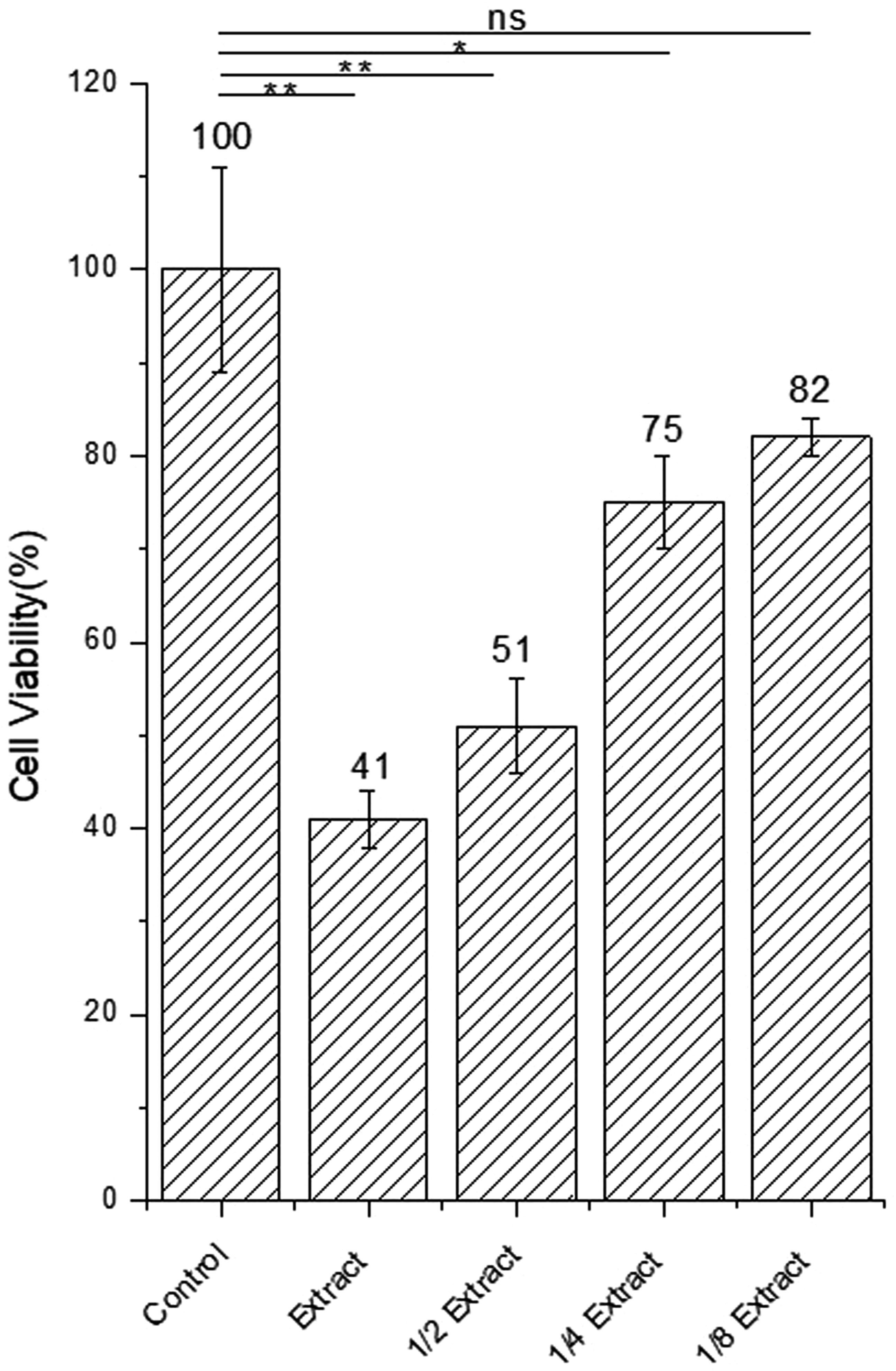
For the 0.15% CaCl2 group (supplemental Figure S2(b)), the undiluted extract resulted in 37% viability, which further decreased to 27% after 1/2 dilution. Although viability increased to 80% and 61% at 1/4 and 1/8 dilutions, respectively, the response was less consistent, possibly due to incomplete crosslinking or residual ionic imbalance.
Based on these results, the intermediate crosslinking concentration of 0.3% CaCl2 was selected for subsequent experiments.
3.3.2. In vitro coagulation analysis
Platelet aggregation was assessed by optical turbidimetry, in which decreased turbidity reflects aggregation.14,15 As shown in Figure 3(a), transmittance (T%) was recorded for the 0.3% Alg/CaCl2 dressing, a commercial dressing, and thrombin. All groups maintained T% values of 99%–100% during the first 6 minutes, indicating minimal aggregation, followed by a marked decline at 7 minutes. (a) Transmittance value (T%) in the in vitro coagulation test. (b) Fibrin formation during platelet activation. Ten fibrin diameter were selected randomly. (c) Each of the selected diameter and the mean diameter was 481±76 nm.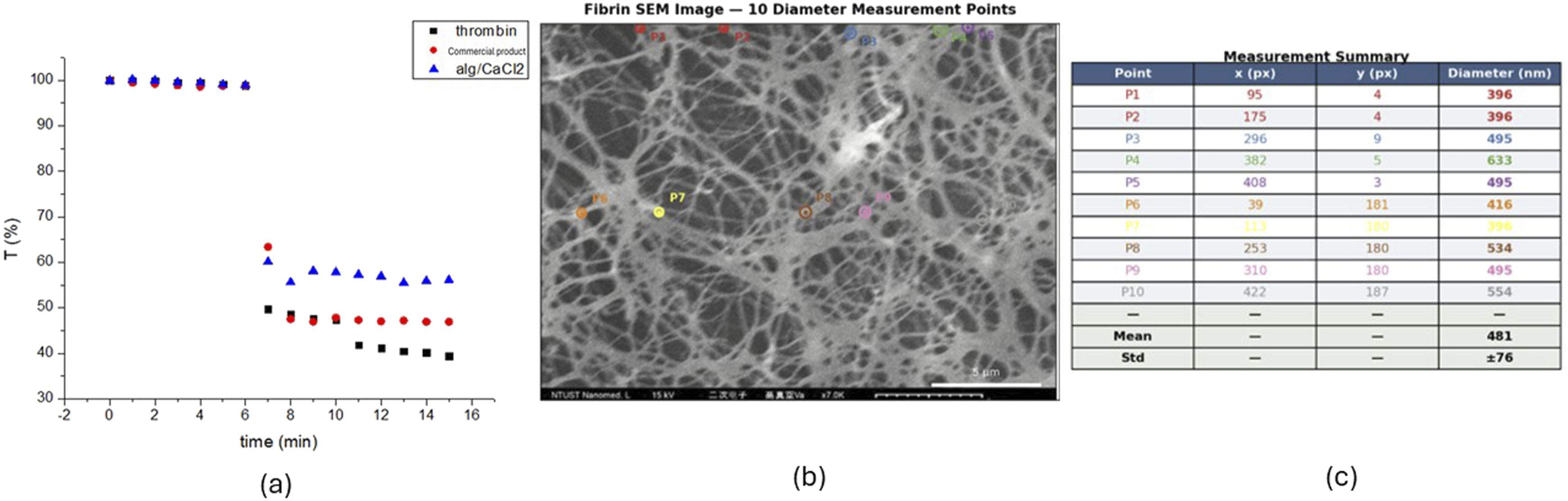
Thrombin produced the greatest reduction in T% (≈40%), whereas commercial dressing showed a moderate decrease (≈46%–47%). The Alg/CaCl2 dressing exhibited the smallest reduction in T%, with final values remaining above 55%, indicating relatively weaker platelet aggregation compared with thrombin and the commercial dressing.
3.3.3. In vitro platelet activation analysis
Activated platelets extend numerous fibrin structures on their surface, such as P-selectin (CD62P), and release granular contents to accelerate the coagulation cascade.16–18 Platelet activation is typically assessed by incubating blood or platelet suspensions with dressing extract solutions and subsequently observing the suspensions via SEM.
Figure 3(b) shows the fibrin networks observed after PRP was exposed to the 0.3% Alg/CaCl2 solution extract. Abundant mesh-like fibers are clearly present, indicating that platelets were successfully activated by the dressing extract, inducing fibrin formation. The diameters of fibrin fibers at ten randomly selected locations in the SEM image shown in Figure 3(b) were measured using ImageJ. The mean fibrin fiber diameter was 487±76 nm in Figure 3(c), while the porosity of the fibrin network was 44.6% as shown in supplemental Figure 5S.
3.4. Results of the in vivo experiments
3.4.1. Allergic response in New Zealand white rabbits: Topical application
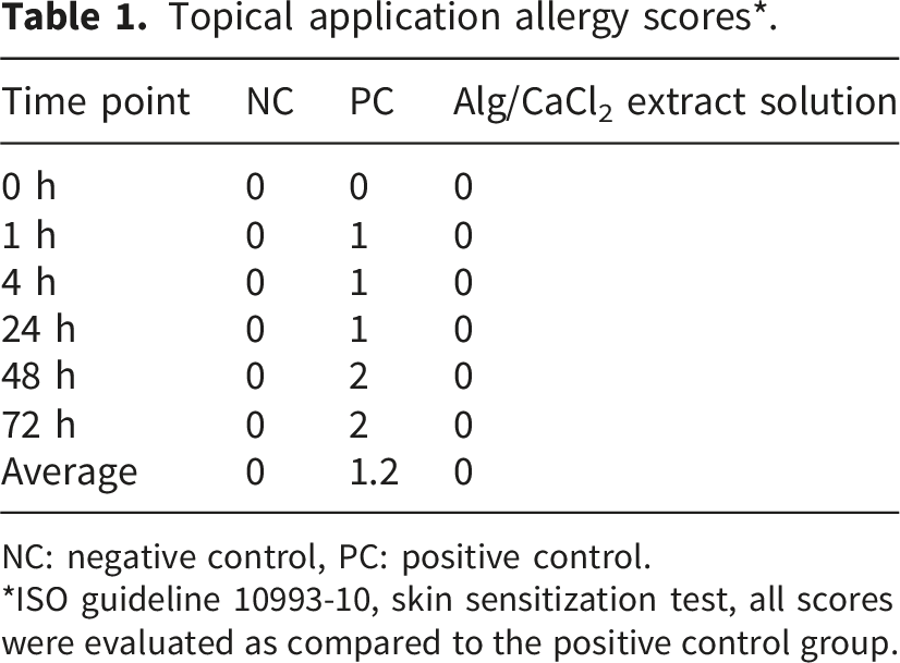
Following ISO 10993-10, skin irritation induced by 0.6% Alg/CaCl2 extract was evaluated in New Zealand white rabbits, with PBS and 1-chloro-2,4-dinitrobenzene as negative and positive controls. Skin reactions were scored at multiple time points.
As shown in Figure 4 and Table 1, the negative control showed no erythema or edema (score 0 throughout), whereas the positive control developed mild erythema that progressed to moderate irritation at 48–72 hours (average score 1.2). The experimental group exhibited transient yellow–brown pigmentation within 24 hours without erythema or edema and did not meet the criteria for skin irritation. Allergic reaction to the topical application of dressing extract in New Zealand white rabbits: (a) 0.6% Alg/CaCl2, (b) NC (1X PBS), and (c) PC (1-chloro-2,4-dinitrobenzene). Topical application allergy scores*. NC: negative control, PC: positive control. *ISO guideline 10993-10, skin sensitization test, all scores were evaluated as compared to the positive control group.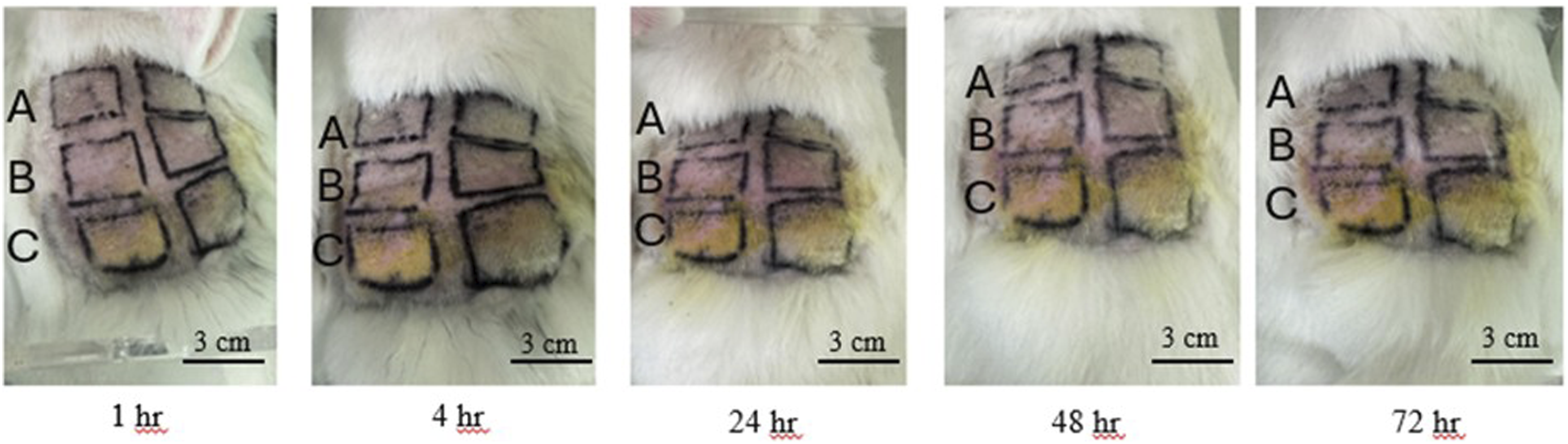
3.4.2. Allergic response in New Zealand white rabbits: Subcutaneous injection
Following ISO 10993-10, subcutaneous irritation induced by Alg/CaCl2 extracts (0.15%, 0.3%, and 0.6%) was evaluated in New Zealand white rabbits, with PBS and 1-chloro-2,4-dinitrobenzene as negative and positive controls. Skin reactions were assessed 72 hours after injection.
As shown in Figure 5, the positive control exhibited marked inflammatory responses, whereas the negative control showed no abnormalities. Sites treated with Alg/CaCl2 extracts appeared largely normal, with only mild transient redness and no swelling, necrosis, or persistent inflammation, meeting the ISO 10993-10 criteria for no to slight irritation.
19
Allergic reaction to subcutaneous injection of dressing extract in New Zealand white rabbits at 72 h: (a) NC (1X PBS), (b) PC (1-chloro-2,4-dinitrobenzene), (c) 0.6% Alg/CaCl2, (d) 0.3% Alg/CaCl2, and (e) 0.15% Alg/CaCl2.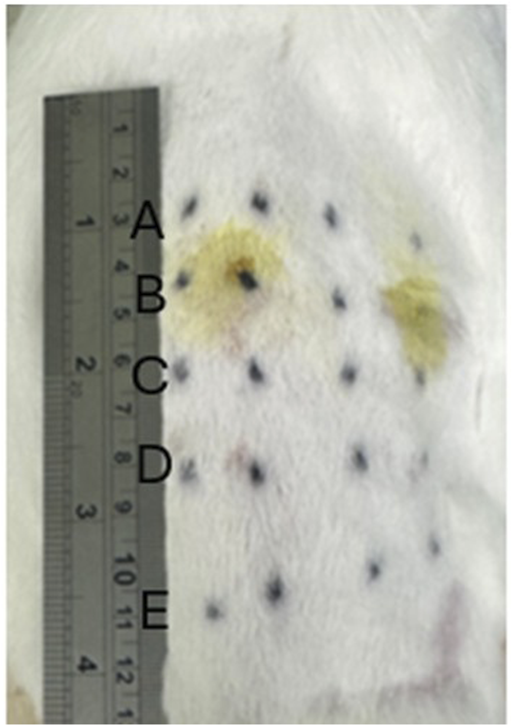
3.5. Pressure hemostasis test in vitro and in New Zealand white rabbits
To evaluate the hemostatic performance of the Alg/CaCl2 dressing, initial compression pressures were measured using a wrist compression device and compared with a device incorporating a commercial dressing. The commercial dressing generated a pressure of approximately 588 mmHg (Supplemental Figure S3), whereas the 0.3% Alg/CaCl2 dressing produced a higher pressure of 981 mmHg (Supplemental Figure S4).
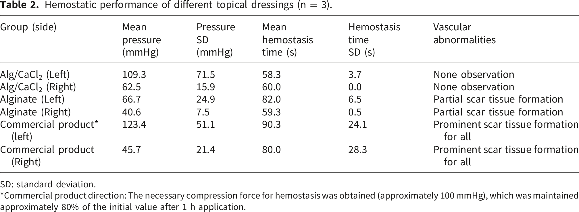
Hemostatic performance of different topical dressings (n = 3).
SD: standard deviation.
*Commercial product direction: The necessary compression force for hemostasis was obtained (approximately 100 mmHg), which was maintained approximately 80% of the initial value after 1 h application.
Hemostatic pressures in the commercial dressing group were highest in the left ear (mean ≈ 123 mmHg) but showed substantial variability. The Alg/CaCl2 group exhibited moderately high pressures (mean ≈ 109 mmHg), whereas the alginate group showed lower pressures in both ears. Although statistical results of pressure and hemostatic time show no significant difference (left ear pressure p=0.499, right ear pressure p=0.808, left ear hemostatic time p=0.303, right ear hemostatic time p=0.569) as compared to the commercial product group; however, the most remarkable finding is that our pressure hemostatic dressing cause no scar tissue formation after application.
On day 10, no abnormal findings were observed in the Alg/CaCl2 group. In contrast, the alginate group exhibited small nodules in several ears, whereas the commercial dressing group showed multiple nodules or incompletely healed wounds.
3.6. Pathology of wounds
To evaluate the effects of the dressings on wound healing, wound tissues were harvested for histological analysis. Figure 6(a)–(c) show hematoxylin and eosin (H&E) staining of wounds treated with the Alg/CaCl2, alginate, and commercial dressings, respectively. Figure 6(a) demonstrated favorable wound healing in the Alg/CaCl2 group, whereas Figure 6(c) showed increased dermal collagen deposition in the commercial dressing group. Histological analysis revealed scar formation only in the commercial dressing group (Figure 6(c)). The scar tissue area accounted for 13,586 pixels out of a total tissue area of 83,153 pixels, corresponding to approximately 16.3%. In contrast, no scar tissue was observed in the Alg/CaCl2 dressing group (Figure 6(a)) or the alginate-only group (Figure 6(b)), yielding a scar area percentage of 0%. H&E staining of a wound after treatment with (a) Alg/CaCl2 dressing, (b) alginate dressing, and (c) commercial dressing. Panel (c) reveals an increased dermal collagen deposit. The marked area was the scar formation area, The scar tissue area accounted for 13,586 pixels out of a total tissue area of 83,153 pixels, corresponding to approximately 16.3%.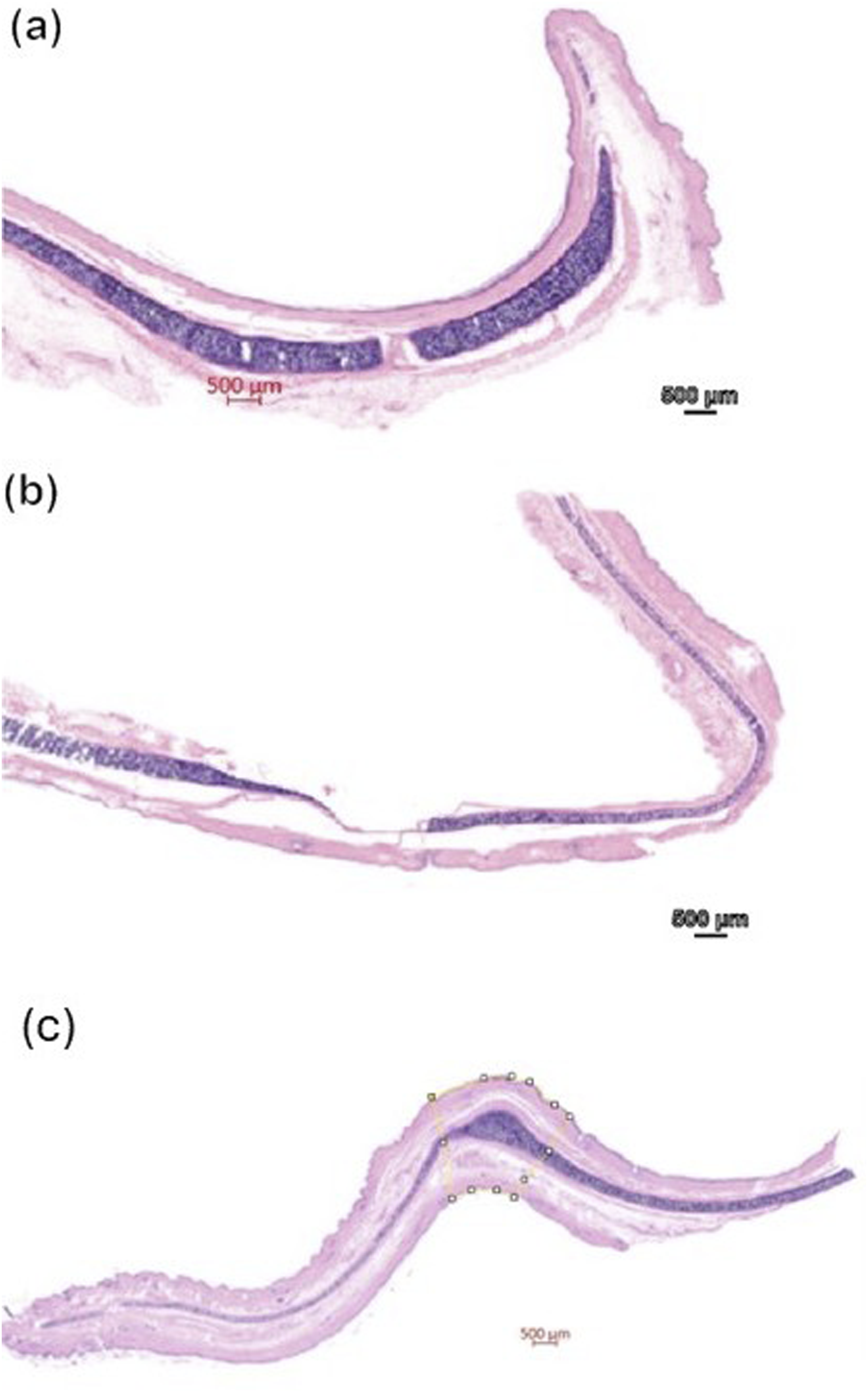
4. Discussion
At different CaCl2 concentrations, although some samples exhibited surface creases or fiber stretching caused by the crosslinking process, no obvious collapse or fracture of the overall structure was observed. The differences in appearance were not significant, indicating that, under the experimental conditions, the calcium chloride concentration in the alcoholic solution had a limited impact on the initial morphology of the final product. This finding may be attributed to the role of alcohol as a solvent, which slows and moderates the crosslinking reaction, thereby helping to preserve the surface structure and overall shape of the dressing.
In the porosity analysis, the uncrosslinked sodium alginate dressing exhibited a porosity of only 9.3%. This dense structure may limit moisture and gas exchange, potentially hindering cell infiltration and wound healing. In contrast, the porosity of the crosslinked dressing reached 92.3%, suggesting that the crosslinking process modified the microstructure and improved its suitability for tissue regeneration. The resulting porous architecture facilitates cell adhesion and proliferation and enhances the exchange of nutrients and metabolic waste, thereby supporting wound healing. The marked increase in porosity following CaCl2 crosslinking may have important implications for hemostatic performance. Highly porous structures can rapidly absorb blood and wound exudates, thereby concentrating platelets, erythrocytes, and coagulation factors at the injury site. This concentration effect may accelerate clot formation and contribute to earlier hemostasis. Furthermore, the interconnected porous network facilitates oxygen diffusion and nutrient transport, both of which are essential for cellular migration and tissue regeneration during wound healing. Similar observations have been reported in other alginate-based biomaterials, where increased porosity was associated with improved fluid management and enhanced tissue integration.
The cytotoxicity results obtained from the three crosslinking concentrations suggest that high-concentration crosslinking may provide advantages in structural stability and Ca2+ incorporation; however, it may also increase the risk of residual Cl- and associated cytotoxicity. In contrast, low-concentration crosslinking may result in reduced Ca2+ release but could allow unreacted components to remain in the extract, potentially contributing to nonspecific toxic effects. The intermediate concentration of 0.3% appears to achieve a balance between crosslinking efficiency and cell viability. Under diluted conditions, this concentration provided a relatively favorable environment for cell culture, indicating improved biocompatibility and supporting its selection for further investigation. Although cell viability below 70% is generally considered indicative of cytotoxicity, the extract from the 0.3% CaCl2-crosslinked dressing required a fourfold dilution to approach this threshold, suggesting that cytotoxicity was evident only at relatively high extract concentrations. Importantly, the experimental conditions of the MTT assay represent a extreme scenario, involving continuous cellular exposure for 72 hours. In contrast, the dressing is intended for temporary clinical application, typically remaining in contact with the wound for only a few hours according to the guidelines of the commercial product. The compression time should be used within 2 hours after application. Therefore, it is impossible to reach this high concentration as undiluted extract solution and exposure time. Furthermore, the dynamic wound environment, characterized by ongoing dilution from blood, wound exudate, and interstitial fluids, is expected to substantially decrease local concentrations of leachable components. Consequently, the cytotoxicity detected in vitro may overestimate the actual biological effects in vivo. This interpretation is further supported by the absence of significant irritation or adverse tissue reactions observed in our animal studies, indicating that the dressing exhibits satisfactory biocompatibility under physiologically relevant conditions.
Our coagulation test suggested that the Alg/CaCl2 extract may reduce direct platelet contact and activation, possibly due to its structural characteristics or controlled Ca2+ release. Calcium functions as coagulation factor IV and is indispensable for multiple enzymatic reactions within both intrinsic and extrinsic coagulation pathways. The presence of calcium facilitates the activation of factors IX and X, assembly of the prothrombinase complex, and conversion of prothrombin into thrombin, ultimately leading to the formation of a stable hemostatic plug. 20
In vitro platelet aggregation tests revealed that our material exhibited weaker aggregation effects than thrombin, which thrombin is used as a positive control to validate the activity of harvested platelet-rich plasma. Although our dressing did not demonstrate a greater reduction in transmittance than the commercial product, it induced platelet aggregation within a comparable timeframe. Given that arterial injury can result in rapid blood loss, the time required to initiate platelet aggregation may be more clinically relevant than the final aggregation percentage. In this study, all three groups demonstrated marked platelet aggregation within 7 minutes. The in vitro platelet assays evaluated chemical coagulation in the absence of mechanical pressure and may therefore not fully replicate the in vivo hemostatic environment. In contrast, the animal model incorporated mechanical compression, suggesting that the synergistic effect of mechanical pressure and chemical coagulation may enhance overall hemostatic performance.
Interestingly, although the Alg/CaCl2 dressing demonstrated weaker platelet aggregation than thrombin and the commercial dressing in vitro, superior hemostatic performance was observed in the rabbit ear artery model. This apparent discrepancy highlights the limitations of conventional in vitro coagulation assays, which evaluate only isolated biochemical interactions under static conditions. In clinical bleeding scenarios, hemostasis is influenced by multiple factors, including blood flow dynamics, tissue compression, fluid absorption, platelet recruitment, and clot stabilization. The favorable in vivo results observed in this study likely resulted from the combined effects of calcium-mediated coagulation activation, rapid fluid absorption, and mechanical compression generated by the dressing. Therefore, the hemostatic efficacy of the Alg/CaCl2 dressing may be better explained by a multifactorial mechanism rather than platelet aggregation alone.
Meanwhile, SEM observations of platelet structure confirmed that the dressing extract could induce platelet activation and fibrin formation. The observed fibrin structures likely resulted from platelet activation, including surface protein expression (e.g., P-selectin) and granule release, which also contributed to the activation of the coagulation cascade. The images revealed fibrin networks with varying degrees of crosslinking and distribution, characterized by fine and evenly dispersed structures.
The presence of fibrin formation indicates potential hemostatic and wound-healing properties. 21 SEM imaging of the fibrin network revealed fiber diameters ranging from 396 to 633 nm (mean: 481 ± 76 nm), consistent with typical fibrin gel morphology (400-600 nm). This demonstrates that our dressing does have a hemostatic effect on the structure. Image analysis demonstrated a porosity of 44.6% as shown in supplemental Figure 5S, indicating a highly interconnected, open-pore network structure favorable for nutrient diffusion and cell infiltration.
The topical and subcutaneous allergic reaction tests indicated that the Alg/CaCl2 extract did not induce any allergic reactions. Although localized pigmentation was noted in the topical application test, it occurred without signs of erythema or edema, suggesting that the color change was due to the extract’s intrinsic color rather than a true irritant response. Thus, the results support the good topical and subcutaneous compatibility of this dressing and its extract, making it suitable for future use in wound dressings or other medical products.
Mechanical compression remains one of the most effective and widely used strategies for controlling acute bleeding. The substantially greater compression force generated by the Alg/CaCl2 dressing compared with the commercial dressing suggests that its structural characteristics may enhance the efficiency of pressure transmission to the injured vessel. Effective compression reduces local blood flow, decreases intravascular pressure at the injury site, and facilitates platelet adhesion and fibrin deposition. The combination of mechanical pressure and biochemical coagulation stimulation may explain the shorter hemostasis times observed in the animal experiments. Such a synergistic mechanism could be particularly useful in involving active arterial bleeding or patients with impaired coagulation function. Moreover, no vascular abnormalities were observed for the Alg/CaCl2 dressing and a full recovery to the original skin condition was obtained after 10 days, indicating that the Alg/CaCl2 dressing possesses desirable repair properties. Histologic evaluation also supported the favorable biologicundefined performance of the Alg/CaCl2 dressing. Compared with the commercial dressing, wounds treated with the Alg/CaCl2 dressing demonstrated complete tissue restoration and fewer abnormal tissue reactions. Excessive collagen deposition observed in the commercial dressing group may reflect prolonged inflammation or delayed remodeling processes. In contrast, the absence of nodules and satisfactory tissue architecture observed in the Alg/CaCl2 group suggest a more balanced healing response. These findings indicate that the dressing may promote rapid hemostasis and provide a microenvironment conducive to subsequent tissue repair.
From a clinical perspective, an ideal hemostatic dressing should combine rapid bleeding control, high fluid absorption capacity, biocompatibility, and ease of application. This Alg/CaCl2 dressing appears to fulfill several of these requirements simultaneously. Because both alginate and calcium salts are widely used in biomedical applications and possess established safety profiles, the proposed dressing may have favorable translational potential. Potential clinical applications include the management of traumatic injuries, postoperative wound care, vascular access procedures, and emergency bleeding control in prehospital settings. Nevertheless, further studies involving larger animal models and clinically relevant bleeding conditions are required before clinical implementation can be considered.
This study has several limitations. First, the animal sample size was small, which may limit statistical power and generalizability, and larger-scale studies are needed to confirm the reproducibility of the findings. Second, the in vitro platelet aggregation assays were conducted under static conditions without mechanical compression and therefore may not fully reflect the physiological hemostatic environment, potentially contributing to the discrepancy between in vitro and in vivo results. Third, variability in platelet-rich plasma preparation and centrifugation may have affected platelet activation status or concentration despite standardized protocols.
In summary, the present study demonstrated that the Alg/CaCl2 dressing possesses a highly porous structure, favorable fluid absorption capacity, acceptable biocompatibility, and effective hemostatic performance. The dressing promoted platelet activation and fibrin formation while releasing biologically active calcium ions that may facilitate coagulation. Furthermore, the combination of rapid fluid absorption and enhanced mechanical compression contributed to superior hemostatic efficacy in the rabbit ear artery model. The absence of significant irritation or adverse tissue reactions further supports its biosafety. Collectively, these findings suggest that the Alg/CaCl2 dressing represents a promising multifunctional hemostatic material with potential applications in wound management and bleeding control. Future studies should focus on larger-scale animal experiments, quantitative coagulation analyses, and clinical validation to further establish its therapeutic value.
Supplemental material
Supplemental material - Development of a Pressure Hemostatic Dressing with the Coagulation Properties of Sodium Alginate as a Base Material
Supplemental material for Development of a Pressure Hemostatic Dressing with the Coagulation Properties of Sodium Alginate as a Base Material by Yu-Ju Lai, Meng-Yi Bai, Ren-Yu Hwang and Yu-Chi Wang in Journal of Applied Biomaterials & Functional Materials.
Footnotes

Funding
The authors disclosed receipt of the following financial support for the research, authorship, and/or publication of this article: Y.C. Wang is grateful for the financial support provided by the Tri-Service General Hospital and National Defense Medical University research programs TSGH-A-115010, TSGH-A-114012, TSGH-A-113014, TSGH-NTUST-113-06, TSGH-NTUST-114-06, TSGH-NTUST-115-07. M.Y.Bai is grateful for the research funding support from an industry-academic collaboration project between Anti-Microbial Savior BioteQ Co. Ltd. And the National Taiwan University of Science and Technology (Contract no.: NTUST-IND-11295, and BME-IND-154). Y.J. Lai is grateful for the financial support provided by the Tri-Service General Hospital, National Defense Medical University research program TSGH-D-113125, and TSGH-D-112171.
Declaration of conflicting interests
The authors declared no potential conflicts of interest with respect to the research, authorship, and/or publication of this article.
Supplemental material
Supplemental material for this article is available online.
References
Supplementary Material
Please find the following supplemental material available below.
For Open Access articles published under a Creative Commons License, all supplemental material carries the same license as the article it is associated with.
For non-Open Access articles published, all supplemental material carries a non-exclusive license, and permission requests for re-use of supplemental material or any part of supplemental material shall be sent directly to the copyright owner as specified in the copyright notice associated with the article.
