Abstract
Background:
Osteochondral injury (OCI) is a known sequela of patellar dislocation (PD), particularly in adolescent patients. Prior studies have identified patella alta and trochlear dysplasia (TD) as potential risk factors for OCI. However, the contribution of anatomic and clinical risk factors to OCI development in this population remains unclear. This study aimed to address this gap by identifying clinical and imaging predictors of PD.
Hypothesis:
The patients with PD and OCI would have a higher prevalence of prior patellar dislocations and a higher prevalence of TD and patella alta compared to patients with PD without OCI.
Methods:
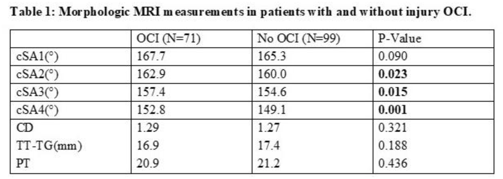
A retrospective review was conducted of patients with MRI-confirmed PD at a single institution. Patients were stratified into two cohorts based on the presence or absence of OCI. Variables collected included age, gender, number of prior patellar dislocations, four axial measurements of cartilaginous sulcus angle (cSA – most proximal, cSA2 –near proximal, cSA3 – near distal, cSA4 – most distal), Caton Deschamps (CD), tibial tubercle–trochlear groove (TT-TG) distance, and patellar tilt (PT). Lesion location and cartilage thickness were recorded for patients with OCI. Independent t-tests were used to compare continuous variables, and chi-squared tests were used for categorical variables.
Results:
A total of 170 participants were included; 71 (57.7% female) had a history of OCI (mean age 14.54 ± 2.53 years), and 99 (60.6% female) had no history of OCI (mean age 15.13 ± 2.20 years). There was no difference in age (p = 0.12) or sex distribution (p = 0.83) between the cohorts. Patients with OCI were associated with elevated sulcus angles at multiple axial levels: cSA2 (p = 0.023), cSA3 (p = 0.015), and cSA4 (p = 0.001) (Table 1). However, there was no difference in cSA1 (p = 0.090), CD (p = 0.321), TT-TG (p = 0.188), or PT (p = 0.426) between cohorts. Patients with OCI (2.69±3.20) had a greater number (p = 0.035) of PDs compared to those without OCI (1.89±1.55). Of the 71 patients with OCI, 17 had injuries to the distal femur, 52 from patella, and 2 from both the patella and femur; 21 of the OCIs were full thickness.
Conclusion:
OCI in patients with PD was significantly associated with a greater number of prior PDs and TD, measured by the cSA. Most lesions were located on the patella, and approximately one-third were full-thickness. These findings support MRI surveillance in patients with frequent dislocations to assess for clinically significant OCI.