
Abstract
In most parts of West Africa and other developing countries, herbal medicines are sometimes used by patients concomitantly receiving conventional drugs, which can result in potentially serious adverse effects. This study examined in vivo cytotoxic effects of Azadirachta indica extracts followed by artesunate administration on some markers of liver and kidney toxicity. Serum ALT, GGT, urea, creatinine, interleukin 1β, tumor necrosis factor α, tissue malondialdehyde and glutathione levels and liver and kidney histology in healthy male Wistar rats administered 100 and 200 mg/kg A. indica for 5 days followed by 10 mg/kg Artesunate for 5 days was determined. Results showed significantly (p < 0.05) higher serum ALT, GGT, urea, creatinine, interleukin 1β and tumor necrosis factor α levels with proportional increase of 16.5, 21.7, 9.2, 6.9, 9.1 and 9.1% respectively when compared to normal control was observed. Malondialdehyde levels were significantly (p < 0.05) higher with a proportional increase of 57.8%, while glutathione levels were significantly (p < 0.05) lower with a proportional decrease of 13.4% in liver homogenates of the treated rats relative to normal control. Histological examination of the liver and kidney of the co-treated rats showed vascular congestion and necrosis. Collectively, the results suggest that administration of A. indica followed by artesunate could predispose to liver and kidney associated cytotoxicity. These findings could have implications for people who habitually use herbal preparations and conventional drugs in sequential fashion.
Keywords
Introduction
Despite significant interventions for disease treatment and control, significant morbidity and mortality cases are recorded in most developing countries prompting locals to try different therapeutic options. 1 For example, it is estimated that at least 90% of reported cases and mortality cases of malaria infection occur in Africa. 2 The burden of disease and the frustration caused by treatment failures, prompts people to try both herbal and conventional therapeutic options. 3 In Nigeria and other parts of West Africa, herbal preparations are used by locals who may feel they are experiencing malaria symptoms, without undergoing necessary laboratory tests. 4,5 Azadirachta indica is a plant widely used in the treatment of malaria. 6,7
Azadirachta indica, commonly called by the name Neem, is a tree belonging to the mahogany family Meliaceae. 8 The tree is native to India and Burma, but now grows in Sub-Sahara Africa due to the remarkable ability of the tree to grow well in tropical and semi-tropical regions. 8,9 Neem extracts contain oxygenated triterpenes, known as limonoids, which are responsible for their bitter taste. 10 The wide usage of A. indica extracts for the treatment of malaria has generated research interest, with results suggesting effectiveness of the extract in treating various diseases including diabetes and cancer. 9 A number of phytocompounds with anti-malaria properties have also been isolated from A. indica. 11
However, despite such acclaimed therapeutic efficacy of A. indica leaf extract, when expected recuperation is not observed, patients often commence the use of conventional malaria drugs. 12 One of such commonly available drug in Nigeria is artesunate. Studies have shown that phytocompounds can alter the metabolism and pharmacodynamics of conventional drugs resulting in unexpected outcomes. 13 Some well-known examples include induction of cytochrome P450 (CYP 3A4) by St. John’s wort plant which renders antiviral treatment less efficient and inhibition of CYP 3A4 by grape fruit juice resulting in CYP 3A4 associated toxicity. 14 Fascinatingly, there is a dearth of information on the safety or otherwise in the use of A. indica extract preparations alongside, before or after conventional drugs. This study examined the effect of A. indica administration followed by artesunate on some indices of toxicity in healthy male Wistar rats. Evidently, our experimental findings could have implications for people who use herbal preparations and conventional drugs arbitrarily while confirming some of the societal confessions as it relates to toxicological complications when the two regimens were applied simultaneously.
Materials and methods
Plant sample collection, identification and extraction
Fresh leaves of A. indica were obtained from trees within Ahmadu Bello University, Zaria and authenticated at the Botany unit of the Department of Biological Science with the herbarium voucher number of 900151 determined. The leaves were shade dried and subsequently made into powdered form using a pertern laboratory mill. Two hundred grams of the powdered leaves was dissolved in a 1 liter pyrex beaker containing 800 ml of hot distilled water for 72 hours. Stirring of the mixture containing the powdered leaves in hot distilled water was carried out periodically. The resulting solution was then filtered using an ashless Whatman filter paper (size 1) and the filtrate obtained from filtering the aqueous suspension of A. indica leaves was freeze dried using a FD 51 ilShin freeze dryer to obtain a deep-brown extract used for the study. The extract obtained was stored in a labeled container.
Experimental animals
Thirty apparently healthy male Wistar rats weighing between 92 g and 128 g were obtained from the animal house of the Faculty of Veterinary Medicine, Ahmadu Bello University (ABU), Zaria. They were randomly allocated into six groups of five rats each. They were acclimatized under the same environmental condition, provided feed and water ad libitum in well-ventilated cages in the animal house of the Department of Biochemistry, ABU Zaria. Administration of the prepared A. indica extract and artesunate (1 ml/100 gram body weight of animal) was done by oral gavage. The A. indica extract was administered for 5 days followed by artesunate for 5 days, amounting to 10 days of treatment. Distilled water was used to dissolve the A. indica extract and artesunate. The doses for A. indica and Artesunate were chosen based on literature reports.
6,15
Group 1: Untreated rats administered normal saline (1 ml/100 g) for 10 days. Group 2: Normal saline for 5 days + A. indica extract (100 mg/kg) only for 5 days. Group 3: Normal saline for 5 days + A. indica extract (200 mg/kg) only for 5 days. Group 4: A. indica extract (100 mg/kg) for 5 days + Artesunate (10 mg/kg) for 5 days. Group 5: A. indica extract (200 mg/kg) for 5 days + Artesunate (10 mg/kg) for 5 days. Group 6: Artesunate (10 mg/kg) for 5 days + normal saline for 5 days.
Collection of samples and tissues
At the end of the experiment, the rats were sacrificed humanely by cervical dislocation and blood collected into clean, nonheparinized tubes and allowed to stand for 2 hours before centrifuging at 10,000 × g for 10 minutes to obtain serum. Liver tissues were excised for oxidative stress analysis using standard protocols. Liver and kidney tissues were excised for histological examination.
Serum analysis
Alanine aminotransferase (ALT)
Serum alanine aminotransferase (ALT) in the experimental rats was determined according to a standard method 16 which monitors the amount of pyruvate hydrazine formed with 2,4-dinitrophenylhydrazine. In the reaction mixture, 0.1 ml of diluted sample, phosphate buffer (100 mmol/l, pH 7.4), L-arginine (100 mmol/l) and α-oxoglutarate (2 mmol/l) were put. The mixture was then incubated for 30 min at 37°C. After that, 0.5 ml of 2,4-dinitrophenylhydrazine (2 mmol/l) was added to the reaction mixture and allowed to stand for 20 min at 25°C. Finally, 5.0 ml of NaOH (0.4 mol/l) was added to stop the reaction and absorbance was read against the reagent blank after 5 min at 546 nm. The corresponding ALT concentration was obtained from a standard curve.
Gamma glutamyltransferase (GGT)
Serum gamma glutamyltransferase (GGT) was determined according to a reported method. 17 Diluted sample (0.1 ml) was mixed with 0.5 ml of tris buffer (100 mmol/l, pH 8.25) and incubated at 25°C. Next, a careful shaking of the mixture was done and 0.5 ml of L-γ-glutamyl-3carboxy-4-nitroanilide (2.9 mmol/l) was added. The reaction mixture was mixed, initial absorbance read, and then absorbance values were recorded after 1, 2 and 3 min at 405 nm wavelength. The equation below was used to determine the GGT concentration in the test samples.

Urea
Urea in serum was determined according to a standard method. 18 Three cuvettes for blank, standard and sample were used. For the blank, 10 µl of distilled water was added to 100 µl of picric acid (35 mmol/l). The cuvettes for standard contained 10 µl of standard and 100 µl of picric acid (35 mmol/l), while the cuvette for sample contained 10 µl of sample and 100 µl of picric acid (35 mmol/l). The three cuvettes were then mixed and incubated at 37°C for 10 min. After that, 2.5 ml of Sodium hydroxide (0.32 mol/l) and Sodium hypochlorite (27 mmol/l) were added to the three cuvettes, mixed and incubated at 37°C for 15 min. Absorbance of sample and standard were then read against the blank at 546 nm. Urea concentration was calculated using the formula below:

Creatinine
Serum creatinine was determined according to a standard method. 19 1 ml of picric acid (35 mmol/l) and Sodium hydroxide (0.32 mol/l) was pipetted into a cuvette and 0.1 ml of sample was added, mixed and absorbance read after 2 and 4 min. The same was done for standard but without addition of sample. All absorbance was read at 492 nm. Creatinine concentration in mg/dl was calculated as change in absorbance of sample/change in absorbance of standard × standard concentration (mmol/l).
Determination of tumor necrosis factor-α and interleukin-1β levels using ELISA kits
ELISA kits for tumor necrosis factor-α and Interleukin-1β antibody purchased from Wuhan Fine Biotech Co., Ltd were used to determine cytokine levels in serum. The plate wells were washed twice before commencing the experiment. Dilution buffer (100 µl) was added to the blank wells. Diluted standards (100 µl) and samples (100 µl) were added to the wells, covered and incubated overnight at 37°C for 90 min. The next morning, all reagents were brought to room temperature before the plate was washed twice with 300 µL wash buffer per well. The residual buffer was then blotted out by firmly tapping the plate upside down on absorbent paper. All subsequent washes were performed similarly. Biotin labeled antibody (100 µl) was added to each well to block non-specific binding. The plate was then covered with a seal and incubated at room temperature for an hour, after which the plate was washed three times with wash buffer and 100 µl of conjugate added to the wells. The plate was then covered, incubated at room temperature for 30 min followed by washing with wash buffer. Diluted detection antibody solution (100 µl) was then added to each well, incubated at room temperature for 1 h with shaking, followed by washing with wash buffer. Diluted HRP-streptavidin conjugate (100 µl) was then added to each well, after which the plates was sealed and incubated at room temperature for 30 min and washed five times with wash buffer. TMB substrate solution (90 µl) was then added into each well, covered with seal and incubated at 37°C for 30 min. Finally, the reaction was stopped by adding 50 µl of stop solution to each well. Absorbance was read at 450 nm and corresponding concentration determined from a standard curve.
Oxidative stress
Excised liver and kidney tissues were rinsed in 1.15% KCl and blotted with filter paper. Next, the tissues were weighed, chopped into bits, followed by homogenization in four volumes of the homogenizing buffer (pH 7.4). The homogenate obtained was centrifuged at 10,000 × g using a cold centrifuge. After 10 min, supernatant was collected and stored for further analysis.
Malondialdehyde
Determination of malondialdehyde (MDA) levels in liver tissue homogenates was done according to a reported procedure. 20 The sample (0.4 ml) was mixed with 1.6 ml of Tris-KCl buffer after which 0.5 ml of 30% TCA was added. Next, 0.5 ml of 0.75% TBA was introduced and placed in a water bath for 45 min at 80°C. After allowing to cool, centrifugation was carried out at 3000 × g. The supernatant collected was used to measure absorbance against a reference blank of distilled water at 532 nm. MDA (mmol/gram) in the tissues was calculated using the formula below (molar extinction coefficient = 1.56 × 10⁻⁵ m−1cm−1).
Reduced glutathione
The levels of reduced glutathione (GSH) in the liver tissue homogenates was determined according to a reported method. 21 The sample (1 ml) was deproteinated by adding 4% sulfosalicyclic acid (1 ml). The mixture was then centrifuged at 4000 × g for 5 min. The supernatant obtained (0.5 ml) was then added to 4.5 ml of Ellman’s reagent. The blank was made of diluted precipitating agent (0.5 ml) and Ellman’s reagent (4.5 ml). Absorbance was read at 412 nm to determine reduced glutathione (µg/gram of tissue).
Superoxide dismutase activity
The activity of superoxide dismutase (SOD) in liver tissue homogenates was carried out using a standard protocol.
22
The sample (0.2 ml) was diluted in distilled water (0.8 ml) and 0.2 ml of the diluted sample was added to 0.05 M carbonate buffer (2.5 ml, pH 10.2). The reaction was started by adding freshly prepared 0.3 mM adrenaline (0.3 ml) to the mixture. Absorbance (480 nm) values were recorded every 30 seconds for 150 seconds. Calculation

where A0 = absorbance after 30 seconds and A3 = absorbance after 150 seconds
One unit of SOD activity was given as the amount of SOD necessary to cause 50% inhibition of the oxidation of adrenaline.
Catalase activity
The activity of catalase (CAT) in liver tissue homogenates was assayed. 23 The sample (0.2 ml) was mixed with 0.8 ml distilled water to obtain a 1:5 dilution. The rection mixture included 2 ml of H2O2 solution (800 µmoles), phosphate buffer (2.5 ml, pH 7.0) and introduced sample (1 ml). The reaction mixture (1 ml) was introduced into dichromate/acetic acid reagent (2 ml) at an interval of 60 sec.
Catalase activity was determined using a standard curve and the concentration of the remaining H2O2 was extrapolated from the curve.

Histological examination
The excised liver and kidney tissues were fixed in 10% buffered formal saline. During tissue processing, the tissues were dehydrated using 100% ethanol, following by clearing using xylene. The tissues were then embedded in paraffin wax. Tissue micro sections (3 µm) were then stained using hematoxylin and eosin and mounted on a mountant according to standard protocol. 24 Histological examination was done using a light microscope at ×400 magnification.
Data analysis
Quantitative data were analyzed using analysis of variance (ANOVA), IBM program (version 20 IBM Corp, Armonk, NY, USA) and expressed as mean ± standard deviation. Duncan Multiple Range Test was used to determine difference between treatment groups. Values of p less than 0.05 (p < 0.05) were considered statistically significant.
Results
Alanine aminotransferase (ALT) levels were significantly (p < 0.05) higher in the group administered A. indica (100 mg/kg) + Artesunate (10 mg/kg) and the group administered A. indica (200 mg/kg) + Artesunate (10 mg/kg). The lowest ALT value of 47.88 UI/l was observed in the control rats administered normal saline only. Gamma glutamyltransferase levels were significantly (p < 0.05) higher in the group administered A. indica (100 mg/kg) + Artesunate (10 mg/kg) and the group administered A. indica (200 mg/kg) + Artesunate (10 mg/kg). The lowest GGT value of 15.3 UI/l was observed in the control rats administered normal saline only (Figure 1).

Alanine aminotransferase (ALT) and gamma glutamyltransferase levels in experimental rats. Results are presented as mean ± standard deviation. *Statistical significance (p < 0.05) compared to untreated control.
Urea and creatinine levels were significantly (p < 0.05) higher in the group administered A. indica (100 mg/kg) + Artesunate (10 mg/kg) and the group administered A. indica (200 mg/kg) + Artesunate (10 mg/kg). The lowest values of 7.09 and 61.02 IU/l for urea and creatinine respectively were observed in the control rats treated with normal saline only (Figure 2)
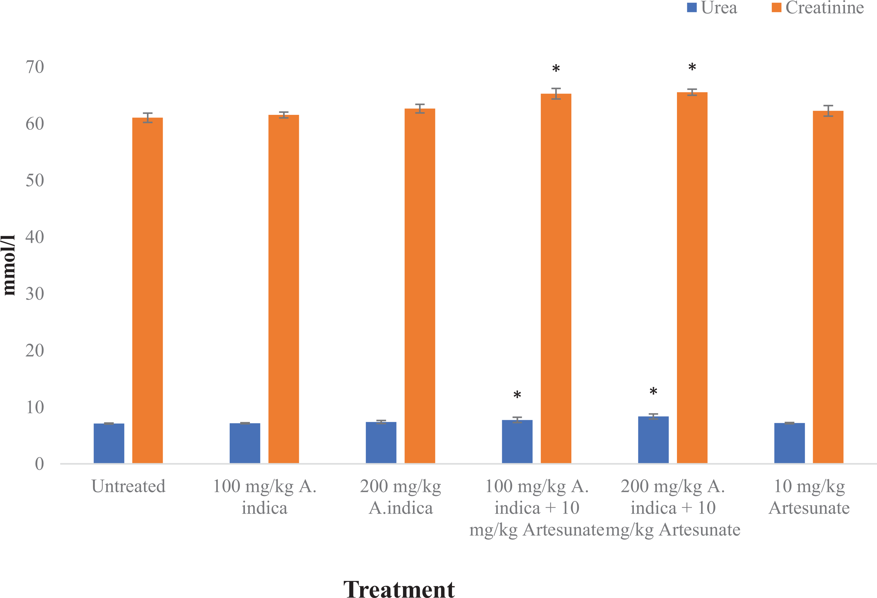
Urea and creatinine levels in experimental rats. Results are presented as mean ± standard deviation. *Statistical significance (p < 0.05) compared to untreated control.
Interleukin 1β and tumor necrosis factor-α levels were significantly (p < 0.05) higher in the group administered A. indica (100 mg/kg) + Artesunate (10 mg/kg) and the group administered A. indica (200 mg/kg) + Artesunate (10 mg/kg). The lowest values of 51.84 and 78.78 ng/ml for interleukin 1β and tumor necrosis factor-α respectively were observed in the control rats treated with normal saline only (Figure 3).
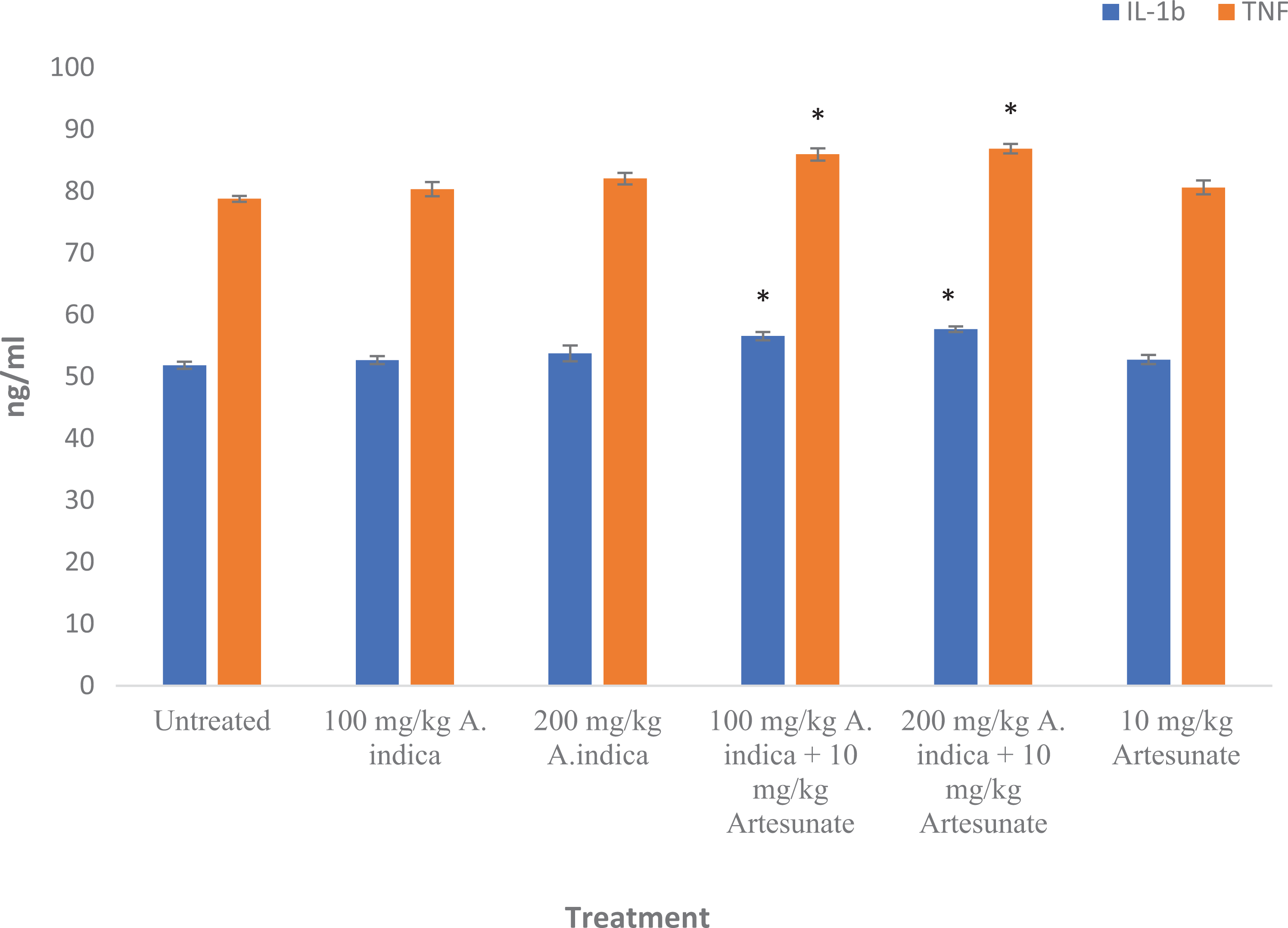
IL-1β and TNF-α levels in experimental rats. Results are presented as mean ± standard deviation. *Statistical significance (p < 0.05) compared to untreated control.
Malondialdehyde levels were significantly (p < 0.05) higher in the group administered A. indica (100 mg/kg) + Artesunate (10 mg/kg) and the group administered A. indica (200 mg/kg) + Artesunate (10 mg/kg). Glutathione levels were significantly (p < 0.05) lower in the group administered A. indica (100 mg/kg) + Artesunate (10 mg/kg) and the group administered A. indica (200 mg/kg) + Artesunate (10 mg/kg) (Figure 4).

MDA (mM/gram of tissue) and GSH (µg/gram of tissue) in experimental rats. Results are presented as mean ± standard deviation. *Statistical significance (p < 0.05) compared to untreated control.
There was no significant difference in catalase (except the 100 mg/kg A. indica + 10 mg/kg artesunate) and superoxide dismutase activity between the groups (Figure 5).

Catalase and superoxide dismutase activity in experimental rats. Results are presented as mean ± standard deviation. *Statistical significance (p < 0.05) compared to untreated control.
Liver histology examination showed slight hepatic necrosis and vascular congestion in the group administered A. indica (100 mg/kg) + Artesunate (10 mg/kg) and the group administered A. indica (200 mg/kg) + Artesunate (10 mg/kg) respectively. The other groups showed normal hepatocyte and vascular features (Figure 6).

Liver histology of experimental rats (magnification ×400). Liver histology shows normal hepatocytes in the untreated, control group (a). Also, liver histology showed normal features in the group administered 100 mg/kg A. indica only (b). For the group administered 200 mg/kg A. indica only, liver histology showed some pyknotic nuclei (c). Slight hepatic necrosis was observed in the group administered 100 mg/kg A. indica + 10 mg/kg artesunate (d). Vascular congestion and slight hepatic necrosis observed in the group administered 200 mg/kg A. indica + 10 mg/kg artesunate (e). For the group administered 10 mg/kg artesunate only, liver histology showed normal features (f).
Kidney histology examination showed slight tubular necrosis and lymphocyte hyperplasia in the group administered A. indica (100 mg/kg) + Artesunate (10 mg/kg) and the group administered A. indica (200 mg/kg) + Artesunate (10 mg/kg) respectively. The other groups showed normal glomerular and tubular features (Figure 7).

Kidney histology of experimental rats (magnification ×400). Kidney histology showed normal tubules and glomerulus in the untreated, control group (a) as well as the group administered 100 mg/kg A. indica only (b) and 200 mg/kg A. indica only (c). Slight tubular necrosis was observed in the group administered 100 mg/kg A. indica + 10 mg/kg artesunate (d). Slight lymphocyte hyperplasia was observed in the group administered 200 mg/kg A. indica + 10 mg/kg artesunate (e). For the group administered 10 mg/kg artesunate only, kidney histology showed normal tubules and glomerulus (f).
Discussion
Reports of resistance or treatment failure has been reported for some endemic diseases among patients in West Africa and other developing countries. 25 This challenge makes people in such areas prone to the use of multiple approaches in the search for therapy. 6 As a result, some people habitually use both traditional herbal preparations and conventional drugs. 6 Artesunate is a semi-synthetic derivative of artemisinin while Azadirachta indica is widely used in traditional health care for the treatment of malaria. Artesunate undergoes metabolism to the active dihydroartemisinin which reacts with heme to generate free radicals that prevent synthesis of macromolecules required for plasmodium metabolism and proliferation. 26 Azadirachta indica extracts has also been reported to contain alkaloids, saponin, tannins, triterpenoids and other phenolics with a wide range of metabolic activities. 27 Physicians who see malaria patients at local hospitals may not be aware of, or be able to elicit history of previously used herbal preparations by their patients.
Serum alanine aminotransferase (ALT) and gamma glutamyl transferase (GGT) are used as markers of liver damage, as damage to hepatocytes during xenobiotic exposure can cause increased serum levels of ALT and GGT. 28 The elevated levels of ALT and GGT in the animals administered A. indica (100 mg/kg) + Artesunate (10 mg/kg) and the group administered A. indica (200 mg/kg) + Artesunate (10 mg/kg) suggests possible liver and kidney damage in the experimental rats. Serum urea and creatinine are used to estimate glomerular filtration efficiency. 29 Physiological alterations that cause damage to nephrons can affect kidney reabsorption, resulting in decreased clearance of urea and creatinine. 30 The serum levels of urea and creatinine observed in this study could be indicative of kidney damage.
Elevated levels of serum cytokines including interleukin 1β (IL-1β) have been associated with impaired renal clearance and increased mononuclear phagocyte production. 31 Increased serum levels of tumor necrosis factor-α (TNF-α) have been associated with development of liver injury due to activation of Kupffer cells. 32 Thus, elevated levels of IL-1β and TNF-α in the group administered A. indica (100 mg/kg) + Artesunate (10 mg/kg) and the group administered A. indica (200 mg/kg) + Artesunate (10 mg/kg) suggests liver and kidney injury in the experimental rats. These results are corroborated by the slight hepatic necrosis and vascular congestion observed from liver histologic examination as well as slight tubular necrosis and lymphocyte hyperplasia observed from kidney histology examination in the group administered A. indica (100 mg/kg) + Artesunate (10 mg/kg) and the group administered A. indica (200 mg/kg) + Artesunate (10 mg/kg) respectively.
Malondialdehyde (MDA) levels are used as a marker of oxidative stress, because MDA is a product of lipid peroxidation. 33 Glutathione is a tripeptide with antioxidant capacity, with cellular levels indicative of antioxidant potency. 34 The relatively higher levels of malondialdehyde and relatively lower levels of glutathione in the group administered A. indica (100 mg/kg) + Artesunate (10 mg/kg) and the group administered A. indica (200 mg/kg) + Artesunate (10 mg/kg) could be indicative of oxidative stress. Malaria pathogenesis is associated with oxidative stress and the mechanism by which artesunate ameliorates malaria could result in oxidative stress. Since healthy rats were used in this study, it is possible that oxidative stress conditions could be exacerbated in malaria patients who sequentially use A. indica and artesunate treatment regimen.
Conclusion
Collectively, the results from the present study suggest some form of liver and kidney damage in the experimental rats subjected to the sequential administration of A. indica and artesunate. Although translating in vivo rodent results to humans can be complicated, it is known that older adult patients who are more likely to suffer from impaired organ function and who are placed on multiple prescriptions are at a higher risk of deleterious drug-herb interactions. Despite the numerous putative benefits of herbal medicines, indiscriminate use of herbal preparations together with conventional prescription drugs may have detrimental effects that must be given attention in the clinical setting. The findings from the present study may then be salient for patients who use herbal preparations and conventional drugs sequentially or concomitantly for the treatment of malaria. Further studies are needed in in vivo models of malaria infection to determine the effect of sequential A. indica and artesunate use on reproductive toxicity, pharmacokinetics of active components, immunotoxicity and response to therapy.
Footnotes
Acknowledgment
The authors wish to acknowledge the assistance of Mr Bamidele and Mr Shailong for histological examination and biochemical analysis, respectively.
Declaration of conflicting interests
The author(s) declared no potential conflicts of interest with respect to the research, authorship, and/or publication of this article.
Ethical approval
Ethical approval was applied for and obtained (Reference number: ABUCAUC/2018/004) from Ahmadu Bello University Ethical Committee in accordance with the ethical standards of the institutional and national research committee in line with the 1964 Helsinki Declaration and later amendments.
Funding
The author(s) received no financial support for the research, authorship, and/or publication of this article.