






Abstract
Kidney disease is common in patients with multiple myeloma. Patients often present with acute kidney failure most commonly the result of light chain cast nephropathy. Presentation can also be in the form of nephrotic syndrome associated with immunoglobulin light chain amyloidosis or monoclonal immunoglobulin deposition disease and tubulopathy as in acquired light chain Fanconi syndrome. We present a case of a 56-year-old male with multiple myeloma who presented with chronic kidney disease and uric acid stones. Chemical analysis of the nephrolithiasis showed it to be entirely composed of uric acid. Fractional excretion of uric acid was elevated at 35.9%. Kidney biopsy was consistent with light chain proximal tubulopathy. Bone marrow biopsy showed 30% kappa light chain–restricted plasma cells. Genetic sequencing identified the light chain to be from the Vk1-33 subgroup. The patient was treated with bortezomib, cyclophosphamide, and dexamethasone, with improvement of his kidney function. This case illustrates a rare presentation of multiple myeloma.
Introduction
Crystalline nephropathy is defined as kidney injury with a distinctive pathological finding of abundant crystals most frequently found in the renal tubules and interstitium regardless of composition. Crystalline nephropathy is a manifestation of multiple myeloma and other dysproteinemias that has been described as a cause of chronic kidney impairment. The most common forms of immunoglobulin-crystalline nephropathy are cast nephropathy, crystal-storing histiocytosis, crystalglobulinemia, and light chain proximal tubulopathy (LCPT). 1 LCPT is uncommon in comparison with other kidney-related complications of dysproteinemias, accounting for just 5% of biopsies. 1 LCPT includes cases with monoclonal light chain (LC) in the proximal tubules with and without evidence of crystalline formation in the proximal tubular cytoplasm.2,3
LCPT can result in alterations of proximal tubule functions leading to Fanconi syndrome characterized by metabolic acidosis, normoglycemic glycosuria, aminoaciduria, hypouricemia, and phosphaturia. 3 The mechanism of proximal tubular dysfunction is thought to involve direct toxicity of the LC on tubular cells in addition to resistance of the LC to enzymatic degradation promoting the crystalline structure formation and accumulation.4,5 Studies also support that decreased acidification of the lysosome further reduces the ability to digest the monoclonal LC, and that reduced endocytosis can be mediated by the pathologic LC. 5 Another rare observation in dysproteinemias is the presence of intra- and extra-cytoplasmic crystalline deposits in the bone marrow. Some suggest that the presence of crystal deposition and site of deposition is more related to the immunoglobulin amino acid substitution and the resultant LC hydrophobicity than the serum level of the immunoglobulin. 6 We describe a case in which a patient presenting with uric acid kidney stones secondary to isolated uric acid wasting in the setting of multiple myeloma with both LCPT and bone marrow LC crystals.
Case report
A 56-year-old man was referred to nephrology for evaluation of recent onset kidney stones and kidney impairment. The patient has a history of hemochromatosis and hypertension. A month prior to presentation, the patient developed left flank pain and was diagnosed with kidney stones. The computed tomography (CT) scan showed a distal left ureteral stone with hydronephrosis and another stone in the collecting system of the left kidney. He was found to have an elevated creatinine of 5.68 mg/dL. Subsequently, he underwent laser lithotripsy with removal of the obstructing ureteral stone. The stones were analyzed and found to be uric acid stones. Despite the relief of ureteral obstruction, the patient’s creatinine remained elevated at 4.2 mg/dL 1 month later. The patient’s creatinine 2 years prior was around 1.9 mg/dL; however, the cause of chronic kidney insufficiency was never determined. No nephrotoxic agent was identified by history.
The physical exam was unremarkable. Diagnostic laboratory workup is shown in Table 1. Serum uric acid level was normal at 4.9 mg/dL with a fractional excretion of uric acid of 35.9% (usually <10%). A diagnosis of partial Fanconi syndrome was made since there was no evidence of aminoaciduria, glycosuria, or hypophosphatemia. Serum protein electrophoresis showed an M-spike in the gamma region which was found to be IgG kappa on immunofixation. Kappa LCs were elevated at 688 mg/dL with kappa/lambda ratio of 529. A follow-up CT scan to assess his kidney stone status showed a non-obstructing 6-mm stone lower pole left kidney without hydronephrosis. Positron emission tomography (PET)/CT did not reveal any boney lesions. He subsequently underwent kidney and bone marrow biopsies.
Diagnostic evaluation for kidney dysfunction.

SPEP: serum protein electrophoresis; UPEP: urine protein electrophoresis; FLC: free light chain; GFR: glomerular filtration rate; eGFR: estimated glomerular filtration rate.
Kidney biopsy
On light microscopy, the kidney biopsy showed distention of the cytoplasm of proximal tubular epithelial cells with intracytoplasmic deposition of crystalline inclusions, which was periodic acid–Schiff (PAS)- and silver-negative and hypereosinophilic on the H&E-stained section. Crystalline casts were seen within rare tubular lumens (Figure 1). There was mild interstitial fibrosis and tubular atrophy. The arteries showed moderate intimal thickening. The glomeruli appeared normal.
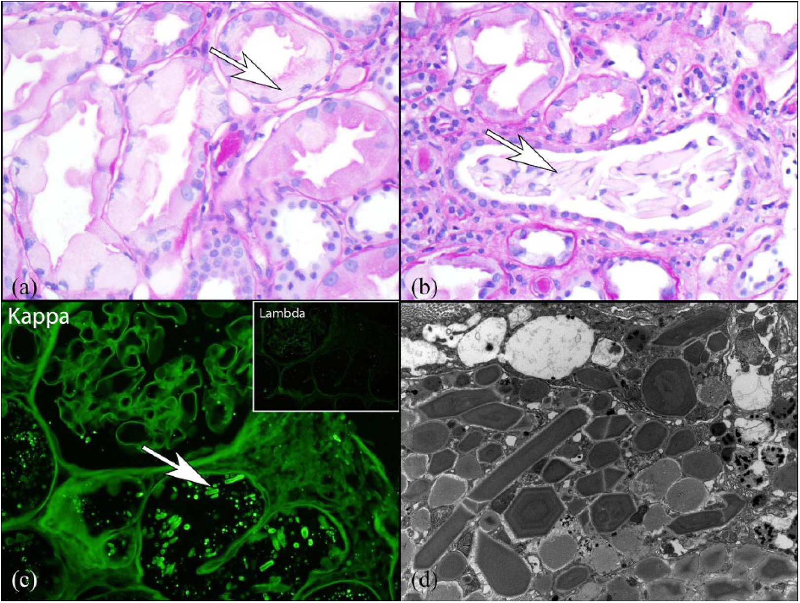
Kidney biopsy: (a) the biopsy showed crystals (arrow) within the proximal tubular epithelial cell cytoplasm by light microscopy (PAS stain at 400×); (b) rare tubules contained crystalline atypical casts (arrow); (c) immunofluorescence on pronase-digested paraffin sections showed positive staining of crystals for kappa light chain and negative staining for lambda light chain; and (d) numerous electron-dense intracytoplasmic crystals were seen within proximal tubular epithelial cells by electron microscopy.
Routine immunofluorescence on frozen sections showed dim linear glomerular and tubular basement membrane staining for kappa LC with negative corresponding staining for lambda LC. The glomeruli were negative for IgA, IgG, C1q, C3, and fibrinogen. Scattered intratubular casts were stained for IgA and kappa and lambda LCs. No intracytoplasmic crystals were identified by routine immunofluorescence. Immunofluorescence staining was also performed on pronase-digested paraffin sections. By this technique, there were numerous tubular intracytoplasmic crystals that stained for kappa LC and showed negative staining for lambda LC. Electron microscopy showed numerous electron-dense crystals within the proximal tubular epithelial cell cytoplasm. The crystals had rhomboid and rectangular shapes.
Bone marrow biopsy
The marrow cellularity was increased at 60%. Erythroid, myeloid, and lymphoid precursors and megakaryocytes were normal. Plasma cells were noted to be increased at 30% of marrow cellularity and had slightly atypical with conspicuous cytoplasmic immunoglobulin deposits and crystals (Figure 2). Extracellular crystals similar to the intracytoplasmic crystals in plasma cells were also noted, consistent with immunoglobulin deposits. The plasma cells expressed monotypic kappa immunoglobulin LCs, CD38 and CD138, and did not express CD19 or CD45 as demonstrated by flow cytometry.

Bone marrow biopsy: (a) the bone marrow aspirate smear (Wright–Giemsa, 1000×) shows a plasma cell infiltrate with mature chromatin, many of which contain intracytoplasmic inclusions that are globular or crystalline-like (arrows). Occasional histiocytes containing numerous intracytoplasmic crystal are also noted (arrow head) and (b) the crystals are present in the cytoplasm of plasma cells and histiocytes (arrow head) as well as in the extracellular space (arrows).
Monoclonal immunoglobulin sequencing study
Total RNA was extracted from bone marrow aspirate and reverse transcribed into cDNA for Sanger sequencing of the Ig kappa variable (V) domains using previously described protocols. 7 Sequences were analyzed using FinchTV software (Geospiza, Inc.; Seattle, WA, USA) and aligned with MultAlin (http://multalin.toulouse.inra.fr/multalin/), and the V subgroup was determined using IMGT/V-QUEST online software (https://www.imgt.org). 8 All sequences (6/6) were identical and derived from the Vk1-33 subgroup with only three amino acid substitutions in the FR1, FR3, and CDR3 regions of the V domain compared to the germline sequence (Figure 3).

Immunoglobulin sequencing.
Diagnosis
Multiple myeloma with crystalline LCPT and focal crystalline cast nephropathy.
Clinical follow-up
The patient was treated with CyBorD (Cyclophosphamide, Bortezomib, and Dexamethasone) chemotherapy. He was also started on potassium citrate for his proximal renal tubular acidosis (RTA). At 1-year follow-up, the patient had no recurrence of stones and a stable serum Cr of 2.1 mg/dL, which is near his previous baseline. At 3 years since diagnosis, kappa free LC was 1.77 mg/dL and creatinine was 2.24 mg/dL.
Discussion
The first case of LCPT with crystals and Fanconi syndrome was published in 1957. 9 It was recognized that the overwhelming majority of patients had a monoclonal kappa LC. 1 Later, LCPT was refined by the presence of monoclonal LC in the proximal tubular cells with or without crystalline inclusions or the acquired Fanconi syndrome.10,11 Cases without crystals often revealed ultrastructural abnormalities in the phagosomes/lysosomes on electron microscopy. Others noted that LCPT cases without crystalline inclusions were more often associated with lambda LC.2,3 Furthermore, Fanconi syndrome was often absent in cases without crystalline inclusions. 3
Patients with LCPT usually present with chronic kidney impairment with proximal tubular dysfunction in the form of a complete or partially acquired Fanconi syndrome. 1 Fanconi syndrome is characterized by urinary wasting of glucose, uric acid, phosphate, amino acid, and bicarbonate. Patients who develop the Fanconi syndrome can present with osteomalacia and fractures. Our patient presented with chronic kidney disease and uric acid stones, which is a rare presentation for patients with multiple myeloma. Only one previous case has been described. 12
The understanding of the cellular mechanisms which lead to proximal tubular dysfunction is increasing. Animal study using a Vk1 LC gene (sequenced obtained from a patient with LCPT and acquired Fanconi syndrome) was capable of causing LC crystal formation in the proximal tubules similar to the human LCPT in transgenic mice. 4 The Vk1 LC produced in these mice showed a complete resistance to proteolytic digestion and yielded a nephrotoxic pattern similar to the human disease. The mice also show morphological alteration of the kidney tubular cells containing rhomboid-shaped crystals and deficiency in lysozyme acidification, proteolysis of immunoglobulin LC, and proximal tubule endocytosis.4,5 Vk subgroups and mutations altering the hydrophobicity and charges are suspected to be involved in the toxicity of LCs in LCPT with crystalline inclusions and acquired Fanconi syndrome. 13 Accordingly, in the present case, the monoclonal LC is composed of a Vk1-33 domain, which, together with Vk1-39, is the most frequent Vk subgroup associated with this disease. However, in contrast with previous observations, the amino acids sequence harbors very few mutations with only one polar to hydrophobic residue change in the CDR3 (D > I) that could impact on the structure and behavior of the protein. This may account for the incomplete Fanconi syndrome observed in the patient. Another rare finding is the association of crystalline LCPT with intracellular and extracellular monoclonal crystal deposits in the bone marrow. Rod, rectangular, and needle-like extracellular bone marrow crystals have been described in cases of multiple myeloma, but since this particular presentation is very rare, its prognostic significance is unknown. 6 Crystal deposition at other sites such as joint and skin has been reported as well in multiple myeloma.
The treatment of LCPT requires the treatment of the underlying cause. In a series of 49 patients, Vignon et al. 11 described the development of end-stage renal disease (ESRD) in 5 out of 41 patients who received aggressive chemotherapy for multiple myeloma as compared to 4 of 8 patients who did not receive treatment. Patients who achieved a very good partial response were more likely to have preservation or improvement of kidney function. In this study, 12% of patients had multiple myeloma and 8% had Waldenström macroglobulinemia. The rest were classified to have monoclonal gammopathy of renal significance (MGRS).14,15 In another series, Stokes et al. 1 observed that the only independent predictor of the final estimated glomerular filtration rate (eGFR) was the initial eGFR, despite hematological response or partial response to chemotherapy/stem cell transplant. This emphasizes the importance of early detection and aggressive treatment. Patients with monoclonal gammopathy and hypokalemia or hypouricemia should undergo more extensive urine testing to look for signs of Fanconi syndrome. Demonstration of aminoaciduria is sufficient for the diagnosis of LCPT. In cases where the electrolyte abnormalities may be hindered by renal impairment or if urinary amino acid testing is not available or negative, a kidney biopsy should be performed to definitely make the diagnosis.
Footnotes
Declaration of conflicting interests
The author(s) declared the following potential conflicts of interest with respect to the research, authorship, and/or publication of this article: N.L. is on an advisory board for Takeda and Aduro.
Funding
The author(s) received no financial support for the research, authorship, and/or publication of this article.
