
Abstract
Keywords
Introduction
Diabetic macular edema (DME) is the most prevalent vision-threatening sequela in patients with diabetic retinopathy (DR), despite improved outcomes after the use of antivascular endothelial growth factor (anti-VEGF). 1 Chronic, low-grade inflammation caused by prolonged hyperglycemia leads to abnormalities in the microvasculature followed by breakdown of the blood–retinal barrier and fluid accumulation beneath or within the retina. 2 The local expression of proinflammatory cytokines and morphologic changes in the endothelial cell tight junctions are thought to initiate macular thickening.2,3 Intravitreal anti-VEGF injections have been shown to be effective in the management of DME; however, they remain associated with a high treatment burden. The effects of shorter lasting treatment result in frequent visits or treatment nonadherence, which can lead to undertreatment and reduced long-term visual outcomes. 4 A subset of patients might also show partial or complete resistance to first-line monotherapies, suggesting a potential limitation in the way in which the complex inflammatory pathways are targeted.
The Diabetic Retinopathy Clinical Research Network (DRCR.net) Protocol T reported persistent edema at week 24 in up to two thirds of eyes despite treatment of up to 6 monthly intravitreal injections of aflibercept, ranibizumab, or bevacizumab. 5 The subsequent DRCR.net Protocol T extension study reported a reduction in visual acuity (VA) from year 2 to year 5 despite continued administration of a median of 4 anti-VEGF intravitreal injections. 6 The relationship between disease chronicity and the complexity of the underlying pathogenesis at play may necessitate the use of adjunctive therapies for disease management. Corticosteroids that target broader inflammatory pathways beyond those involving VEGF are being implemented for their ability to improve clinical outcomes for ME irrespective of disease duration. Intravitreal implants that deliver sustained-release corticosteroids offer a potential solution for alleviating the high treatment burden associated with disease management while simultaneously targeting recalcitrant or recurrent edema that has previously shown a suboptimal response to current first-line monotherapies. 7
The US Food and Drug Administration approved the use of the corticosteroid fluocinolone acetonide implant (Iluvien, Alimera Sciences) in 2014 for the clinical indication of DME in eyes without clinically significant increases in intraocular pressure (IOP). Fluocinolone acetonide is available as a sterile, nonbiodegradable intravitreal implant for the treatment of DME secondary to DR, providing sustained release of 0.2 µg/day into the vitreous for up to 36 months. 8 The pharmacokinetic profile of the lipophilic implant allows for preferential tissue delivery and absorption compared with other available steroidal implants.9,10
The pivotal phase 3 Fluocinolone Acetonide in Diabetic Macular Edema (FAME) studies A and B showed the efficacy and durability of the 0.19 mg fluocinolone acetonide intravitreal implant in a controlled setting. A significant improvement in best-corrected VA (BCVA) (15 letters or more on the Early Treatment Diabetic Retinopathy Study [ETDRS] chart) was seen in 28.7% of patients with a low-dose fluocinolone acetonide implant and in 28.6% of those with a high-dose implant. 11 Significant improvements were also seen in patients with chronic edema of 3 or more years over the 36-month period, as indicated by 34% of patients having a 15-letter gain compared with 13.4% in the sham control group. 12
Real-world longitudinal studies of the 0.19 mg fluocinolone acetonide intravitreal implant in a noncontrolled setting are emerging but remain largely limited. In this study, we present a 36-month analysis of the use of the 0.19 mg fluocinolone acetonide intravitreal implant for the treatment of DME secondary to DR in a real-world patient population.
Methods
This retrospective single-center noncontrolled study comprised patients who received a 0.19 mg fluocinolone acetonide intravitreal implant for DME from January 2016 to August 2021. Institutional review board (Pro00064827) exemption was obtained. This study adhered to the tenets of the Declaration of Helsinki and the guidelines of the US Health Insurance Portability and Accountability Act.
Eyes with any stage of DR and center-involving ME that were followed for 12 or more months before fluocinolone acetonide delivery and up to 36 months after fluocinolone acetonide delivery were included. Patients with a follow-up shorter than 6 months were excluded. All included eyes were assessed at baseline and at months 3, 6, and 12 at a minimum; a subset of patients was followed for a maximum of 36 months.
Patients who received the 0.19 mg fluocinolone acetonide intravitreal implant for the clinical indication of DME secondary to DR were identified and anonymized into a single dataset. A 12-month period before implant administration was identified for each patient so that ocular treatments could be standardized for further analysis.
Data collection included the recording of Snellen BCVA, central subfield thickness (CST) using spectral-domain optical coherence tomography, and DME-related treatments administered by the treating physician. Treatments were defined as any intravitreal anti-VEGF injection, intravitreal steroid, or laser photocoagulation for the management of DME. Safety outcomes related to treatment-induced increases in IOP above threshold, the initiation or addition of IOP-lowering medication(s), IOP-lowering procedures or surgical intervention, lens status, and implant migration were also recorded and assessed. IOP-lowering medications containing a combination of active agents were reported and analyzed as a single entity.
Statistical Analysis
Data were analyzed using GraphPad Prism software (version 9.0, GraphPad Inc) or SAS software (version 9.4, SAS Institute). Statistical significance was assessed accounting for the intrasubject correlation for patients contributing a second eye. The assessment of means was completed using a repeated-measures model with a compound symmetry covariance structure, and a rate comparison was completed using bootstrap analyses resampling at the patient level. VA was assessed using a Snellen chart and converted to ETDRS letters for subsequent analysis. VA measurements of counting fingers and hand motions were converted to 20/800 and 20/1600, respectively. Statistical significance was defined as a 2-sided P value of 0.05 or less. Mean values are ±SD. Results after fluocinolone acetonide administration are based on data of patients available (ie, not lost to follow-up) at a specific timepoint.
Results
The study included 148 unique eyes of 115 patients (24-month completion, n = 97).
Visual Acuity and Central Subfield Thickness
The VA remained unchanged throughout the follow-up. The mean baseline BCVA was 56.8 ± 16.0 letters in 20.6% of patients. The mean BCVA was 56.0 letters at month 24, representing a 0.8-letter decrease (Figure 1, A and C). The proportion of patients with a BCVA of 70 letters (20/40 Snellen equivalent) or more was 20.6% at baseline and 23.7% 24 months after fluocinolone acetonide administration (Figure 1B).
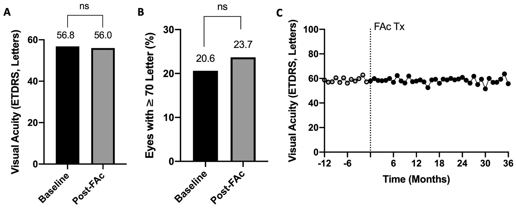
Best-corrected visual acuity (Early Treatment Diabetic Retinopathy Study [ETDRS] letters). (A) BCVA at baseline compared with 24 months after fluocinolone acetonide (FAc) treatment (Tx). (B) Percentage of patient eyes with a BCVA of 70 ETDRS letters (Snellen equivalent 20/40) or more at baseline and at 24 months. (C) Mean BCVA from 12 months before FAc treatment to 24 months after based on available data by month.
The baseline CST was 300 μm or less (mean, 380.65 ± 122.4 μm) in 30.9% of eyes (Figure 2). The mean reduction in CST ranged from −40.7 μm (month 24) to −108.0 μm (month 18). Analysis of the last available measurement through 24 months showed a mean change from baseline of −59.2 μm (P < .001). At month 24, 58.7% of eyes had a CST of 300 μm or less (P < .001).
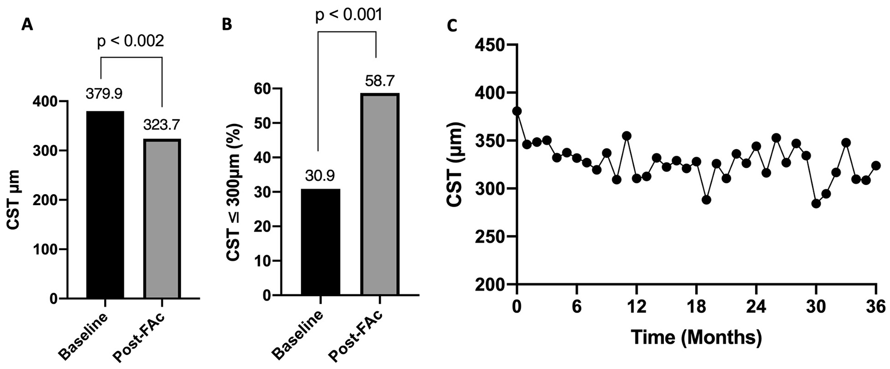
(A) Mean central subfield thickness (CST) of eyes with a 24-month follow-up (n = 97) after fluocinolone acetonide (FAc) treatment. Significance was determined using the Student 2-tailed t test (P < .05). (B) Percentage of eyes with a CST of 300 μm or less 24 months after FAc treatment. Significance was determined by the Z test on normal distribution (P < .05). (C) Mean CST at baseline (month 0) and at each month after FAc treatment.
Treatment Burden
Most patients had intravitreal anti-VEGF injections, most commonly bevacizumab and aflibercept, within 12 months before fluocinolone acetonide administration. The mean number of DME-related treatments required per year decreased from 4.9 before implant placement to 1.5 after (P < .001), a 69.4% decrease overall (Figure 3A). The Kaplan-Meier time-to-rescue analysis found that adjunct treatment was not required until month 17 after fluocinolone acetonide administration. Figure 3C shows the frequency distribution of eyes receiving DME-related treatments in the 12 months before and 24 months after fluocinolone acetonide administration. The proportion of eyes requiring 0 to 2 DME-related treatments per year after fluocinolone acetonide increased from 20.3% to 74.3% (P < .001), while the proportion of eyes requiring 3 to 5 DME-related treatments per year decreased from 37.2% to 17.6% (P < .006). Eyes requiring more than 5 DME-related treatments per year decreased from 42.6% to 8.1% after fluocinolone acetonide administration (P < .001).
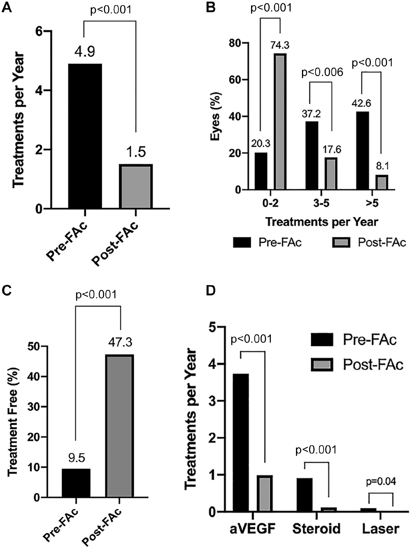
(A) Mean DME treatments per year 12 months before and 24 months after FAc treatment. (B) Percentage of eyes that were treatment free for DME management in the 12 months before and 24 months after FAc treatment. (C) Frequency distribution of eyes receiving DME treatments in the 12 months before and 24 months after FAc treatment. (D) Treatments per year by DME treatment class in the 12 months before and 24 months after FAc treatment. Significance was determined by the 2-tailed paired Student t test. Rate comparisons were completed using bootstrap analyses resampling at the subject level.
Safety Outcomes and Adverse Events
Throughout 24 months, the change in the mean IOP from baseline was not statistically significant (P ≥ .0771 for all months) (Figure 4). The mean change from baseline through 24 months was 0.4 mm Hg (P = .39). IOP elevations greater than 25 mm Hg and greater than 30 mm Hg occurred in 16.9% of all eyes and 9.5% of all eyes, respectively. The proportion of patients who required the initiation or addition of IOP-lowering medication throughout the follow-up was 33.1%. Of those, 59.2%, 20.4%, and 20.4% required 1, 2, or 3 or more IOP-lowering medication(s), respectively. Six eyes (4.0%) required incisional IOP-lowering surgery, and 1 eye (0.68%) required laser trabeculoplasty. Of the 6 eyes requiring incisional IOP-lowering surgery, 1 had uveitic glaucoma and 1 neovascular glaucoma.
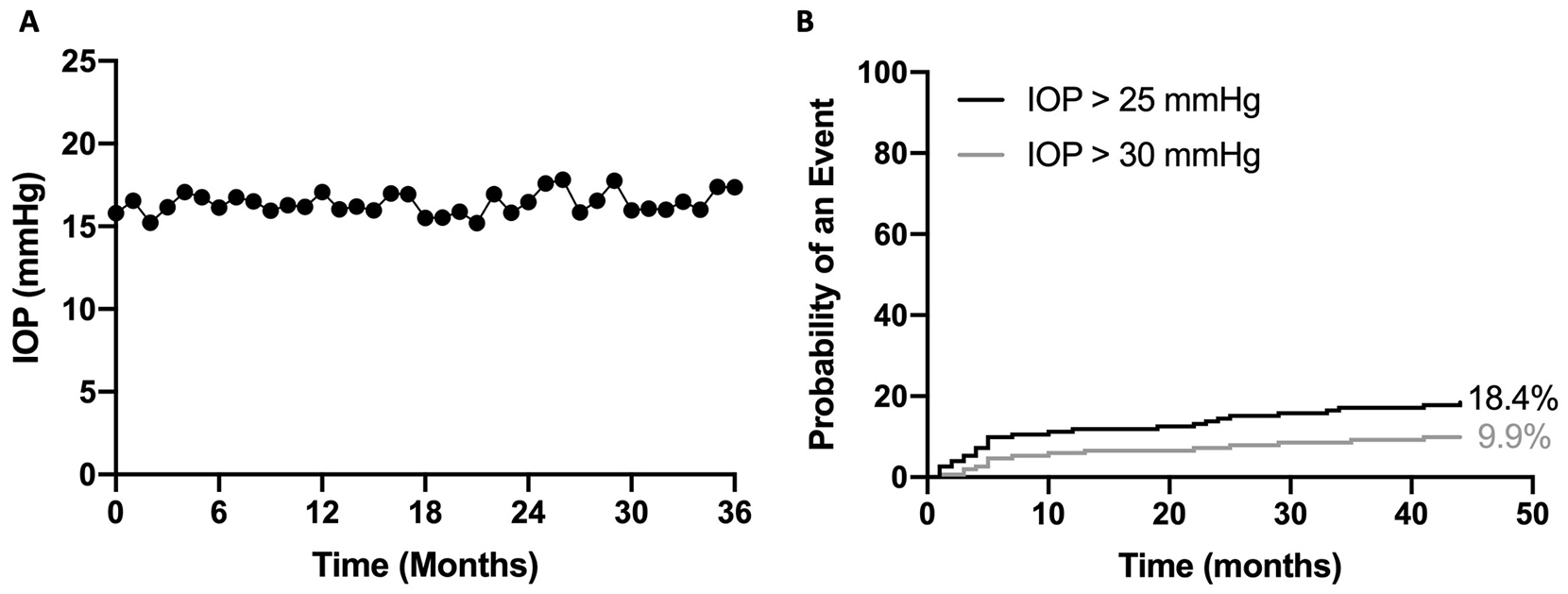
Intraocular pressure (IOP) at baseline and after fluocinolone acetonide treatment for diabetic macular edema. (A) The mean IOP at baseline (month 0) and each month up to 24 months after fluocinolone acetonide treatment. (B) Kaplan-Meier curve of the likelihood of a spike in IOP of more than 25 mm Hg and more than 30 mm Hg.
Thirteen eyes were lost to follow-up before month 6 and were excluded from the analysis. No eye in this cohort had an IOP spike above 25 mm Hg or 30 mm Hg. Of the 13 excluded eyes, the mean recorded IOP was 14.23 mm Hg at baseline and 16.38 mm Hg at the last recorded visit.
Most eyes (69.6%) were pseudophakic at the time of implant insertion. Of the 45 phakic eyes at baseline, 34 required cataract extraction at some point during the follow-up. Implant migration to the anterior chamber occurred in 2.0% of eyes (n = 3), 67% (n = 2) of which had previous vitrectomy and required removal of the implant. None of the eyes developed endophthalmitis post-injection.
Conclusions
The results in the current study support the existing body of evidence that corticosteroid-microdosing intravitreal implants stabilize VA and the macular anatomy in eyes with DME while significantly reducing the treatment burden. In our study, there was a 0.8-letter decrease in BCVA from baseline to 24 months after placement of a 0.19 mg fluocinolone acetonide intravitreal implant, despite patients receiving fewer treatments per year after implant delivery. The proportion of eyes with a BCVA of 70 ETDRS letters (20/40 Snellen equivalent) or more was 20.6% at baseline and 23.7% at month 24.
Previous studies, including the USER 13 and PALADIN, 14 also found improved or maintained BCVA at levels similar to those before fluocinolone acetonide administration. The visual outcomes in these studies were worse than in the FAME trial; however, in general, noncontrolled retrospective studies tend to show worse visual gains than controlled or randomized clinical trials. 11 Furthermore, a large proportion of eyes in the current study had chronic refractory DME with a presumed degree of limited potential for VA improvement. We also found reductions in the CST, as did previous studies. The proportion of patients with a CST of 300 μm or less was 24.6% in the USER trial, 13 which is significantly lower than the 58.7% in our study. However, our results represent the proportion at the last recorded measurements for available patients.
Notable improvements were observed in the frequency of supplemental treatments after fluocinolone acetonide administration. An average of 1.4 supplementary treatments were administered per year after implant placement, which approximated 4.2 treatments for the duration of implant efficacy. The percentage of eyes that required 2 or fewer treatments increased significantly, from 20.3% to 74.3%, while the time-to-rescue analysis showed that additional treatment was not required until month 17 after fluocinolone acetonide implant placement. The significant reduction in treatment burden provided by the fluocinolone acetonide implant was confirmed in several previously published reports. The PALADIN study 14 found a 70.5% reduction in overall treatment burden, which mirrors the 69.5% reported in the current study. Greater improvements were observed in the USER study, 13 in which an overall reduction of 80% was observed and treatments were administered every 14.3 months after fluocinolone acetonide delivery.
It is well known that corticosteroid use is associated with IOP elevations, cataract progression, and the potential for implant migration to the anterior chamber. 7 In our study, the change in the mean IOP from baseline using available data by month was not statistically significant (P ≥ .08 for all months). The last available mean change in IOP from baseline through 24 months was 0.4 mm Hg (P = .39). Although 33.1% of our entire cohort required initiation or addition of IOP-lowering medication, only 4.0% required surgical intervention and 0.68% required laser trabeculoplasty. This indicates that elevations above the threshold were relatively frequent but were managed with the use of topical medication. Steroid-induced IOP elevations above the threshold can be mitigated by posing a steroid challenge in which topical medication is administered 3 to 4 weeks before implant delivery to ensure there is no response to the steroid.
Nonbiodegradable corticosteroid intravitreal implants pose a threat to eyes with disruptions in the iris–lens diaphragm, including those with aphakia, zonular dehiscence, an anterior chamber intraocular lens (IOL), or a scleral-fixated posterior chamber IOL. 15 Dexamethasone intravitreal implants (Ozurdex, Allergan Inc) are associated with corneal endothelium toxicity and anterior migration, which can result in irreversible corneal edema that requires urgent surgical removal of the implant. 16 However, the fluocinolone acetonide implant has several advantages in cases of anterior chamber migration. It is nontoxic and smaller than the dexamethasone implant; thus, it will cause less trauma to the corneal endothelium. Fluocinolone acetonide also has the potential to be sclerally fixated, which prevents anterior migration in eyes at high risk. 15
There were several limitations related to the retrospective nature of this study. Data were unavailable for all timepoints for a notable portion of the patient population (ie, lost to follow-up). Data collection was largely limited to patient chart recordings, and subsequent analyses relied on the complete and accurate documentation of patient findings. In addition, supplemental therapies for disease management were administered at the treating physician’s discretion. The treatment burden was defined as supplemental treatment and did not account for treatment-free physician visits. Despite these limitations, these results remain consistent with those in previous noncontrolled studies and capture the nuances of disease management and the advantages of the 0.19 mg fluocinolone acetonide intravitreal implant in a real-world clinical setting in which treatment nonadherence is common.
Footnotes
Authors’ Note
Presented in part at the annual meeting of the Association for Research in Vision and Ophthalmology, New Orleans, Louisiana, USA, April 24–27, 2023.
Ethical Approval
Ethical approval for this study was waived by the institutional review board (Pro00064827) given the retrospective nature of the study. This study adhered to the tenets of the Declaration of Helsinki. Data collection and analysis were performed in accordance with US Health Insurance Portability and Accountability Act guidelines.
Statement of Informed Consent
Individual informed consent was not obtained because this study does not contain personal information that could lead to the identification of patients.
Declaration of Conflicting Interests
The author(s) declared the following potential conflicts of interest with respect to the research, authorship, and/or publication of this article: Dr. Tabandeh: Alimera Sciences (stock) and Coherus BioSciences (stock, public).
Dr. Boyer: 4D Molecular Therapeutics, Achillion Pharma, Adverum Biotechnologies, AiViva Biopharma, Alcon, Aldeyra Therapeutics, Alimera, Alkahest, Allegro, Allergan, Allgenesis, Amydis, Annexon Biosciences, Apellis Pharmaceuticals, Applied Genetec Technologies Corp, AsclepiX Therapeutics, Ashvattha Therapeutics, Aviceda Therapeutics, Bausch + Lomb, Bayer, Biovisics Medical, Boehringer-Ingelheim Pharma, Cell Care Therapeutic, Chengdu Kanghong Biotechnology, Clearside Biomedical, Curacle Co Ltd, Delsitech, EyePoint Pharmaceuticals, Genentech, Glaukos, jCyte Inc, Iveric Bio, Kriya Therapeutics, Kyowa Kirin, Inc, Lineage Cell, LumiThera, Inc, Nanoscope Therapeutics, NGM Biotherapeutics, Novartis Ophthalmics, Ocugen Inc, Ocular Therapeutix, Oculis SA, Ocuphire Pharma, OcuTerra Therapeutics, Ocutrx Vision Technologies, Opthea, Optigo Biotechnology, Optos, Oxurion NV, Palatin Technologies Inc, Pfizer, Regeneron Pharmaceuticals, RetinAI Medical AG, Ripple Therapeutics, Roche, Sanofi, Santen, Shenyang XingQi Pharma, Smilebiotek Zhuhai Limited, Stealth BioTherapeutics, Surrozen Inc, Syneos, Thea Laboratories, Unity Biotech, Vanotech Corp, Verseon Corp, Vitranu Inc, Vitro Biopharma, and Viva Vision Biotech (consultant and/or, speakers’ bureau); Allegro and DigiSight (Verana Health) (stock).
Dr. Dayani: EyePoint Pharmaceuticals (speaker).
J. Kasper is an employee of Alimera Sciences, Alpharetta, GA, USA.
Dr. Rahhal: Alcon (consultant); BVI (advisor); ReVana Therapeutics, AivoCode, Outlook Therapeutics, Vantage Therapeutics, and Coherus (advisory board); and ExSight Ventures (equity).
None of the other authors declared potential conflicts of interest with respect to the research, authorship, and/or publication of the article.
Funding
The author(s) received no financial support for the research, authorship, and/or publication of this article.