
Abstract
Keywords
Introduction
Scleral buckling was pioneered by Ernst Custodis for the management of rhegmatogenous retinal detachment (RRD). 1 Despite the advances in microincision vitrectomy surgery, scleral buckling remains the preferred means of managing uncomplicated RRD in phakic eyes and pediatric eyes.
Although the preference for pars plana vitrectomy (PPV) has grown over the past 2 decades, scleral buckling has certain advantages over PPV. It is an extraocular surgery with enhanced postoperative photoreceptor realignment, has a high single-surgery success rate, and obviates the risk for cataract formation and the need for postoperative positioning.2,3 Noncontact chandelier-assisted scleral buckling and 3-dimensional digitally assisted vitreoretinal surgery have renewed surgeons’ interest in scleral buckling.
The purpose of this study was to analyze the clinical presentation and baseline fundus parameters, visual outcomes, and complications of scleral buckling performed in a large cohort at a tertiary eye care institution. The study also assessed the presenting factors that determine postoperative visual outcomes.
Methods
This retrospective study comprised a consecutive series of patients with RRD who had scleral buckling from January 2018 to December 2022 with a minimum follow-up of 6 months at a tertiary eye care center after a thorough evaluation. The study received institutional ethics committee approval.
Patients with coexisting ocular pathology (cataract, glaucoma, diabetic retinopathy, macular degeneration) and those who did not attend follow-up visits were excluded. Figure 1 shows the selection criteria for determining which cases of RD would be scheduled for scleral buckling or for PPV. Data collected included the presenting best-corrected visual acuity (BCVA), lens status, number of predisposing lesions/breaks, predominant quadrant of the detachment, and macular status (on or off). Preoperative identification of breaks and diagrammatic representation on the Amsler Dubois chart were performed after an indirect ophthalmoscopic examination with scleral indentation.
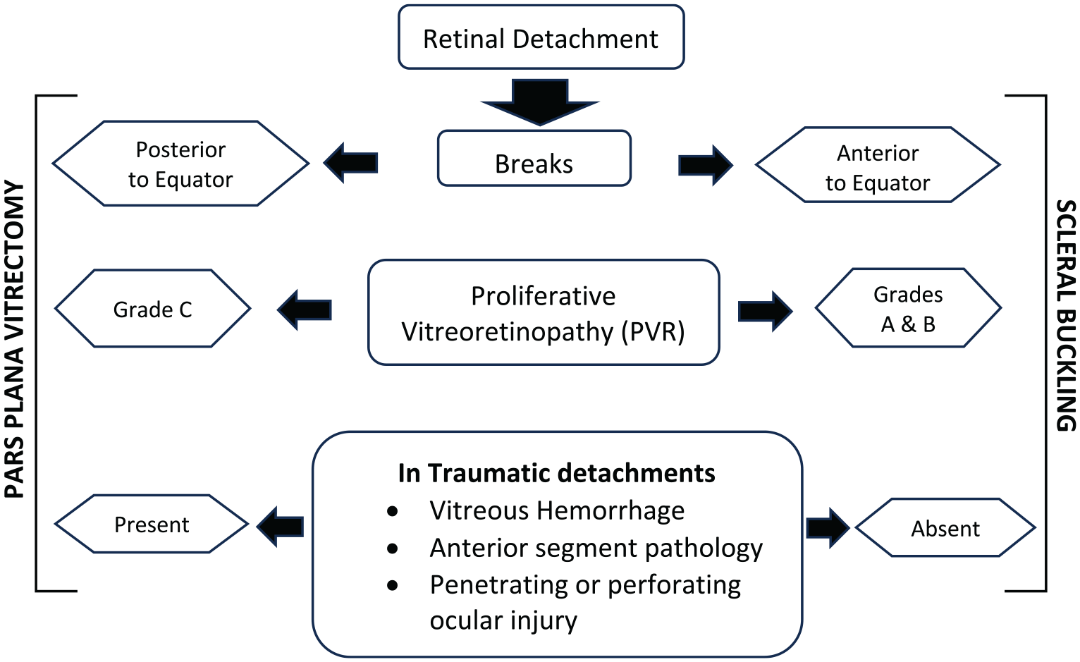
Flow chart of the decision-making process in the choice of surgical procedure (pars plana vitrectomy vs scleral buckling) for cases of retinal detachment.
The surgeries were performed by 1 of 3 consultants from the Department of Retina and Vitreous at the eye care center. Each consultant had more than 10 years of in-house experience as an independent vitreoretinal surgeon. All patients underwent scleral buckling with cryotherapy to the retinal breaks and drainage of subretinal fluid (SRF). Under peribulbar anesthesia (or general anesthesia for pediatric and uncooperative patients), conjunctival peritomy and recti muscle bridling were performed. Cryotherapy was applied to the breaks in the retina until an adequate gray-to-white reaction was obtained. The breaks were supported by silicone buckle implants.
The type of buckle used was based on the clinically determined type of break(s). As a general guideline, a 287 convex symmetrical band was used for dialyses and a 277 symmetrical concave band was used in cases of lattices with holes. A 276 asymmetrical band was used to support horseshoe tears. An encircling buckle was used in cases with large tears and/or multiple breaks in different quadrants. A 240 band for encirclage was used in conjunction with tires. A 42 or 240 band for encirclage was used alone in cases in which the indent produced by encirclage was deemed to be adequate.
The SRF was drained by making a scleral puncture at the most dependent site using a 26-gauge needle. The surgeon ensured that the site of SRF drainage was supported by the buckle elements.
The postoperative BCVA at 1 day, 1 month, 3 months, and 6 months was noted. The postoperative complications, subsequent management, and visual gain were recorded. These parameters continued to be assessed for 6 months after the final surgical intervention.
Results
Preoperative and Intraoperative Characteristics
The study comprised 307 eyes of 290 patients who had scleral buckling. The mean patient age at presentation was 32.10 years (range, 10-77); 72.41% of patients (n = 210) were men, and 17.00% had bilateral RD. Table 1 shows the baseline characteristics of the eyes. Of the eyes, 54.40% were emmetropic, 43.32% were myopic, 6.19% had high myopia (more than −6.00 D), and 2.28% had hyperopia. Forty-two eyes (14.95%) presented with traumatic RRD and 32 eyes (10.42%) with macula-on detachments. Most eyes (97.39%) were phakic. The quadrant in which the RRD was located was inferior in 48.21% of eyes, superior in 20.20%, temporal in 18.89%, and nasal in 2.61%. An RRD involving all quadrants was present in 10.10% of eyes.
Baseline Parameters of Eyes.
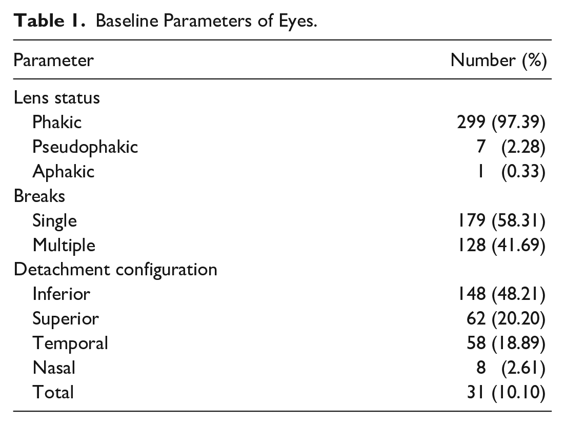
Table 2 shows the type of predisposing lesion(s). A single predisposing lesion (break/hole) was present in 8.31% of the detachments, while the rest had 2 or more predisposing lesions. Lattice with a hole was the most common predisposing lesion followed by an atrophic hole and horseshoe tear. Other lesions included dialysis, giant retinal dialysis, and cystic tufts. Of the cases, 16.94% had multiple types of lesions.
Type of Predisposing Lesion(s).
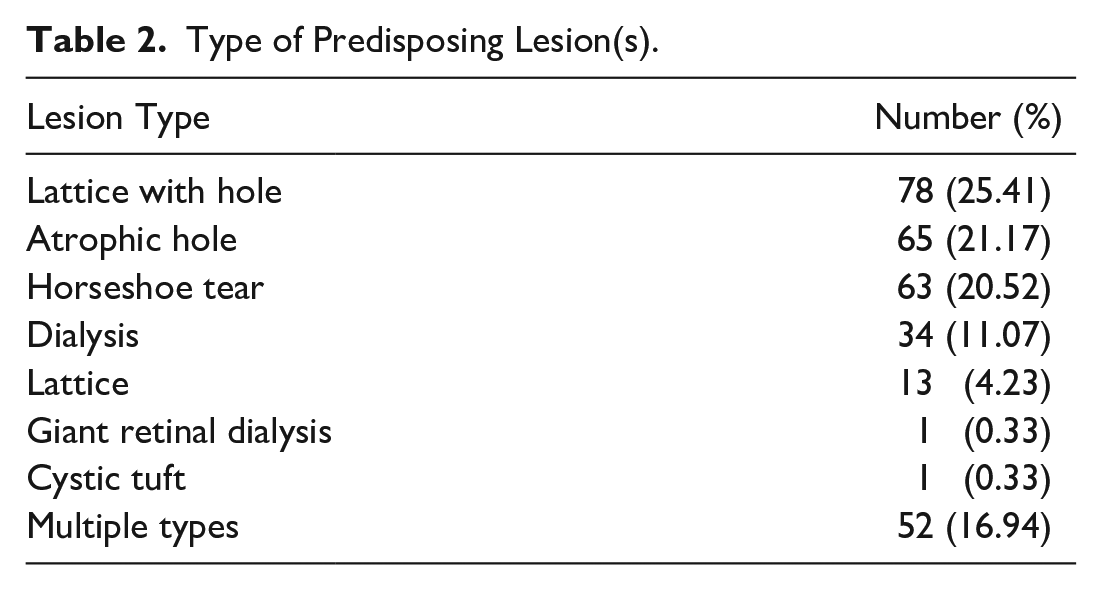
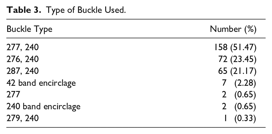
Table 3 shows the type of buckle used. The most common was a 277 band with 240 encirclage (51.47%) followed by a 276 band with 240 encirclage (23.45%) and a 287 band with 240 encirclage (21.17%).
Type of Buckle Used.
Primary anatomic attachment was defined as retinal reattachment maintained for 6 months after primary scleral buckling surgery. This was achieved in 288 eyes (93.81%).
Visual Outcomes
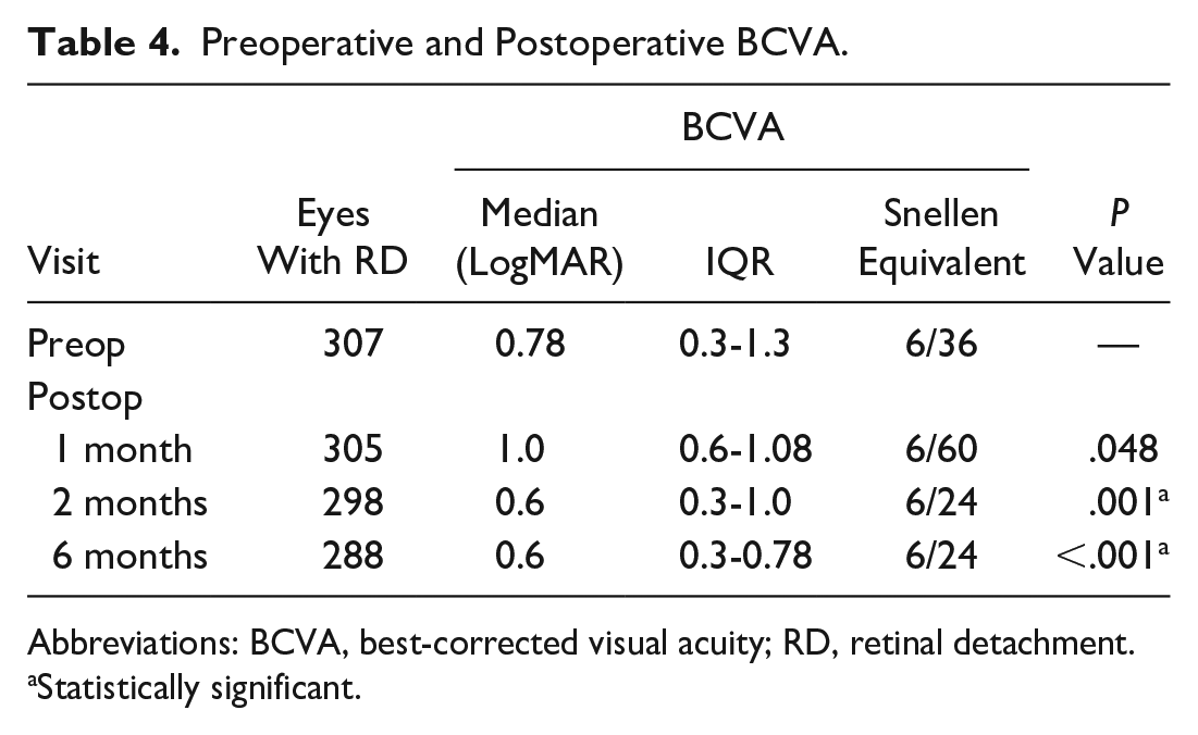
Table 4 shows the preoperative and postoperative BCVA. The mean preoperative Snellen BCVA was 6/36 (logMAR equivalent, 0.78; IQR, 0.3-1.3 logMAR). At the 6-month follow-up, the mean Snellen BCVA was 6/24 (logMAR equivalent, 0.6; IQR, 0.3-0.78 logMAR). The improvement in logMAR BCVA from preoperatively to 3 months and 6 months postoperatively was statistically significant (P = .001 and P < .001, respectively).
Preoperative and Postoperative BCVA.
Abbreviations: BCVA, best-corrected visual acuity; RD, retinal detachment.
Statistically significant.
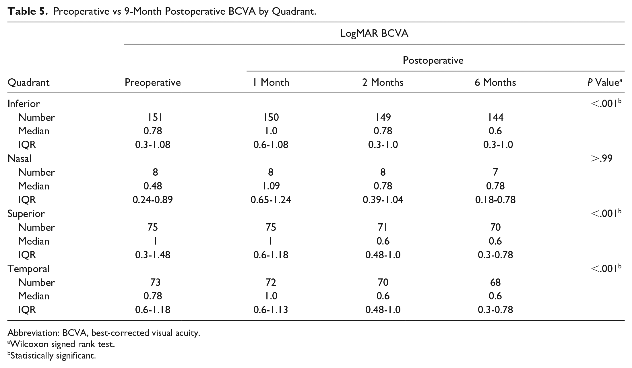
Table 5 shows a comparison of the logMAR VA between 6 months postoperatively and preoperatively by quadrant. There was a statistically significant improvement in VA in cases in which the RRD involved the inferior, superior, or temporal quadrant.
Preoperative vs 9-Month Postoperative BCVA by Quadrant.
Abbreviation: BCVA, best-corrected visual acuity.
Wilcoxon signed rank test.
Statistically significant.
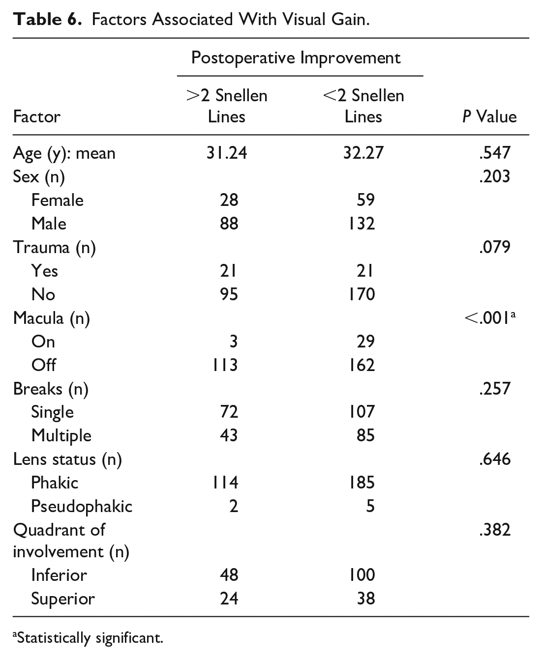
Tables 6 and 7 show the factors associated with a significant visual gain, defined as an improvement in BCVA of 2 Snellen lines or more postoperatively vs the presenting BCVA. Macula-on detachment had a significant relationship with visual gain (P < .001, χ2 test). No other factor had a significant relationship.
Factors Associated With Visual Gain.
Statistically significant.
Refractive Error Associated With Visual Gain. a

P = .568.
Complications
Recurrent detachment, defined as redetachment within the first 6 months of scleral buckling surgery, occurred in 19 eyes (6.19%). Of these eyes, 1 had a macula-on RD at the time of presentation; 1 was pseudophakic, with the rest being phakic; 10 had a single break; and 9 had 2 or more breaks. Six eyes had inferior detachments, 4 had superior detachments, and 9 had predominantly temporal detachments. All cases subsequently underwent PPV with silicone oil tamponade followed by oil removal 3 months later. No statistically significant gain in VA was noted after PPV in cases with a redetachment, although the final anatomic success rate (ie, an attached retina at the end of 6 months after PPV) was 100%.
Five cases (1.68%) developed secondary increased intraocular pressure (IOP), which was successfully managed with antiglaucoma medication. None of these eyes required surgical intervention to lower the IOP.
The buckle was removed in 2 cases (0.67%) because of a buckle infection secondary to buckle exposure. The infection was diagnosed 6 months after scleral buckling surgery in 1 case and 2 years after scleral buckling surgery in the other case. Topical and oral antibiotics were started before buckle removal. These cases did not develop redetachment during the 6 months after buckle removal.
One eye developed suture-related conjunctival granuloma close to the temporal limbus, necessitating excision because the patient reported chronic irritation and for cosmetic purposes. Another developed subretinal hemorrhage over the buckle implant and had PPV with drainage of the hemorrhage and sulfur hexafluoride tamponade. The final BCVA of this patient was 6/18, and the retina remained attached through the end of the follow-up period.
Conclusions
This study assessed the clinical characteristics of patients who had scleral buckling surgery and the impact of surgery on the final visual outcomes. It also determined the primary attachment rate after scleral buckling surgery, the complications, and subsequent management.
Scleral buckling has classically been advocated as a mainstay of management in phakic eyes with uncomplicated RRD. In the current study, 97.39% of eyes were phakic, 7 (2.28%) were pseudophakic, and 1 (0.33%) was aphakic.
The Scleral Buckling versus Primary Vitrectomy in Rhegmatogenous Retinal Detachment Study, 4 a randomized control trial that enrolled 416 phakic patients and 265 pseudophakic patients, compared scleral buckling and vitrectomy in the management RRDs of medium complexity, defined as detachments with large breaks (ie, 1 to 2 clock hours, multiple breaks, marked vitreous traction, centrally extending breaks, and superior bullous in nature). The study found better BCVA in phakic eyes that had scleral buckling than in eyes that had PPV.
A study by van Leeuwen et al 5 of 4447 patients who had RD surgery in 2016 found an increase in the incidence of RD of 44% over the incidence in 2009. The study also found an increase in the prevalence of mild, moderate, and severe myopia. It was postulated that a simultaneous myopic shift in the population may be associated with an increase in the incidence of RD.
In a study by Kim et al of 1599 eyes with RRD, 6 the highest peak incidence of RD occurred at an age of 55 to 59 years, with a second peak at 25 to 29 years. Myopia-induced early vitreous detachment appeared to be a major mechanism of RRD in young patients, whereas senile vitreous liquefaction and detachment were the main mechanisms in elderly patients.
In our study, the mean age at presentation was 32.10 years; 43.32% of eyes were myopic, with 6.19% having high myopia. Recently, awareness of the need for evaluation of the peripheral retina in eyes with myopia has grown. Also, our study was based at a tertiary eye care hospital, which in the past decade has seen an increase in the number of young patients with myopia opting for keratorefractive surgery. In the global setting of an increasing incidence of myopia and RD, further studies are needed to determine the role of fundus screening and prophylactic barrage treatment of breaks in myopic eyes in reducing the overall incidence of RD.
The principles of scleral buckling surgery are the closure of retinal breaks with the elimination of vitreoretinal traction and formation of chorioretinal adhesion. Placement of buckle elements helps achieve the former, and the choice of the type of buckle must be appropriate based on the configuration of retinal breaks. 7 A radial buckle is most effective for a single horseshoe tear, whereas a circumferential explant is effective for retinal dialysis and atrophic retinal holes.
A 277 buckle with or without 240 encirclage is the most common type used in scleral buckling surgery, 8 as was the case in our study. In our study, lattice with a hole was the most common predisposing lesion (25.41% of cases). A 277 buckle with a 240 band was used in cases of atrophic holes, horseshoe tears, and combinations of different types of lesions.
Numerous studies have determined factors that affect visual outcomes after scleral buckling surgery. Kobashi et al 9 found that a preoperative macula-off status played a significant role in determining the visual outcomes. Heussen et al 10 compared the surgical outcomes of scleral buckling surgery in phakic eyes and pseudophakic eyes; the common prognostic factor in both subgroups was the number of breaks. In a study by Salicone et al, 11 the preoperative factors associated with better visual outcomes were macula-sparing RRD, a smaller extent of RRD, and a lower degree of preoperative myopia. In a study by Wolfensberger, 12 complete foveal reattachment occurred earlier in eyes with a macula-off RRD that had PPV, whereas a subclinical persistence of subfoveal fluid was observed for several months after scleral buckling surgery.
In our study, a macula-on RRD was significantly associated with better postoperative visual outcomes, which is consistent with results in the literature. However, other factors, such as phakic status, the number of breaks, and the location and extent of the detachment, were not significantly associated with visual outcomes.
The reported rates of recurrent detachment after scleral buckling range from 7% to 14%.13,14 In our study, the primary anatomic attachment rate was 93.81% and the final anatomic success rate was 100%. Eyes that had PPV for recurrent detachment did not have a significant improvement in visual outcomes vs the improvement after primary scleral buckling surgery. Other complications included secondary glaucoma, buckle infection, conjunctival granuloma, and subretinal hemorrhage, all of which occurred infrequently and were successfully managed.
In a retrospective study by Radice et al of 135 eyes undergoing scleral buckling, 15 the primary anatomic success rate was 94% and the final anatomic success rate was 100% after a 12-month follow-up. Azad et al 16 found a primary reattachment rate of 80.6% in phakic patients and a final anatomic success rate of 100% at the end of 6 months; 16% had residual SRF that required PPV. An increase in IOP occurred in 6% of eyes and was controlled with antiglaucoma therapy. One eye (3%) had a buckle-related infection.
The study by Radice et al 15 found that the visual outcomes at the 1-week and 1-month follow-up were significantly better in the scleral buckling group than in the PPV group, with the latter having an increased risk for postoperative cataract formation. A meta-analysis by Sun et al 17 showed similar outcomes.
In a study of 169 eyes with phakic RRD by Ryan et al, 18 the single-surgery anatomic success rate was highest for scleral buckling followed by scleral buckling along with PPV and last by PPV. Scleral buckling yielded significantly better visual outcomes than PPV or combined scleral buckling with PPV for macula-on or macula-split cases, even after controlling for cataract.
However, a study by Park et al 19 of 72 phakic eyes of patients older than 35 years found that PPV performed using a wide-angle viewing system had a higher primary success rate than scleral buckling. Although the final success rate was 100% in both groups, the rate of persistent SRF was statistically significantly higher in eyes that had scleral buckling.
A report published by the European Vitreo-Retinal Society based on a retrospective review of 7678 cases with RRD found that the final failure rate in phakic eyes with uncomplicated RRD was lower with scleral buckling than with vitrectomy, while the opposite was true in pseudophakic eyes. 20 A study of 50 pseudophakic eyes with RRD that were divided into a scleral buckling group or a PPV group found a primary reattachment rate of 76% and 84%, respectively; the final anatomic reattachment rate was 100% in both groups. 21
In contrast, in a study by Ahmadieh et al, 22 225 pseudophakic eyes and aphakic eyes with RRD were randomly assigned to scleral buckling or PPV. There were no statistically significant differences in the single-surgery retinal reattachment rate between the 2 groups.
Unlike in these studies, we could not make a direct correlation between phakic eyes and pseudophakic eyes because the phakic eyes vastly outnumbered the pseudophakic eyes. The retrospective nature of the study and selection bias resulting from a general inclination toward scleral buckling in phakic eyes are contributory factors to the larger number of phakic eyes. However, there was no statistically significant difference in significant visual gains based on phakic status. Also, of the 19 eyes that developed a redetachment, only 1 was pseudophakic.
In this series, 6.19% of cases developed redetachment and 68.42% had multiple retinal breaks. More cases had predominantly inferior detachments. The visual outcomes remained poor despite additional surgery. Other complications were well managed, indicating that scleral buckling remains a surgical procedure with a high anatomic success rate and good visual outcomes with few complications, all of which were manageable.
The limitations of our study include its retrospective nature, short follow-up, a lack of an adequate number of pseudophakic eyes and aphakic eyes, and selection bias that might have had an effect on visual outcomes.
In conclusion, this study analyzed a large cohort of eyes that had scleral buckling surgery at a fellowship-training institute, the findings of which will be helpful in an era in which there has been a marked paradigm shift to PPV. Most eyes that had scleral buckling were phakic with a single predisposing lesion. However, multiple breaks, inferior detachments, and a traumatic etiology did not appear to adversely affect the visual outcomes; thus, we believe that scleral buckling can be considered a surgical option in the same.
Macular status played a significant role in the visual prognosis. This finding will help surgeons determine the visual potential after surgery as well as counsel patients and modify their expectations accordingly. Evaluation of the peripheral retina and prophylactic barrage treatment of breaks in myopic eyes may play a role in decreasing the overall incidence of RD. Despite being a demanding procedure in terms of precision, scleral buckling is far from being an outdated procedure. Adequate exposure to performing scleral buckling, along with PPV procedures, may provide a more rounded approach in retinal surgery training.
Footnotes
Ethical Approval
This study was conducted in accordance with the Declaration of Helsinki. The collection and evaluation of all protected patient health information were performed in a US Health Insurance Portability and Accountability Act–compliant manner.
Statement of Informed Consent
Informed consent, including permission for publication of all photographs and images included herein, was obtained before the procedure was performed.
Declaration of Conflicting Interests
The author(s) declared no potential conflicts of interest with respect to the research, authorship, and/or publication of the article.
Funding
The author(s) received no financial support for the research, authorship, and/or publication of this article.