






Abstract
Introduction
Soft drusen can cause visually significant metamorphopsia for patients with age-related macular degeneration (AMD). 1 These soft drusen can disappear spontaneously; however, such changes are often associated with retinal pigment epithelium (RPE) atrophy and progression to advanced AMD. 2 In AREDS Report Number 28, eyes with drusenoid pigment epithelial detachments (PEDs) were shown to develop geographic atrophy (GA) and choroidal neovascularization (CNV) at 5 years in 19% and 23% of cases, respectively. 3 Laser photocoagulation has been shown to induce drusen disappearance but without improvement in the rates of CNV or GA or improvement in visual acuity (VA). 4 However, the mechanisms involved in spontaneous or laser-induced drusen regression remain unclear.
Few cases of spontaneously regressing drusen without functional consequences have been reported.5–8 Drusen disappearance has also been reported after surgery for full-thickness macular hole (FTMH) repair9–11 and retinal detachment (RD) repair.12,13 These previous reports have shown this phenomenon with color photographs but not with optical coherence tomography (OCT). Using multimodal imaging, we present a case of a subfoveal drusenoid PED that quickly resolved after RD repair.
Case Report
An 83-year-old woman presented with blurred vision in the left eye first noted 1 month earlier. Her ocular and medical history were notable for intermediate AMD, pseudophakia, hypertension, hyperlipidemia, and endometrial cancer. She had no history of smoking. On initial presentation, the VA in the left eye was 20/60 and there was a prominent nasal choroidal detachment and an inferior RD (Figure 1, A and B). OCT showed drusenoid PEDs in the left eye (Figure 1C). The macula remained attached. The patient wished to delay the repair until she had assistance with her recovery. Three days later, a repeat OCT showed shallow subretinal fluid (SRF) in the inferior macula extending to the inferior perifoveal area (Figure 1, D and E). However, the area involving the primary subfoveal PED remained attached.
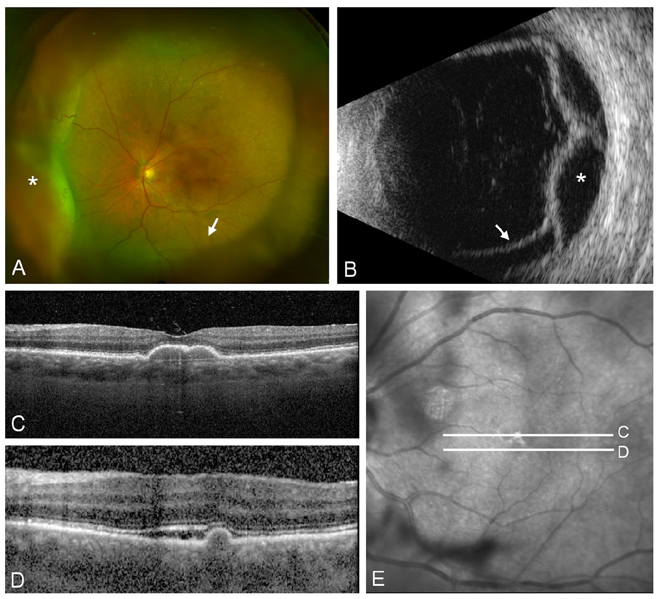
(A) Fundus photograph of the left eye shows drusenoid pigment epithelial detachments (PEDs), nasal choroidal detachments, and an inferior retinal detachment (RD). (B) The B-scan of the left eye shows prominent nasal serous choroidal detachment and an inferior RD in the nasal transverse view. Asterisks indicate choroidal detachments and arrows show the RD. (C) Optical coherence tomography (OCT) shows subfoveal drusenoid PEDs in the left eye on initial presentation. (D) Three days later, OCT of the left eye shows shallow subretinal fluid (SRF) involving the inferior perifoveal macula. (E) En face OCT shows the location of the subfoveal drusenoid PEDs (C) and the superior extent of SRF (D).
Four days later, the patient underwent nasal choroidal drainage, 25-gauge pars plana vitrectomy, drainage retinotomy, fluid–air exchange, endolaser application, and gas tamponade with 14% octafluoropropane. One month postoperatively, the retina was attached and the choroidal detachments had resolved. The VA in the left eye improved to 20/40, and the drusenoid PEDs remained (Figure 2A). Serial OCT images showed a reduction in the size of the drusenoid PEDs 1 month postoperatively compared with preoperative imaging (Figures 1C and 3A). Two months postoperatively, there was complete resolution of macular drusen with the presence of trace RPE irregularities (Figure 3B). Six months postoperatively, the VA improved to 20/25 and fundus photographs and autofluorescence showed resolution of the drusenoid PEDs (Figure 2, B and C). In addition, there were no drusen or RPE irregularities on OCT; however, a few hyperreflective foci were noted in the outer retina (Figure 3C).

(A) One month postoperatively, optical coherence tomography shows smaller subfoveal drusen. (B) Two months postoperatively, the drusen have resolved and trace retinal pigment epithelium (RPE) irregularities remain. (C) Six months postoperatively, the drusen and RPE irregularities have resolved.

(A) Fundus photograph shows macular drusenoid pigment epithelial detachments (PEDs) (arrow) 1 month postoperatively. The gas meniscus is visible in the superior macula. Complete resolution of the drusenoid PEDs was noted 6 months postoperatively as shown on (B) fundus photography and (C) autofluorescence.
There was no evidence of RPE atrophy or CNV in the postoperative period. The patient noted significant improvement in the left eye metamorphopsia. Interestingly, she inquired about having the same procedure to treat the metamorphopsia in the right eye, which had similar subfoveal drusenoid PEDs. Of note, the patient also had extramacular drusen within the area of detachment that persisted 6 months postoperatively (Figure 4).

(A) Fundus photographs 6 months postoperatively show extramacular drusen within the area of detachment (white box), with higher magnification in the inset. (B) Autofluorescence images highlight the extramacular drusen, with higher magnification in the inset.
Conclusions
Drusen disappearance after RD repair was previously described in 2 case reports using fundus photographs. Margolis et al 13 described a macula-involving RD repaired with pneumatic cryopexy that showed improvement in subfoveal confluent soft drusen within the area involving shallow SRF. Lim et al 12 described a chronic near-total RD repaired with vitrectomy in which extramacular drusen resolved postoperatively. In both cases, improved VA and lack of atrophy indicated restoration of the RPE–Bruch complex. Multimodal imaging, including OCT, in our case directly showed complete resolution of the drusen and restoration of the RPE–Bruch complex. Moreover, subfoveal drusen within areas of the macula not involving SRF also disappeared. Unlike Lim et al’s case, the extramacular drusen in our patient persisted in areas where there clearly had been SRF, despite the complete resolution of macular drusen and drusenoid PED (Figure 4). The extramacular deposits were more consistent with hard drusen, whereas the macular deposits were soft drusen, indicating that the composition of drusenoid material may influence their metabolism and resolution.
Drusen resolution has also been documented after vitrectomy surgery for FTMH repair,9–11 with 1 case reporting CNV 3 months postoperatively 11 and another case describing the use of autologous platelet concentrate without membrane peeling. 10 In these cases, the drusen disappeared between 2 months and 12 months postoperatively; however, these preoperative and postoperative findings were not compared using OCT. Both our report and that of Margolis et al 13 describe rapid drusen resolution within 1 month, and both reports were associated with RDs.
Several mechanisms have been posited for the disappearance of drusen after vitreoretinal surgery. The presence of SRF could allow for dissolution of drusen material. In the current case, the fluid was present for approximately 4 days before RD repair and only partly involved the drusenoid PEDs. Another possible mechanism includes the stimulation of inflammatory and immune responses caused by the RD. 14 The hyperreflective foci noted 6 months postoperatively (Figure 3C) may indicate residual inflammatory infiltrates. Additional factors promoting inflammation may include gas tamponade or even cryotherapy or laser photocoagulation to the peripheral retina. The resolution of soft drusen may also be spontaneous and has been shown to occur in approximately 3.5% of eyes with soft drusen. 5 In the current case, the factors responsible for resolution appear to be local to the eye with the detachment because the patient’s contralateral eye had similar drusenoid PEDs that did not improve during this period.
A more systematic investigation of drusen disappearance after vitreoretinal surgery and characterization of the sequelae of drusen resolution, including RPE atrophy and CNV, would be useful. A better understanding of the mechanisms involved may also provide greater insights into the pathogenesis of AMD.
Footnotes
Ethical Approval
The case report was conducted in accordance with the Declaration of Helsinki. The collection and evaluation of all protected patient health information were performed in a US Health Insurance Portability and Accountability Act–compliant manner.
Statement of Informed Consent
Informed consent, including permission for publication of all photographs and images included herein, was obtained before the procedure was performed.
Declaration of Conflicting Interests
The authors declared no potential conflicts of interest with respect to the research, authorship, and/or publication of the article.
Funding
The authors received no financial support for the research, authorship, and/or publication of this article.
