
Abstract
Introduction
Ocular manifestations of HIV were first reported by Holland et al 1 in 1982. Retinal microangiopathy and cytomegalovirus (CMV) retinitis can be seen in almost 30% to 40% of patients with HIV; however, the advent of the highly active antiretroviral treatment (HAART) regimen has led to a significant reduction in the number of patients with these manifestations. Although the incidence of HIV-associated microangiopathy was 15% to 30% in the era before HAART, the presence of these signs during treatment indicates therapeutic failure from drug-related or compliance-related issues. 2
In addition, HIV is an independent risk factor for retinal vascular occlusions. 3 Although large-vessel involvement is less frequently noted, few isolated case reports involving central retinal artery occlusion (CRAO) have been reported in the literature.4,5 In a retrospective chart review of patients with HIV and retinal vascular occlusion, only 1 eye of 2484 patients with HIV was found to have CRAO. 6 Vascular occlusions with concurrent retinitis have also been described.7–11
It is unclear whether the vascular occlusion precedes the episode of retinitis or is an independently occurring entity. Our case is unique in that it shows the sequential development of infective retinitis after an episode resembling ocular ischemic syndrome in 1 eye and the subsequent development of the syndrome in the fellow eye.
Case Report
A 40-year-old woman presented with a sudden, painful decrease in distance and near vision in the right eye of 1-month duration. On examination, the best-corrected visual acuity (BCVA) was 20/800 OS. Disc pallor, arteriolar attenuation, and blot hemorrhages were seen in the midperiphery of the fundus of the right eye (Figure 1A). The left eye was unremarkable, with a BCVA of 20/20.
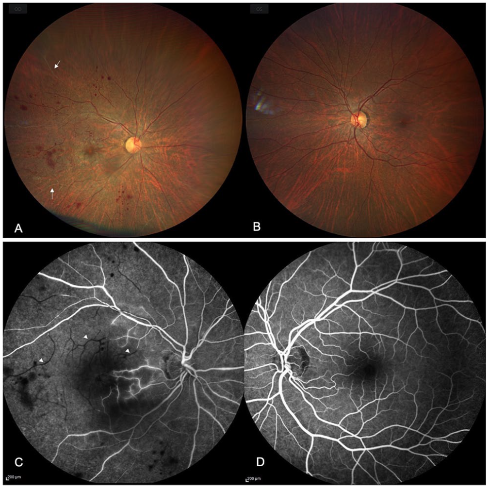
(A) Fundus photograph of the patient’s right eye shows sclerosed arterioles (arrow) and patchy retinal whitening at the macula. (C) Fundus fluorescein angiography shows delayed arm-to-retina time and poor filling of the arterioles (arrowhead). (B and D) The left eye was normal.
Fundus fluorescein angiography (FA) of the patient’s right eye showed a delayed arm-to-retina time of 36 seconds along with delayed filling of the retinal arterioles (Figure 1B). FA of the left eye was normal. A diagnosis of ocular ischemic syndrome in the right eye was made. Ancillary investigations, including a complete blood count, peripheral smear, random blood sugar, serum homocysteine, and lipid profile, were within the normal range except for an elevated erythrocyte sedimentation rate. The patient was advised to have a carotid Doppler, 2D-echocardiography, and a thorough systemic evaluation.
The patient was lost to follow-up but presented 4 months later with painless decreased vision in the left eye for a 10-day duration. The BCVA was no light perception OD and 20/250 for distance OS. The fundus examination of the right eye showed extensive retinitis patches (Figure 2A), and a cherry-red spot, boxcarring, and multiple sclerosed arterioles were seen in the left eye (Figure 2B). Optical coherence tomography (OCT) of the left eye showed hyperreflective inner retinal layers with intraretinal fluid (Figure 2C), and OCT angiography showed severe macular ischemia (Figure 2D). A carotid Doppler examination showed bilateral intima-media hyperplasia in the common carotid and the external and internal carotid arteries.
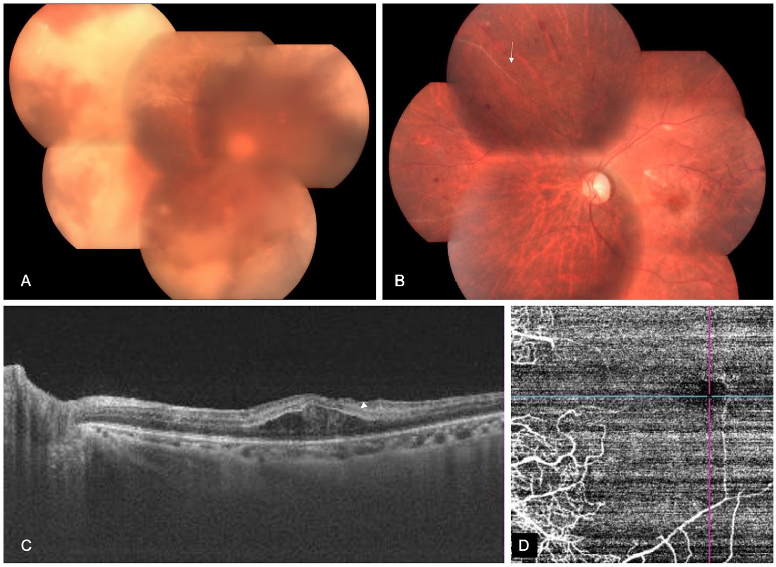
(A) On follow-up, the patient’s right eye shows extensive retinitis lesions. (B) The left eye shows multiple sclerosed vessels with patchy retinal whitening around the fovea. (C) Optical coherence tomography (OCT) shows hyperreflectivity of the inner retinal layers (arrowhead) with intraretinal fluid pockets in the outer retina. (D) OCT angiography shows extensive macular ischemia.
Further investigation revealed that the patient was positive for HIV serology. She later disclosed that she had known about her HIV status but was not on any medication. She was advised to initiate HAART and oral valganciclovir and to consult a cardiologist for systemic management. However, a few months after presenting to our clinic, she succumbed to systemic complications of HIV. Figure 3 shows a summary of the patient’s timeline.
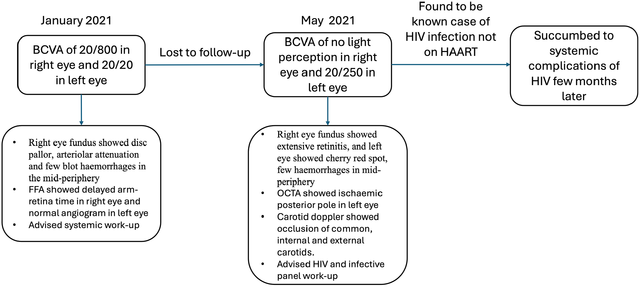
The patient’s timeline, from presentation to the clinic to follow-up.
Conclusions
Noninfectious microvascular abnormalities, also referred to as HIV retinopathy or background AIDS retinopathy, are the most common ocular findings described in HIV retinopathy and are associated with focal capillary closure, nerve fiber layer ischemia, and stasis of axoplasmic flow. 2 HIV-associated retinal macrovasculopathy is less commonly reported. Veins are more frequently involved than arteries and thus far have been described in association with viral retinitis, lymphoproliferative disorders, or as isolated case reports.
In the single largest retrospective chart review of patients with HIV presenting with vascular occlusions, Dunn et al 6 identified no consistent risk factors. Patients were younger than 50 years and did not have cardiovascular disease. The study found a strong association between microvasculopathy and retinal vein occlusion in addition to a significant association with advancing age. Hypertension and thrombotic disease were found in higher proportions in patients with macrovasculopathy; however, there were no cases of ocular ischemic syndrome. An interesting outcome in the Dunn et al study was that there was no association between CD4 counts and macrovasculopathy.
Barring isolated case reports, no major retrospective or prospective studies have examined additional risk factors for arterial occlusions in individuals with HIV. Lenci et al 5 described an unusual case of a young patient who developed CRAO after starting HAART. The authors concluded that elevated lipids secondary to the treatment regimen could have led to carotid atherosclerosis and subsequently CRAO. Branch retinal artery occlusion and vein occlusion have also been reported in patients with HIV8,9,11; however, these reports describe the occurrence of occlusion in eyes with preexisting retinitis. Kim and Lee 12 reported 1 case of CMV retinitis that was preceded by an ocular ischemic syndrome–like event, similar to our case. The patient in that report was immunocompromised as a result of lymphoma, indicating that CMV can cause vaso-occlusive manifestations independent of HIV.
CMV can reach the retina hematogenously, where it infects the vascular endothelium first, before the breakdown of the blood–retinal barrier, and by spreading into the surrounding retinal tissue. Histologically, HIV can also change the vascular media via the scattered chronic inflammatory cell infiltrates, calcification, smooth muscle cell damage, and fibrosis. 13
In our patient’s case, although we could not obtain her CD4 counts or confirm the diagnosis of CMV because she was noncompliant to instructions and did not have regular follow-ups, the possibility of CMV could not be ruled out. Furthermore, severe carotid occlusion has been described in patients with HIV, 13 which itself can be a risk factor for ocular ischemic syndrome. These could have been reasons a vaso-occlusive retinopathy was seen before the onset of retinitis. Whether CMV or HIV was the cause behind the ocular ischemic syndrome–like event or whether it was secondary to the carotid occlusion requires further study. This raises an important question regarding whether HIV screening should be included in the workup of young patients presenting with this potentially blinding condition.
In conclusion, unilateral or bilateral ocular ischemic syndrome can be seen in patients with HIV, even without concurrent retinitis, and could represent a coexisting severe life-threatening systemic vasculopathy. Moreover, viral retinitis could potentially be preceded by ocular ischemic syndrome. Our case highlights the importance of assessing systemic parameters and performing HIV screening in young patients presenting with ocular ischemic syndrome. Whether initiating HAART at the earliest in individuals with HIV would prevent macrovasculopathy events and acute retinal necrosis must be validated in the future.
Footnotes
Ethical Approval
This case report was conducted in accordance with the Declaration of Helsinki.
Statement of Informed Consent
The patient consented to publication of the clinical history, findings, and images.
Declaration of Conflicting Interests
The authors declared no potential conflicts of interest with respect to the research, authorship, and/or publication of the article.
Funding/Support
Hyderabad Eye Research Foundation provided funding.