
Abstract
Keywords
Introduction
Retisert (fluocinolone acetonide, 0.59 mg, Bausch + Lomb) is a commercially available surgically implanted device for the treatment of chronic noninfectious posterior uveitis when topical or systemic therapy is ineffective or poorly tolerated. It is designed to deliver fluocinolone acetonide to the posterior segment, providing sustained drug delivery over a period of up to 30 months.
We report a case of a persistent wound leakage and endophthalmitis occurring 3 years after intravitreal (IVT) implantation of fluocinolone acetonide (0.59 mg) in a patient treated for serpiginous choroidopathy. Although endophthalmitis has been reported as a potential complication of fluocinolone acetonide IVT implants, its occurrence 3 years after implantation is highly unusual. To our knowledge, this is the first reported case of a complication of this nature.
Case Report
An 18-year-old man presented with 4 days of progressively worsening vision and pain in his left eye. His history included serpiginous choroidopathy in both eyes and IVT implantation of fluocinolone acetonide (0.59 mg) in the left eye elsewhere 3 years before presentation. He denied any ocular injury, recent eye surgery, or systemic infections. His past ocular history was significant for steroid-responsive glaucoma in both eyes. Baseline best-corrected visual acuity was 20/20 in the right eye and 20/200 in the left eye due to foveal scarring secondary to macular serpiginous lesions. At presentation, visual acuity (VA) in the left eye was light perception (LP), and intraocular pressure was 0 mm Hg. Examination revealed periocular edema, conjunctival injection, 4+ anterior chamber cells, posterior synechiae, fibrin on the intraocular lens implant, and 4+ vitreous haze (Figure 1A). Endophthalmitis was suspected, and emergency pars plana vitrectomy (PPV) was scheduled for the same day.
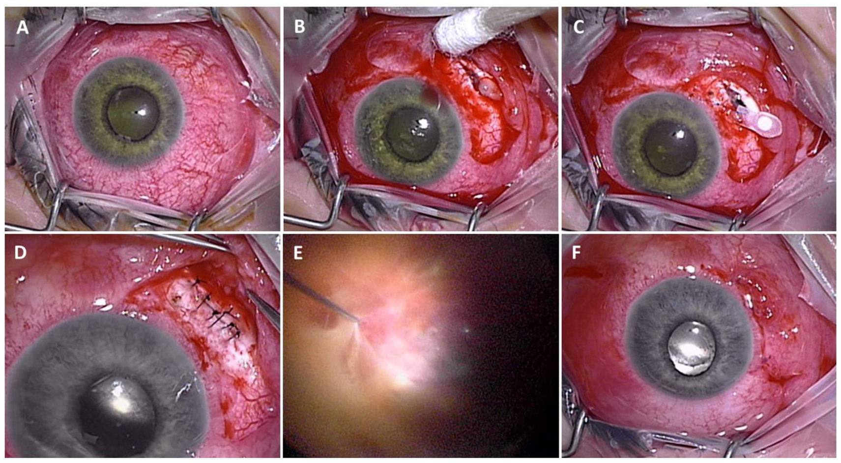
(A) Preoperative photograph of the left eye shows marked conjunctival injection without an obvious conjunctival defect. (B) Intraoperative photograph after limited conjunctival peritomy reveals an open sclerotomy site with prolapsed vitreous and loose and broken sutures. (C) Explanted fluocinolone acetonide insert after removal from the sclerotomy site. (D) Intraoperative photograph shows closure of the original sclerotomy using 6 interrupted 8-0 nylon sutures. (E) Intraoperative view during 25-gauge pars plana vitrectomy demonstrates removal of dense vitreous membranes and vitritis secondary to endophthalmitis. (F) Appearance of the left eye at the conclusion of surgery.
Intraoperative examination of the conjunctiva revealed no obvious defects or wounds. A limited conjunctival peritomy was performed in the inferonasal quadrant, where prolapsed vitreous and 3 polypropylene sutures were found spanning an open sclerotomy incision measuring 4.5 mm in length (Figure 1B). The scleral wound did not extend beyond the sutures spanning it, remaining patent with clean surgical margins through its entire length and no evidence of necrosis. One of the sutures was broken, and the other 2 were loose. The sutures were excised, and the fluocinolone acetonide implant was easily explanted (Figure 1C). The prolapsed vitreous was excised and the sclerotomy closed using 6 interrupted 8-0 nylon sutures (Figure 1D). A standard 3-port, 25-gauge PPV was then performed, removing densely opacified vitreous and membranes (Figure 1E). A single large retinal tear was found superiorly and was barricaded with endolaser. A partial fluid–air exchange was performed to facilitate the administration of IVT ceftazidime (2.25 mg in 0.1 mL) and vancomycin (1.0 mg in 0.1 mL). The conjunctiva was closed using interrupted 7-0 polyglactin sutures (Figure 1F).
Vitreous cultures grew Haemophilus influenzae that was sensitive to both ceftazidime and vancomycin. At 1 month, VA improved to 20/200; however, the patient’s eye remained hypotonous, with an intraocular pressure of 4 mm Hg, presumably due to cyclitic membranes or ciliary body shutdown. Subsequently, an inferior macula-involving retinal detachment with proliferative vitreoretinopathy developed and was treated with vitrectomy, membrane peeling, and silicone oil tamponade. The retina remained attached under silicone oil, but the eye remained hypotonous, and VA decreased to LP. The patient was referred to a rheumatologist for a systemic workup, which was negative for autoimmune or inflammatory conditions.
Conclusions
IVT fluocinolone acetonide implants have proven effective for the treatment of chronic noninfectious posterior uveitis and cystoid macular edema. The device is surgically implanted in the posterior segment, while fluocinolone acetonide (0.19 mg, Iluvien, Alimera Sciences) and fluocinolone acetonide (0.18 mg, Yutiq, Alimera Sciences) are injected into the vitreous cavity. Despite their ability to control noninfectious uveitis, IVT fluocinolone acetonide implants are associated with several complications, including cataract progression, elevated intraocular pressure, glaucoma, choroidal detachment, temporary decrease in VA, exacerbation of intraocular inflammation, retinal detachment, vitreous hemorrhage, vitreous loss, wound dehiscence, and endophthalmitis.1–3
Persistent scleral wound leakage after IVT corticosteroid therapy has been associated with previous ocular surgery and autoimmune disease. A case of scleral melt with active leakage after fluocinolone acetonide IVT implantation was reported; however, the scleral melt was attributed to psoriatic arthritis rather than the implant itself. 4 In another report, a patient required a scleral patch graft after a second implantation, although the etiology of the wound leak was not specified. 5
Persistent wound leakage may occur due to sclerotomy incompetence after IVT injections or sutureless vitrectomy. It has been theorized that scleral thinning after surgical wounds can lead to persistent wound leakage. IVT fluocinolone acetonide implantation may also contribute to scleral thinning, particularly in cases of reimplantation and those with long-term follow-up. 6 Furthermore, recent reports have suggested that IVT dexamethasone injections may be associated with wound leakage, particularly in patients with systemic exposure to corticosteroids or a history of past vitrectomies or repeated injections at the same site. 7 Because the sclera does not heal by primary intention but through fibrous tissue repair, repeated incisions or injections may affect wound integrity, predisposing to leakage. Patch grafts, including human pericardium, may reduce the likelihood of suture erosion or implant extrusion in cases of compromised scleral or conjunctival integrity. 8
Cases of hypotony and wound leakage have been reported after trabeculectomy or vitrectomy. A case series on glaucoma aqueous shunt implantations described 4 instances of persistent wound leaks, which resulted in severe complications including endophthalmitis, hypotony, suprachoroidal hemorrhage, and corneal decompensation. 9 An increased risk of sclerotomy leakage after vitrectomy has also been associated with younger age at the time of surgery, history of past vitrectomies, and vitreous base dissection. 10
Spontaneous dissociation of the IVT fluocinolone acetonide implant is another late complication of surgical implantation that may be attributable to suture breakage. In 1 such case, a patient presented with a new shadow in his vision 5 years after IVT fluocinolone acetonide implantation, and the drug pellet was found freely floating in the vitreous cavity. The pellet was suspected to have spontaneously separated from the anchoring strut, requiring removal via PPV. 11 Although poor wound healing was the most likely primary cause of the open sclerotomy in the present case, suture breakage may have contributed to the loss of structural integrity, thereby increasing the risk of delayed sclerotomy reopening.
Corticosteroid implantations such as fluocinolone acetonide (0.59 mg) are associated with a higher risk of endophthalmitis compared with other IVT injections. 12 It has been hypothesized that wound healing may be inhibited by local and systemic immunosuppression, thereby increasing susceptibility to persistent wound leakage and delayed endophthalmitis. In 1 report, a patient developed endophthalmitis 12 days after an IVT dexamethasone injection (0.7 mg, Ozurdex, Allergan) due to a nonhealing conjunctival defect at the injection site. The endophthalmitis was attributed to a full-thickness scleral defect that was discovered after conjunctival dissection. 13
Prolonged hypotony has been reported after IVT fluocinolone acetonide implantation performed with a keratome blade. 14 Persistently open sclerotomies can increase the risk of endophthalmitis, and implementing techniques such as closing the wound with multiple permanent sutures may help minimize such risks. 15
Wound leaks in the conjunctiva and cornea are traditionally diagnosed in the office using the Seidel test due to their superficial location. 16 However, in cases of hypotony, there may be insufficient aqueous flow to produce a positive Seidel test without applying pressure directly to the eye, which is generally not recommended. The patient in the present case had no obvious conjunctival defects or subconjunctival fluid to suggest the presence of a scleral wound leak. Exploratory peritomy was therefore required to identify the open scleral incision, which was hidden under the severely injected conjunctiva. Microbreaks in the conjunctiva may have allowed bacterial entry into the open sclerotomy with subsequent intraocular dissemination. Vitreous prolapse plugging the sclerotomy likely prevented the flow of detectable fluid into the subconjunctival space. The vitreous prolapse may also have contributed to the superior retinal tear. Only 3 permanent sutures were found at the large sclerotomy site, and these were either broken or loose. Poor wound healing related to corticosteroid exposure, elevated intraocular pressure from steroid-induced glaucoma, and an insufficient number of sutures used to close the large incision may all have contributed to the sclerotomy’s reopening.
To prevent reopening of the sclerotomy, vitreous prolapse, and subsequent endophthalmitis, we recommend creating a smaller sclerotomy or using additional permanent sutures to ensure tight wound closure. Although tight scleral closure may induce astigmatism, this effect may lessen over time. 17 The IVT fluocinolone acetonide implant measures 3 mm × 2 mm × 5 mm 18 and is typically inserted through a 3.0-mm to 3.5-mm incision. 19 If a larger sclerotomy is created, additional permanent sutures should be used to secure the wound and prevent delayed reopening.
Early detection of endophthalmitis is critical for effective management and visual prognosis. 20 Preoperative counseling before IVT fluocinolone acetonide implantation should include not only the risk of acute endophthalmitis but also a discussion about the possibility of delayed endophthalmitis occurring years after implantation. Patients should be informed of the warning symptoms of endophthalmitis, including decreased vision, redness of the eye, and pain. 21 In particular, in cases involving fluocinolone acetonide or other corticosteroid implants, it is important to counsel patients about the potential risk of delayed or impaired wound healing and advise against rubbing the eye, even long after the implantation procedure.
IVT fluocinolone acetonide implants are effective for managing noninfectious uveitis, but their use is associated with significant risks, particularly in cases requiring reimplantation. Continued refinement of implantation techniques and the development of alternative treatment strategies are essential to mitigate these risks and optimize patient outcomes.
Footnotes
Ethical Approval
This case report was conducted in accordance with the Declaration of Helsinki. The collection and evaluation of all protected patient health information was performed in a Health Insurance Portability and Accountability Act–compliant manner.
Statement of Informed Consent
Informed consent was not required for the publication of this paper due to the use of de-identified patient data.
Declaration of Conflicting Interests
The authors declared no potential conflicts of interest with respect to the research, authorship, and/or publication of this article.
Funding
The authors received no financial support for the research, authorship, and/or publication of this article.