






Abstract
Keywords
Introduction
Central retinal artery occlusion is a rare form of ischemic stroke, with an estimated incidence of 1.3 to 1.8 per 100 000 person-years. 1 The cilioretinal artery, a branch of the posterior ciliary artery, supplies the fovea in approximately 15% to 30% of the global population. 2 While these events are typically associated with cardiovascular risk factors, we report a case of an isolated non-arteritic cilioretinal artery occlusion in a perimenopausal woman following the initiation of subcutaneous testosterone pellet therapy.
Case Report
A 48-year-old woman presented to the emergency department with an acute central “gray spot” in her left eye that began 6 hours before presentation. Her past medical history was significant for retinal migraines, hormonal acne, and perimenopausal low testosterone levels. Her medications included spironolactone and subcutaneous testosterone pellets started 1 month before presentation. Visual acuity (VA) was 20/20 OU, and intraocular pressures were normal in both eyes. Pupillary examination revealed a sluggish pupil with a relative afferent pupillary defect in the left eye. Confrontation visual field testing demonstrated a scotomatous defect in the left eye, while color vision was intact in both eyes. No focal neurologic deficits were identified.
Anterior segment examination was unremarkable in both eyes. Dilated fundus examination of the right eye was normal. In the left eye, intraretinal whitening of the inferior macula was observed along the distribution of the cilioretinal artery, with foveal sparing. Optical coherence tomography (OCT) of the right eye was normal, while OCT of the left eye demonstrated thickening and hyperreflectivity of the inner retinal layers in the inferior juxtafoveal region (Figure 1).

Initial optical coherence tomography of the left eye demonstrating thickening and hyperreflectivity of the inner retinal layers in the inferior juxtafoveal region.
A diagnosis of non-arteritic cilioretinal artery occlusion of the left eye was established, and the patient was admitted to the Neurology service for a comprehensive stroke work-up. She was not a candidate for thrombolytic therapy due to presentation outside the therapeutic window. Neuroimaging, including contrast-enhanced magnetic resonance imaging of the brain, contrast-enhanced magnetic resonance angiography of the neck, and transthoracic echocardiography, was unremarkable. Laboratory evaluation, including complete blood count, comprehensive metabolic panel, prothrombin time, international normalized ratio, activated partial thromboplastin time, hemoglobin A1c, erythrocyte sedimentation rate, lipid panel, thyroid-stimulating hormone, lupus anticoagulant panel, and homocysteine, was within normal limits. An additional hypercoagulability work-up, including plasminogen activator inhibitor antigen, factor V Leiden mutation, and factor II (prothrombin G20210A) mutation, was negative. C-reactive protein was mildly elevated at 1.7 mg/dL. Serum testosterone was elevated at 228.0 ng/dL (normal range, 8.4–48.1 ng/dL for adult women aged 20–49 years).
In the absence of other identifiable stroke risk factors, the Neurology service attributed the cilioretinal artery occlusion to the patient’s recent testosterone supplementation. The Gynecology department was consulted regarding removal of the subcutaneous pellets; however, removal was not possible. The patient was therefore discharged on medical management, including aspirin, antiplatelet therapy, and a high-intensity statin, for the anticipated duration of spontaneous pellet dissolution (approximately 6 months).
At the 10-day follow-up, VA remained 20/20 OU, with subjective improvement in the left eye scotoma. Dilated fundus examination of the left eye demonstrated decreased intraretinal whitening (Figure 2, A and B). OCT of the left eye showed decreased hyperreflectivity and edema of the inner retinal layers (Figure 2C). Fundus autofluorescence (FAF) of the right eye was normal, while FAF of the left eye showed inferior juxtafoveal hyperautofluorescence (Figure 2, D and E). Fluorescein angiography of the left eye demonstrated mild leakage along the cilioretinal artery distribution without a persistent perfusion defect (Figure 3).
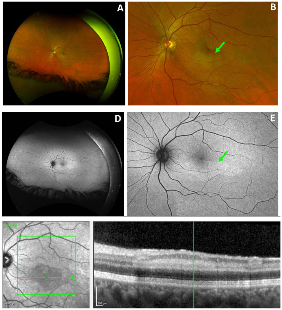
(A) Fundus photograph at 10-day follow-up demonstrating decreased intraretinal whitening in the left eye. (B) Magnified view highlighting residual intraretinal whitening of the inferior macula along the cilioretinal artery, with sparing of the fovea. (C) Optical coherence tomography of the left eye demonstrating reduced edema and persistent hyperreflectivity of the inner retinal layers in the inferior juxtafoveal region. (D, E) Fundus autofluorescence of the left eye demonstrating inferior juxtafoveal hyperautofluorescence along the distribution of the cilioretinal artery.

Fluorescein angiography of the left eye demonstrating (A) early-phase and (B, C) late-phase images with mild leakage along the cilioretinal artery distribution, without evidence of a persistent perfusion defect.
At the 6-week follow-up, VA remained 20/20 OU. The retinal whitening in the left eye had completely resolved, and OCT demonstrated further improvement in inner retinal hyperreflectivity.
Conclusions
Overall, cilioretinal artery occlusions are rare, comprising 5.3% to 7.1% of all retinal artery occlusions. 3 Cilioretinal artery occlusions have been further classified into 3 categories: non-arteritic cilioretinal artery occlusion occurring in isolation, non-arteritic cilioretinal artery occlusion associated with central retinal vein occlusion (CRVO), and arteritic cilioretinal artery occlusion associated with giant cell arteritis. 4
Testosterone replacement therapy is used for a wide variety of conditions, including hypogonadism, gender-affirming care, and low testosterone levels. 5 During menopause, declining ovarian and adrenal function leads to reduced androgen production. In this setting, testosterone replacement therapy has become increasingly used in peri- and postmenopausal women for symptoms associated with low testosterone, including female sexual dysfunction. 6 Reported adverse effects of testosterone therapy include erythrocytosis, decreased high-density lipoprotein cholesterol, and secondary polycythemia.5,7,8
The relationship between testosterone replacement therapy and thromboembolic risk remains inconclusive, with differing evidence for venous and arterial events. Venous thromboembolism includes deep vein thrombosis and pulmonary embolism, whereas arterial events encompass myocardial infarction, stroke, and retinal artery occlusion (RAO). Systemic reviews and meta-analyses, including those by Cannarella et al,9–11 suggest that randomized clinical trial data do not demonstrate an increased risk of arterial thrombosis, stroke, myocardial infarction, venous thromboembolism, pulmonary embolism, or mortality associated with testosterone therapy.
In contrast, observational studies have reported an association between testosterone replacement therapy and a reduced risk of arterial thrombotic events, myocardial infarction, venous thromboembolism, and mortality. 9 However, these findings are difficult to extrapolate to the present case, as existing studies have been conducted exclusively in male populations. Data regarding retinal vascular occlusions and hormone therapy in women are limited and have largely focused on oral contraceptives, which have not demonstrated an increased risk. 12 The risk profile in women, particularly those receiving testosterone supplementation for perimenopausal symptoms, remains poorly studied.
Select case reports have described venous thromboembolisms in patients on testosterone replacement therapy in the setting of underlying thrombophilia–hypofibrinolysis, including factor V Leiden heterogeneity, lupus anticoagulant, and elevated lipoprotein(a). 13 Notably, venous thrombosis (including CRVO) has been observed in both female and male patients on testosterone replacement therapy and previously undiagnosed thrombophilia–hypofibrinolysis.14–16 One proposed mechanism involves the aromatization of testosterone to estradiol. Glueck et al 15 hypothesized that estradiol-induced thrombophilia can exacerbate underlying familial thrombophilia, thereby increasing the risk of venous thromboembolism.
To date, few cases of arterial thromboses secondary to testosterone replacement therapy have been reported. Review of the literature revealed cases of amaurosis fugax, renal artery thrombosis, myocardial infarction, and small-vessel thrombosis of the dermal and epidermal arteries.14,17 All reported cases involved male patients in the third to fifth decades of life receiving either recreational or prescribed testosterone therapy. Hypercoagulability evaluation was unrevealing in 3 cases, while the patient with amaurosis fugax was found to have heterozygosity for multiple thrombophilias. 14
Notably, cilioretinal artery occlusions may occur concurrently with CRVO, suggesting that a primary arterial thromboembolic event may not always be required for their pathogenesis. A transient venous thromboembolic event causing elevated venous pressure can result in secondary hypoperfusion of the cilioretinal artery, given the relatively lower relative perfusion pressure of the choroidal vascular system from which it originates. 18
Overall, the European Academy of Andrology recommends assessing both personal and family history of venous thromboembolism and arterial disease before initiating testosterone replacement therapy. 19 The guidelines further emphasize screening for hypercoagulable states based on individualized risk stratification. In addition, transdermal formulations (eg, gels) are favored over pellet implants owing to their reversibility and lower risk of extrusion and infection. 19
In this patient, a negative stroke and hypercoagulability work-up suggested that recent testosterone pellet supplementation may have contributed to her presentation. Her history of retinal migraine may have acted synergistically, as several large studies have demonstrated a significantly increased risk of retinal vascular occlusion, including RAO, in patients with migraine.20–22 Notably, migraine with aura carries a greater risk than migraine without aura. 21 While existing literature suggests weak associations between testosterone replacement therapy and thrombotic events, this case highlights a potentially increased risk in female patients, particularly those with additional vascular risk factors, such as retinal migraine.
In conclusion, this report describes a novel case of testosterone-associated cilioretinal artery occlusion in a perimenopausal woman with a negative thrombophilia–hypofibrinolysis work-up. Before initiation of testosterone therapy, patients should be counseled and appropriately screened for hypercoagulable states and a history of migraine, given the potential for increased risk of both venous and arterial thromboembolic events.
Footnotes

Ethical Approval
Institutional review board approval was not required for this case report in accordance with our institutional policies. Informed and written consent was obtained from the patient described in this case report.
Statement of Informed Consent
Written informed consent was obtained from the patient for the publication of this case and any accompanying images. The patient reviewed the manuscript, confirmed understanding, and provided voluntary consent for participation. All identifying information has been omitted to protect patient confidentiality.
Declaration of Conflicting Interests
The authors declared no potential conflicts of interest with respect to the research, authorship, and/or publication of this article.
Funding
The authors received no financial support for the research, authorship, and/or publication of this article.