
Abstract
Keywords
Introduction
Sclerochoroidal calcification is an uncommon, typically benign ocular condition characterized by calcium deposition within the sclera, with secondary involvement of the choroid. First described in a patient with pseudohypoparathyroidism, this entity marked the initial recognition of its association with systemic disturbances of calcium and phosphate metabolism. 1 Since then, several systemic associations have been identified, primarily involving disorders that lead to metastatic or dystrophic calcification, including primary or secondary hyperparathyroidism, chronic kidney disease, Gitelman syndrome, Bartter syndrome, hypomagnesemia, and other abnormalities affecting calcium–phosphate homeostasis. 2 However, the majority of cases are often bilateral, idiopathic, and discovered incidentally in older adults during routine evaluation. 2
Case Report
A 66-year-old man who had been followed in our retina service for over 3 years for moderate nonproliferative diabetic retinopathy was incidentally found to have bilateral sclerochoroidal lesions during a routine visit. His medical history was extensive and notable for hypertension, diabetes mellitus diagnosed at age 40, prostate cancer treated with prostatectomy at age 50, renal cysts, and chronic kidney disease. His family history was notable for a son with renal cysts diagnosed at birth and later with maturity-onset diabetes of the young at age 19. Genetic testing in the son had revealed a stop codon variant (p.Trp171X) in the hepatocyte nuclear factor-1β gene. As part of this prior family-based evaluation, the patient himself had undergone genetic testing approximately 10 years earlier, which had confirmed the same pathogenic hepatocyte nuclear factor-1β variant.
Anterior segment examination was unremarkable, with best-corrected visual acuity of 20/20 in both eyes. Fundus examination revealed bilateral, well-circumscribed, mildly elevated, placoid-appearing yellow lesions located in the superotemporal region of both eyes (Figure 1, A and B). Multiple microaneurysms and dot blot hemorrhages were observed in the retinal periphery, consistent with nonproliferative diabetic retinopathy. Fundus autofluorescence demonstrated corresponding hyperautofluorescent foci (Figure 1, C and D), while optical coherence tomography through the lesions demonstrated irregular hyperreflective scleral elevations with overlying focal ellipsoid zone loss (Figure 1, E and F). No signs of choroidal neovascularization were present, and the lesions remained stable on serial imaging over a 2-year follow-up period.
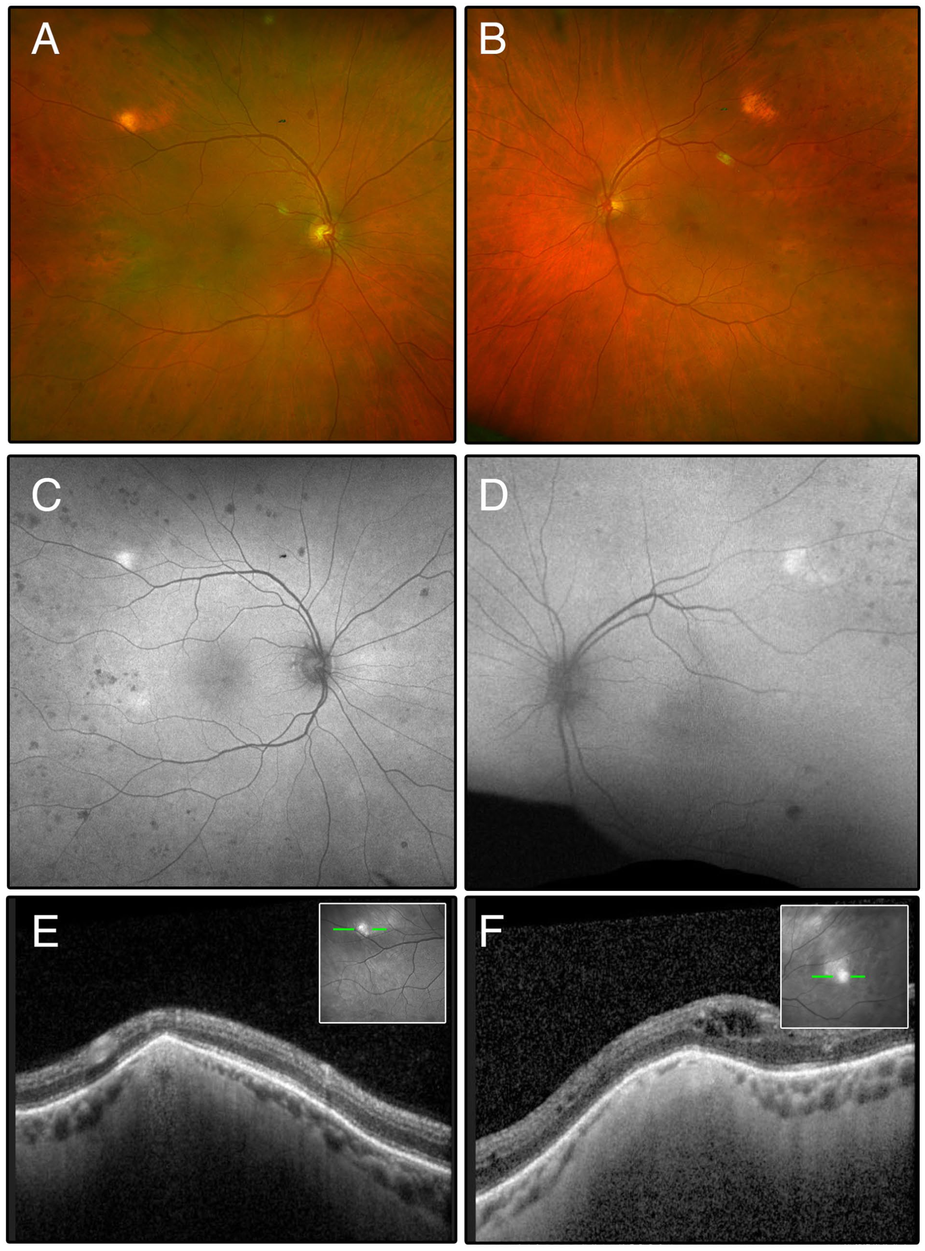
Multimodal imaging findings of bilateral sclerochoroidal calcification. (A, B) Pseudocolor fundus photographs demonstrating bilateral, well-circumscribed, placoid yellow lesions in the superotemporal fundus. (C, D) Fundus autofluorescence demonstrating corresponding hyperautofluorescent foci. (E, F) Optical coherence tomography through the lesions revealing irregular, hyperreflective scleral elevations with overlying focal ellipsoid zone disruption.
Conclusions
Hepatocyte nuclear factor-1β encodes a transcription factor expressed early in the development of the kidney, pancreas, liver, bile ducts, genital tract, and other organ systems. 3 Mutations in hepatocyte nuclear factor-1β may result in a broad spectrum of clinical manifestations, including renal malformations, tubulointerstitial disease, electrolyte wasting, early-onset diabetes, hyperuricemia, and genitourinary anomalies. 3 Although the clinical presentation is variable, renal dysfunction and electrolyte abnormalities, especially hypomagnesemia, are widely recognized as core features.3,4 Several studies have suggested hypomagnesemia as a useful biomarker to prompt genetic evaluation in adults with unexplained renal disease or cystic changes, with reported detection rates of hepatocyte nuclear factor-1β mutations approaching 19%.3,4 The mechanism underlying the hypomagnesemia is thought to involve dysregulation of the FXYD2 gene, which encodes a subunit of the Na⁺/K⁺-ATPase that is critical for magnesium reabsorption in the renal tubule. 3
Notably, hepatocyte nuclear factor-1β mutations have been associated with a Gitelman-like tubulopathy, reflecting overlapping electrolyte disturbances. 5 Given the established association between sclerochoroidal calcification and Gitelman or Bartter syndrome, a hepatocyte nuclear factor-1β-related tubulopathy represents a similar biologically plausible mechanism for the development of sclerochoroidal calcification. Hypomagnesemia may promote calcium pyrophosphate deposition by reducing its solubility, while chronic kidney disease with secondary hyperparathyroidism further predisposes patients to metastatic calcification within ocular tissues.4,5 Beyond its role in developmental and metabolic disorders, loss-of-function variants in HNF1B have also been implicated in the pathogenesis of prostate cancer, underscoring the gene’s broad functional importance. 6
We acknowledge that sclerochoroidal calcification has previously been associated with chronic kidney disease and metabolic derangements, both of which were present in this patient. However, in this case, these abnormalities occurred in the context of a well-characterized monogenic disorder known to directly cause renal dysfunction, electrolyte wasting, and early-onset diabetes mellitus through established molecular mechanisms. Notably, the patient’s diabetes is a recognized manifestation of hepatocyte nuclear factor-1β deficiency rather than an independent risk factor. In this regard, hepatocyte nuclear factor-1β deficiency syndrome may be considered analogous to other inherited renal tubulopathies, such as Gitelman and Bartter syndromes, which are known systemic associations of sclerochoroidal calcification due to shared downstream disturbances in electrolyte homeostasis.
Ocular manifestations of hepatocyte nuclear factor-1β deficiency syndrome have been infrequently reported but appear to involve multiple ocular structures. Prior reports have described associations with strabismus, nystagmus, myopia, coloboma, and Purtscher-like retinopathy. 7 In addition, microvascular retinal abnormalities, including diabetic retinopathy, have been observed in patients with early-onset diabetes mellitus. 7 The pathogenic hepatocyte nuclear factor-1β variant identified in our patient and shared with his son is characteristic of hepatocyte nuclear factor-1β deficiency syndrome and provides a unifying etiology for his renal cystic disease, metabolic derangements, diabetes mellitus, and prior genitourinary malignancy.
Thus, in patients presenting with bilateral sclerochoroidal calcification in the setting of early-set diabetes mellitus, renal cystic disease, or otherwise unexplained electrolyte or renal abnormalities, consideration should be given to multidisciplinary evaluation, including nephrology and genetics, for underlying renal tubulopathies such as hepatocyte nuclear factor-1β deficiency syndrome.
Sclerochoroidal calcification generally follows a fairly benign clinical course, with the majority of lesions remaining stable over time.2,4 Visual acuity is typically preserved unless lesions involve the macula or are complicated by choroidal neovascularization.2,4 Accordingly, routine observation with periodic imaging is appropriate for most patients, with closer follow-up warranted in cases involving the macula or demonstrating progression. Although many cases are idiopathic, systemic evaluation may be considered at the time of diagnosis, particularly in patients with bilateral disease or younger age at presentation, to assess for underlying disorders of calcium and electrolyte metabolism, including renal and endocrine conditions. 2
In summary, we report the first case linking sclerochoroidal calcification with hepatocyte nuclear factor-1β deficiency syndrome, a genetic disorder characterized by renal cystic disease, tubulopathy, electrolyte wasting, genitourinary abnormalities, and early-onset diabetes mellitus. This case expands the spectrum of extrarenal manifestations of hepatocyte nuclear factor-1β syndrome and underscores the importance of detailed systemic assessment and a multidisciplinary approach in the management of patients with sclerochoroidal calcification.
Footnotes
Ethical Considerations
Ethical approval was not required for this case report in accordance with institutional policy.
Consent for Publication
Informed consent, including permission for publication of all photographs and images included herein, was obtained before the procedure was performed.
Consent to Participate
Written informed consent for publication of clinical details and images was obtained from the patient.
Author Contributions
All authors meet the criteria for authorship, contributed to the conception and design of the work, drafting, and critical revision of the manuscript. All authors approved the final version for submission.
Funding
The authors received no financial support for the research, authorship, and/or publication of this article.
Declaration of Conflicting Interests
The authors declared no potential conflicts of interest with respect to the research, authorship, and/or publication of this article.