
Abstract
Keywords
Introduction
Retinopathy of prematurity (ROP) is a leading cause of visual impairment in preterm infants. Approximately 10% of infants are born prematurely worldwide, 1 and the incidence of ROP has been reported to be as high as 73% in infants born before 27 weeks of gestation. 2 Management depends on disease severity, with laser ablation of the avascular retina remaining a commonly used treatment for type 1 ROP. 1 Although cryotherapy was introduced in the 1980s for retinal ablation, it has largely been replaced by transpupillary laser photocoagulation due to improved structural and functional outcomes and fewer adverse effects.3–5 However, serous retinal detachment has been described as a complication following retinal ablative therapy with both cryotherapy 4 and laser photocoagulation.3,6–9 The reported incidence of this complication is approximately 4%. 10 Prophylactic topical steroids and cycloplegic agents are commonly used to mitigate this risk. 10 While prior studies have described the use of systemic corticosteroids to hasten the resolution of persistent serous retinal detachment following laser treatment for ROP, the optimal route of administration and recommended safe dosage remain unclear.9,11 In contrast, systemic intravenous corticosteroids are well-established in neonatal care for the management of bronchopulmonary dysplasia, with clearly defined dosing regimens.12,13 Herein, we report 2 cases (4 eyes) of premature infants who developed serous retinal detachments following laser therapy for ROP and describe the use of a standardized intravenous corticosteroid regimen that led to their rapid resolution.
Case Reports
Two consecutive cases of serous retinal detachment following laser therapy for ROP, initially managed by 2 pediatric ophthalmologists and subsequently treated by a single provider (MJW) at a large vitreoretinal surgical referral center, were identified. Institutional review board approval was not required for reporting these cases. The clinical course, management, and treatment outcomes are described.
Both infants developed bilateral serous retinal detachments after laser therapy for type 1 pre-threshold ROP. The mean gestational age at birth was 25 weeks, and the mean birth weight was 715 g. The mean postmenstrual age at laser photocoagulation was 47.5 (range, 40–55) weeks. Following the development of serous retinal detachments, both patients were treated with systemic corticosteroid therapy.
Case 1
An infant was born at 24 weeks and 4 days of gestation with a birth weight of 570 g. Initial ROP screening at 40 weeks’ postmenstrual age demonstrated zone II, stage 2 disease with pre-plus in both eyes. The infant was subsequently screened at 1- to 2-week intervals according to the Early Treatment for Retinopathy of Prematurity Study guidelines. 14 At 42 weeks’ postmenstrual age, progression to type 1 pre-threshold ROP was noted in both eyes, and bilateral intravitreal bevacizumab (0.625 mg) injections were administered. Screening examinations were continued until 55 weeks’ postmenstrual age, when reactivation of ROP was identified. Laser therapy was performed in the areas of nonperfused retina. Postoperatively, topical prednisolone acetate 1% and cyclopentolate hydrochloride 1% were initiated 4 times a day in both eyes.
One week later, bilateral serous retinal detachments were detected, more pronounced in the right eye. The patient was admitted, and intravenous dexamethasone therapy was initiated at a rate of 0.6 mg/kg every 6 hours under the close supervision of the neonatology intensive care unit team. Topical prednisolone acetate 1% and cyclopentolate hydrochloride 1% were continued at 4 times a day. Intravenous corticosteroid therapy was administered for 1 week, followed by an oral prednisone taper. Topical prednisolone was gradually tapered by 1 drop per week, and cyclopentolate was discontinued. Complete resolution of the serous retinal detachments was observed within 2 weeks after starting systemic corticosteroid therapy.
Case 2
An infant was born at 25 weeks’ gestation with a birth weight of 860 g. The infant underwent serial ROP screening at 1- to 2-week intervals in accordance with the Early Treatment for Retinopathy of Prematurity Study guidelines. 14 At 40 weeks’ postmenstrual age, the infant was found to have type 1 pre-threshold ROP in both eyes, and laser therapy was performed in the areas of nonperfused retina. Postoperatively, topical prednisolone acetate 1% and cyclopentolate hydrochloride 1% were initiated 4 times a day in both eyes.
One week later, bilateral vitreous hemorrhages developed, obscuring the view of the posterior segment. Upon referral to the vitreoretinal surgery service, B-scan ultrasonography demonstrated bilateral serous retinal detachments without evidence of vitreoretinal traction or rhegmatogenous components (Figure 1, A and B). The patient was admitted, and intravenous dexamethasone therapy at a rate of 0.6 mg/kg every 6 hours was started under close supervision of the neonatology team. Topical prednisolone acetate 1% and cyclopentolate hydrochloride 1% were continued at 4 times a day.
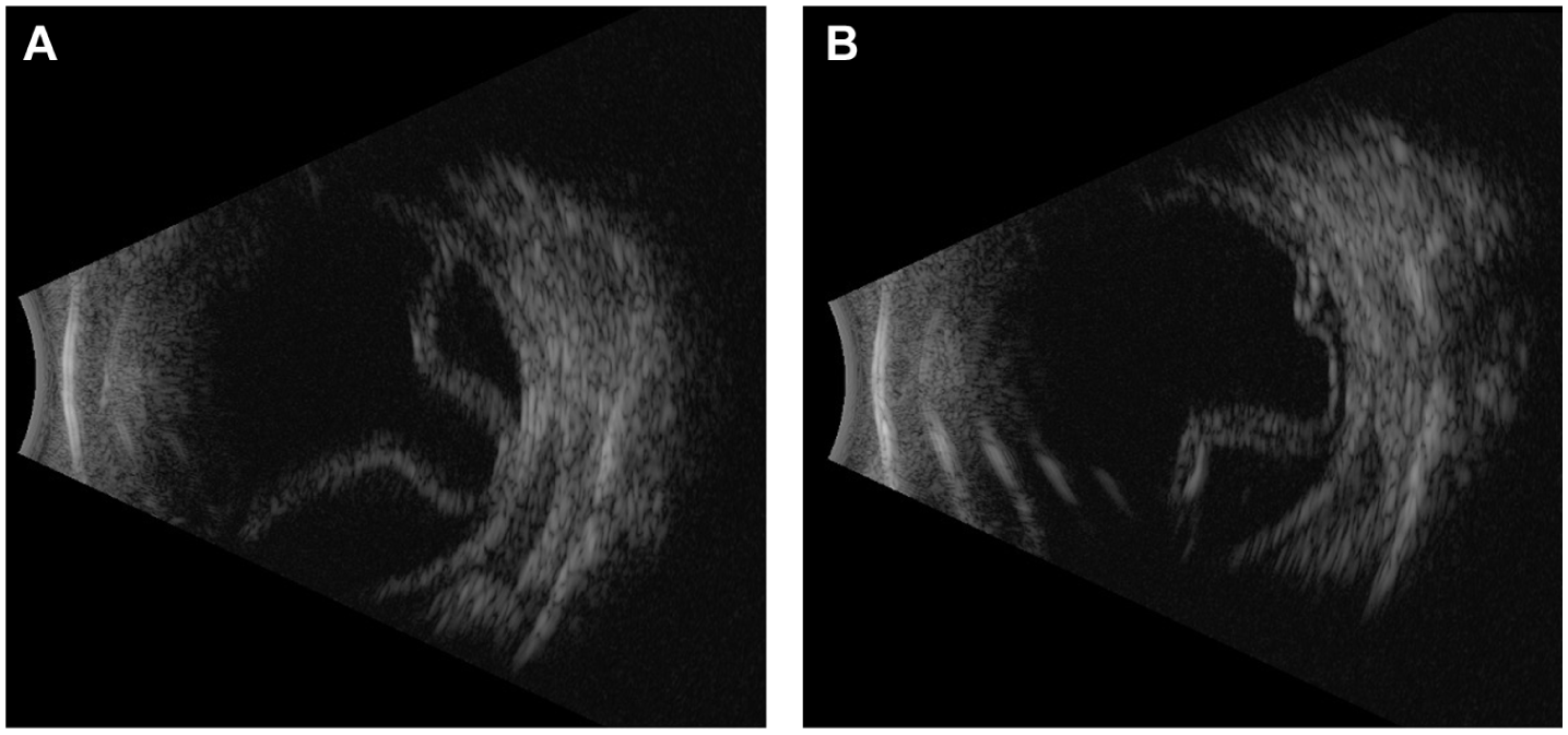
(A) B-scan ultrasonography of the right eye demonstrating serous retinal detachment following retinal laser ablation in the patient described in Case 2. (B) B-scan ultrasonography of the left eye demonstrating serous retinal detachment following retinal laser ablation in the same patient.
Intravenous corticosteroid therapy was administered for 1 week, followed by transition to an oral prednisone taper. Topical prednisolone was gradually tapered by 1 drop per week, and topical cyclopentolate was discontinued. Complete resolution of the serous retinal detachments was achieved within 4 weeks of starting corticosteroid therapy (Figure 2, A and B).
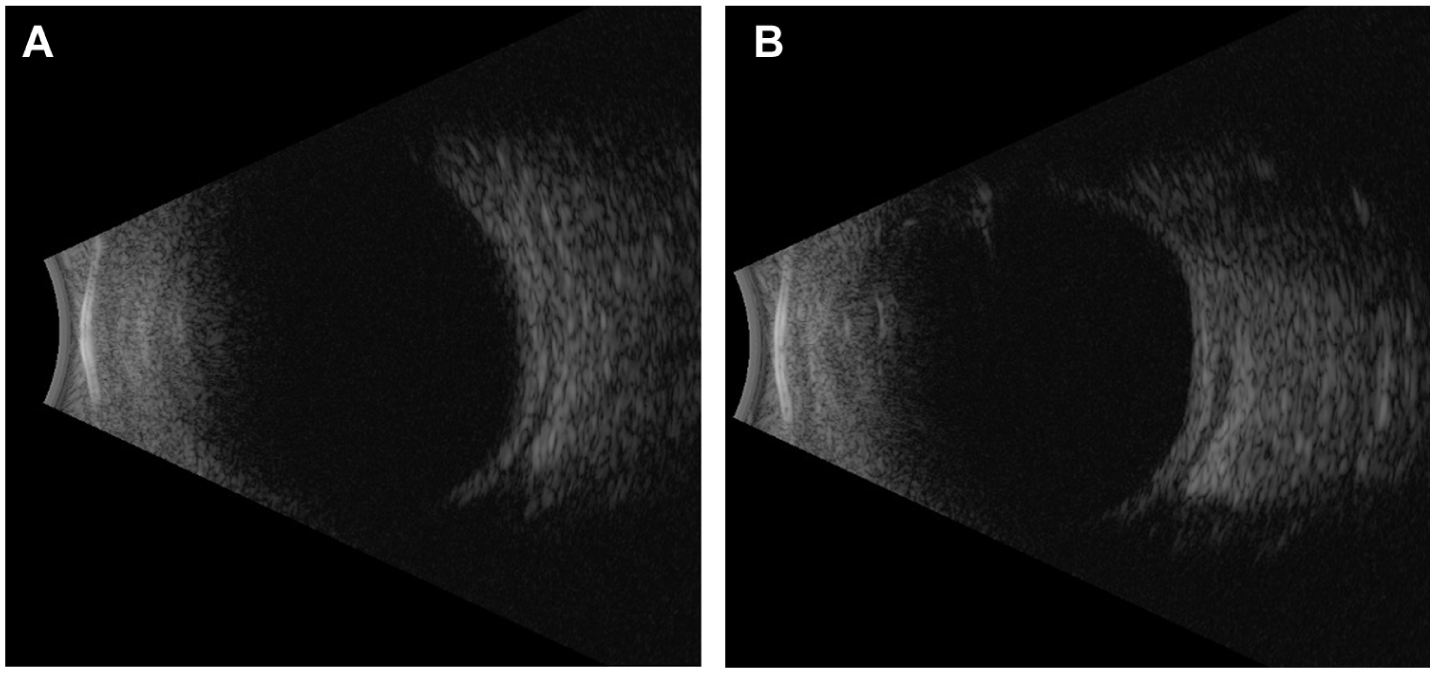
(A) B-scan ultrasonography of the right eye demonstrating resolution of serous retinal detachment following intravenous dexamethasone therapy in the patient described in Case 2. (B) B-scan ultrasonography of the left eye demonstrating resolution of serous retinal detachment following intravenous dexamethasone therapy in the same patient.
Conclusions
Serous retinal detachment and uveal effusion following retinal laser ablative therapy have been well described in the literature.15–21 The proposed mechanism involves transient breakdown of the blood–retinal barrier by the laser, which exceeds the energy-absorbing capacity of the retinal pigmented epithelium (RPE). Additionally, delayed choroidal perfusion induced by the laser may impair the ability of the choroidal circulation to effectively clear fluid. 8 This leads to accumulation of transudative fluid in the subretinal space, overwhelming the fluid transport functions of both the RPE and the choroid.8,15
The extent of exudative fluid accumulation appears to be dependent on the amount of laser delivered in each treatment session. The distribution of laser applications over multiple sessions has been associated with a decreased incidence of complications, such as retinal detachment, choroidal detachment, and angle-closure glaucoma. 19 As conventional ablative laser treatment for prethreshold and threshold ROP requires energy levels comparable to those used in proliferative diabetic retinopathy,14,22 infants undergoing retinal laser ablation are at an elevated risk of developing serous retinal detachment following treatment. These detachments are often subclinical and may resolve spontaneously without intervention.6,7,23 In fact, Zhang et al 24 reported prompt resolution of serous retinal detachments with observation or topical corticosteroid therapy alone. However, cases that fail to resolve may progress to chronic subretinal fluid accumulation, leading to pigmentary maculopathy, photoreceptor attenuation, macular scarring, and irreversible vision loss.6,9,11,23,24
Corticosteroids, administered either intravitreally or systemically, have demonstrated effectiveness in hastening the resolution of serous retinal detachments following laser ablative procedures.9,15,18,25 Although Moshfeghi et al 9 reported the effectiveness of systemic dexamethasone for this indication, they did not specify the dose administered. In contrast, Blair et al 11 described the use of oral prednisone at 1 mg/kg/day in combination with topical corticosteroids, with resolution observed after 4 weeks.
The use of dexamethasone for the prevention of bronchopulmonary dysplasia in premature infants has been well documented in the literature, with typical dosing ranging from 0.3 to 1.0 mg/kg/day for 3 to 10 days, followed by an appropriate taper.12,13 In the present cases, both infants were treated with intravenous dexamethasone at a dose of 0.6 mg/kg every 6 hours for 7 days under the careful supervision of a neonatal intensivist, followed by an oral prednisone taper. Neither patient experienced immediate adverse effects, and both demonstrated complete and rapid resolution of their serous retinal detachments.
In conclusion, we present 4 eyes of 2 premature infants who developed serous retinal detachments after laser ablation for type 1 pre-threshold ROP. While these detachments may resolve spontaneously, persistent cases can lead to permanent macular changes and irreversible vision loss. In our experience, systemic dexamethasone administered at 0.6 mg/kg every 6 hours for 7 days, followed by an appropriate taper, was associated with rapid and complete resolution of serous retinal detachments without immediate adverse effects. This regimen may represent a useful therapeutic option; however, larger studies are needed to establish its safety and efficacy.
Footnotes
Ethical Considerations
This case report was conducted in accordance with the Declaration of Helsinki. The collection and evaluation of all protected patient health information was performed in a Health Insurance Portability and Accountability Act–compliant manner.
Statement of Informed Consent
Informed consent was not required for reporting this case series in accordance with institutional policy and because no identifiable patient information is included.
Funding
The authors received no financial support for the research, authorship, and/or publication of this article.
Declaration of Conflicting Interests
The authors declared no potential conflicts of interest with respect to the research, authorship, and/or publication of this article.