
Abstract
Functional connectivity magnetic resonance imaging (fcMRI), a specific form of MRI imaging, quantitatively assesses connectivity between brain regions that share functional properties. Functional connectivity magnetic resonance imaging has already provided unique insights into changes in the brain in patients with conditions such as depression and pain and symptoms that have been reported by patients with psoriasis and are known to impact quality of life. To identify the central neurological impact of psoriasiform inflammation of the skin, we applied fcMRI analysis to mice that had been topically treated with the Toll-like receptor agonist, imiquimod (IMQ) to induce psoriasiform dermatitis. Brain insula regions, due to their suggested role in stress, were chosen as seed regions for fcMRI analysis. Mouse ear and head skin developed psoriasiform epidermal thickening (up to 4-fold, P < .05) and dermal inflammation after 4 days of topical treatment with IMQ. After fcMRI analysis, IMQ-treated mice showed significantly increased insula fc with wide areas throughout the brain, including, but not limited to, the somatosensory cortex, anterior cingulate cortex, and caudate putamen (P < .005). This reflects a potential central neurological impact of IMQ-induced psoriasis-like skin inflammation. These data indicate that fcMRI may be valuable tool to quantitatively assess the neurological impact of skin inflammation in patients with psoriasis.
Introduction
Although the clinical features of psoriasis have been known for centuries, the focus of research has mainly been on mechanisms of disease and the treatment of psoriatic skin and joint disease. Recently, the systemic comorbidities of psoriasis, including cardiac and metabolic abnormalities, have been more fully appreciated. 1 Objective assessment of skin pathological conditions (eg, psoriasis area and severity index scores) by physicians and researchers does not include assessment of quality of life (QoL), which is clearly compromised in patients with psoriasis. 2 -4 In particular, the impact of psoriasis on the central nervous system in terms of stress, depression, and other neuro-emotional factors are not taken into account in most clinical studies of psoriasis treatment.
Functional magnetic resonance imaging (fMRI) is an emerging technique for objectively measuring brain and neuronal activity by quantitatively assessing increased blood flow and blood oxygenation in confined regions of neural activation. 5 These 2 physiological changes alter the MR signal by changing the distribution of the magnetic field in the brain. Functional connectivity MRI (fcMRI), a frequently used fMRI technique, is defined as the temporal correlation between spatially remote neurophysiological events and is measured by correlating the neuroimaging time series in distributed brain areas. 6 In a number of disease states, including depression and Alzheimer disease, fcMRI has been used to gain new insights into neurological dysfunction. 7 -10 Although limited fMRI research in psoriasis has been conducted, one study used fMRI methods to show that the insular (Ins) cortex could play a role in differential visual image processing in patients with psoriasis. 11
Murine models of psoriasiform dermatitis such as those induced by interleukin-23 12 or imiquimod (IMQ) 13 have been instrumental in understanding the immunological mechanisms underlying the development of psoriasis in humans and would be ideal for serial examination of brain changes during the course of development of dermatitis. Technical challenges, chiefly the difficulty of achieving sufficient resolution in the much smaller brain of mice compared to human, are only now being overcome with the advent of MRI devices with sufficient magnetic field strength. This prompted us to determine whether advanced fcMRI methods could be applied to the objective measurement of brain changes in the skin of mice that were stimulated to develop psoriasiform changes through topical application of IMQ. Here, we show that striking fcMRI changes that are reversible by effective treatment are found in the brain of mice that have psoriasiform dermatitis even when histologic changes are not readily apparent. Moreover, fcMRI changes correlate strongly with pruritus as measured by scratching behavior, suggesting that MRI methods may be amenable to objectively measuring QoL parameters in clinical testing.
Methods and Materials
Animals and Physiological Monitoring
Experiments were performed on 24, naive, adult (24 ± 3 g), female, age-matched C57BL/6 mice (Charles River, Chicago, Illinois). All experimental procedures and protocols were approved by the Institutional Animal Care and Use Committee of the Medical College of Wisconsin (Milwaukee, Wisconsin). Animals were randomly divided into 4 groups (n = 6 each): wild-type control group, receiving 4 daily morning and 2 daily afternoon vehicle cream treatment; IMQ 1 day group, receiving only 1 morning IMQ treatment; IMQ 4 days group, receiving 4 daily morning IMQ treatment and 2 daily afternoon vehicle cream treatment; and IMQ 4 days + clobetasol group, receiving 4 daily morning IMQ treatment and 2 daily afternoon clobetasol treatment. All treatments described above were applied to the skin of both ears and part of the shaved head as demonstrated in Figure 1.
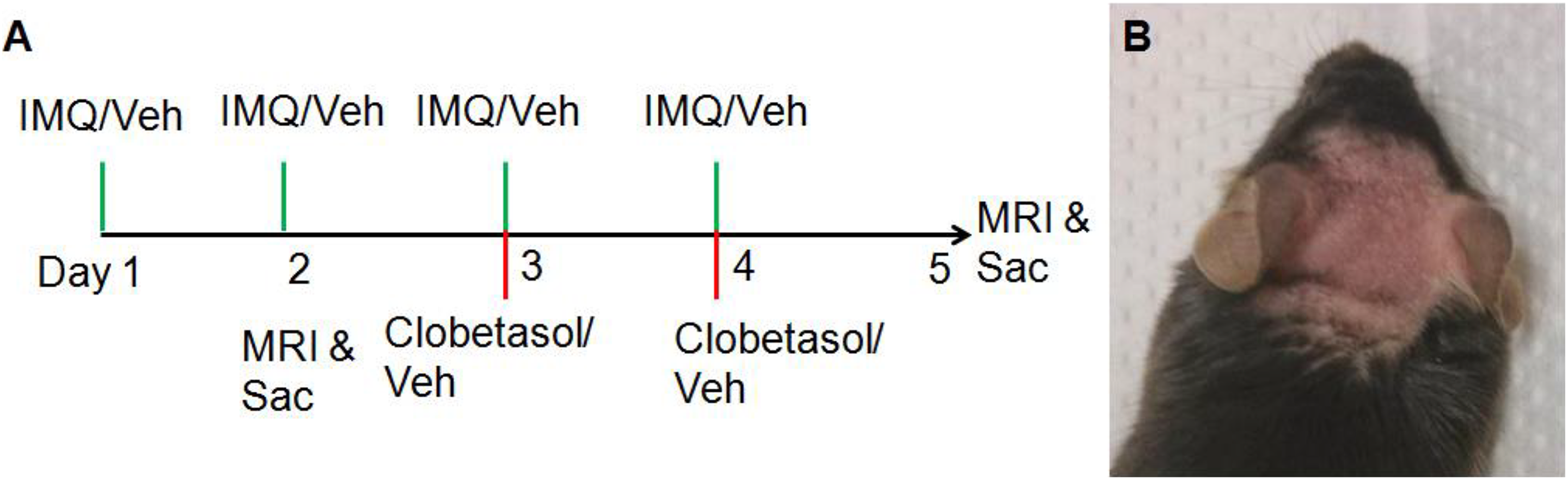
A, Time line of experimental design. Control group mice were treated with vehicle (Vanicream, Minnesota); IMQ 1 day group were treated with 1 daily topical application of IMQ (
On the scan day (day 2 for IMQ 1 day group and day 5 for all other groups), anesthesia was first induced with 2% isoflurane in an O2 and medical air mixture (3:7) followed by a polyurethane tube (PE50) placed subcutaneously (SC) on the back of animal for delivery of anesthetics during scans. Each mouse was then transferred into the scanner and placed on a custom-designed cradle fabricated with G-10 fiberglass material, with a magnetic susceptibility similar to air. The head of the animal was stabilized with a Bruker mouse MRI cradle, equipped with a nosecone and 3-point fixation system (tooth-bar and ear-plugs). During MRI data acquisition, as no stimulation was involved and anesthesia was adjusted to maintain a stable respiration pattern with rate within a tight range of 60 ± 10/min, animal head movement was minimal. Animal scans with movement of greater than 1 mm/2° in any frame were discarded. An MR-compatible small rodent air heater system (SAI Instruments Inc, Stony Brook, New York) was used to warm the animals inside the scanner. The animals’ core temperatures were monitored with a rectal thermometer and maintained at 37°C ± 0.5°C. A small animal monitoring system (model 1025; SA Instruments Inc) was used to monitor core temperature and respiratory rate. All physiological variables were kept close to normative levels throughout the duration of the experiment protocol and acquired using WINDAQ software (DATAQ Instruments, Akron, Ohio).
Magnetic Resonance Imaging Hardware Preparation and Imaging Protocol
All MRI data were acquired on a Bruker 9.4 T (Biospec Avance 94/31; Bruker, Karlsruhe, Germany) scanner with a Bruker linear transmission coil (T10325). Signal acquisition was performed using a self-designed 10 mm receive coil with low-noise amplifier and high sensitivity for signal detection.
All mice retained the ability to breathe spontaneously throughout the imaging session. Isoflurane was decreased to 0.5% 5 minutes after the SC bolus infusion of dexmedetomidine 0.1 mg/kg, and anesthesia was maintained with sedative dose of dexmedetomidine SC continuous infusion of 0.3 mg/kg/h throughout the rest of the scan. Image acquisition began once mouse vital signs reached steady state (as confirmed by physiological parameters). After a localization scan with a fast low angle shot (FLASH) 14 sequence, a relaxation enhancement rapid acquisitions (RARE) pulse sequence was applied to acquire the high-resolution anatomical scan as 15 contiguous coronal slices (center of the eighth slice in alignment with animal midline) with the following acquisition parameters: repetition time (TR) of 5000 millisecond, echo time (TE) of 8 millisecond, number of averages = 2, RARE factor of 8, field of view of 20 × 20 mm2, matrix size of 256 × 256, and slice thickness of 0.5 mm. After the high-resolution anatomical scan, high-order shimming was performed before high-resolution functional scans. Two sessions of functional scans were taken with a single-shot echo-planar imaging sequence. Using the same geometry as that of the anatomy, the scan parameters of each functional resting session were as follows: TR of 1000 millisecond, TE of 18 millisecond, single average, 600 repetitions, and matrix size of 96 × 96. In-plane resolution for functional scan was 208 × 208 μm. A B0 field map was always acquired before fc echo-planar imaging signal acquisition for each animal. All time series were temporally registered to the 6th frame for motion correction before being coregistered to the template image for group analysis. Scans with motion greater than 1 mm/2° in any frame were discarded.
The first 5 frames were always skipped in data processing to allow the MRI signal to stabilize. All data were coregistered to 1 set of representative mouse brain images, and regions of interest were hand-delineated with reference to the Allen brain mouse online atlas: http://atlas.brain-map.org/atlas?atlas=1#atlas=1&plate=100960260&structure=95&x=5280&y=3744.0003051757812&zoom=-3&resolution=16.75&z=5.
Pruritus Behavior Assessment
Additional mice (n = 6 for each group) were assessed for pruritic behavior. Instead of undertaking MRI scans, on day 2 for wild-type and IMQ 1 day groups and on day 5 for IMQ 4 days and IMQ 4 days + clobetasol groups, mice were housed individually and videotaped continuously for 20 minutes, during which the time spent on skin scratching with either front or hind paws of each mouse was manually recorded with a stopwatch.
Data Analysis and Statistics: fMRI
The Analysis of Functional NeuroImages (AFNI) software package (NIH, Bethesda, Maryland) was used for data analyses. The anatomical image set of 1 mouse was chosen as a reference template. After slice-timing correction and motion correction, the registration tool, FLIRT, 15 within the FSL software package (V5.0, FMRIB, Oxford, UK) was used to register all mouse fMRI images onto the reference template.
All fMRI data were slice time difference corrected, motion corrected, detrended, and frequency filtered (0.01-0.1 Hz) before the fc computation. Functional connectivity was assessed using seed-based analysis. Brain Ins regions, due to their suggested role in pain, emotion, stress, and self-awareness, were chosen as seed regions for fcMRI analysis. To obtain fc values as described previously, 16 the averaged preprocessed blood oxygen-level dependent signal time course from each seed region was used as reference that was cross-correlated (Pearson r) with each voxel’s preprocessed time course across the whole brain and represented as fc (r value) maps for individual animals. All r values in the individual subject map were transformed to Fisher Z scores as follows: the results were first subjected to Fischer R to Z transformation, then converted the standard Z scores after statistical analysis. Finally, all Z maps were clustered with minimum cluster size of 16 voxels (clusterwise P < .05, simulated result with AlphaSim command in AFNI). Group fc maps were generated using a 1-sample t test to examine both negative and positive values. Significantly correlated fc was identified using a threshold of P < .005.
Results and Discussion
Following daily topical application to skin, IMQ, a Toll-like receptor-7 agonist, induces T helper 17 cell pathway–dependent psoriasiform dermatitis in mice. 13 To determine whether IMQ-treated mice show changes in fc in the brain, we treated the shaved head region of mice with either topical IMQ or vehicle cream (Vanicream, Pharmaceutical specialties Inc, Rochester, MN) alone according to the time line illustrated in Figure 1A. An additional group of mice received topical clobetasol propionate, a potent topical steroid used clinically for human psoriasis, during the last 2 days of IMQ treatment as illustrated (Figure 1A). Compared to mice treated with vehicle alone, the skin of IMQ-treated mice developed erythema and scaling over the course of treatment (Figure 1B). Histological examination of the skin of treated mice revealed marked epidermal acanthosis, parakeratosis, and an inflammatory infiltrate in the dermis (Figure 2E-H). As expected, clobetasol treatment during the last 2 days of treatment reversed many of these histological features. Of note, however, 1 day of IMQ treatment did not induce apparent histological skin changes compared to vehicle alone treatment. Thus, daily topical IMQ treatment induced expected psoriasiform changes in treated mice within 4 days as previously reported by us and others.
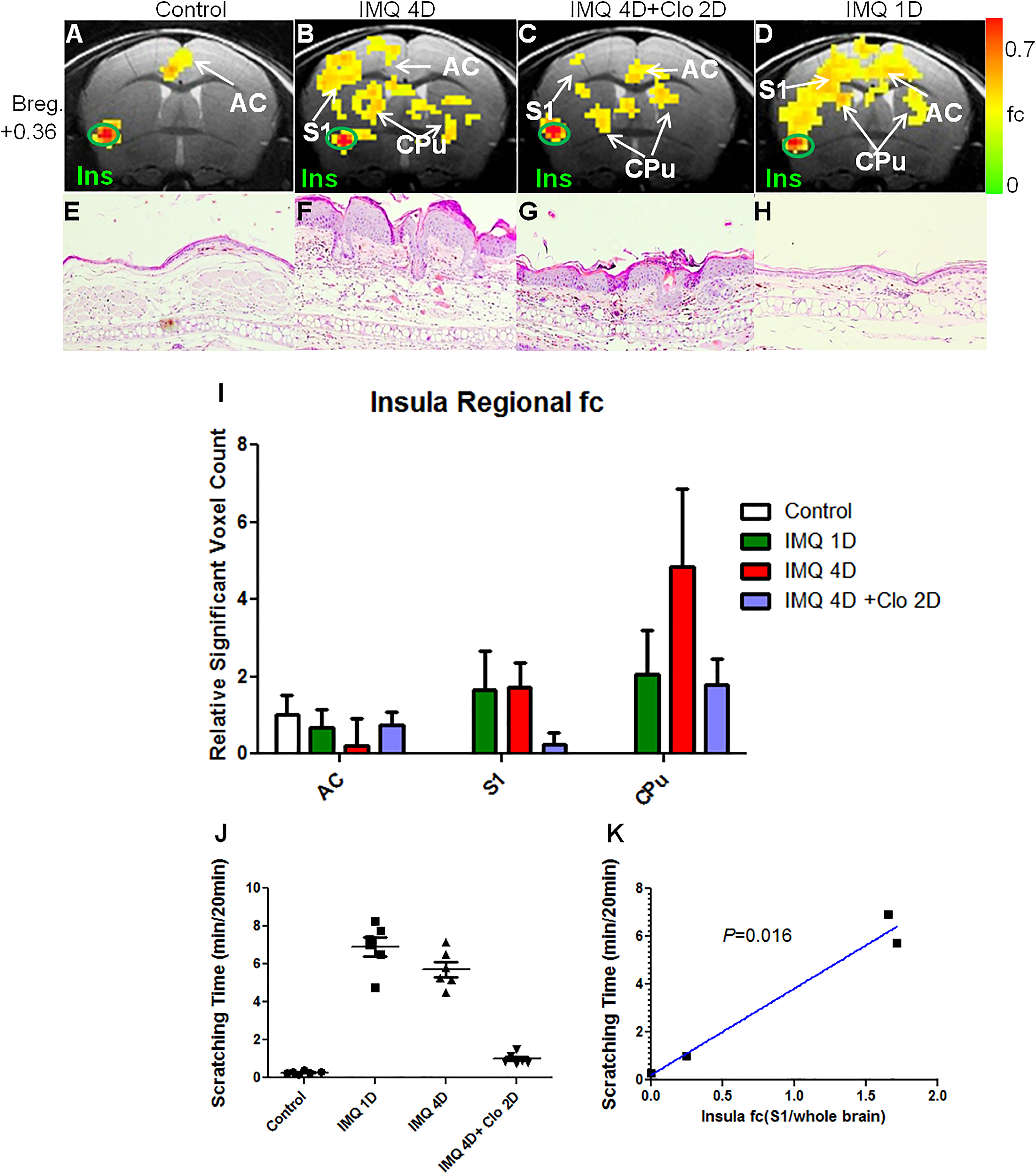
Mouse brain functional connectivity (fc) maps of the insular cortex. A, In control mice, Ins (green crosshair) has significant fc in anterior cingulate (AC). B, In IMQ 4 day (D) treated mice, Ins has significant fc in AC, primary somatosensory cortex (S1), and caudate putamen (CPu). C, In IMQ 4 day and clobetasol (Clo) 2 day treated mice, Ins has significant but decreased (compared to B) fc in AC, primary somatosensory cortex (S1), and caudate putamen (CPu). D, In IMQ 1 day treated mice, Ins has significant but decreased (compared to B) fc in AC, primary somatosensory cortex (S1), and caudate putamen (CPu). In Figures A-D, composite images from n = 6 mice are generated to show fcMRI changes in the regions of the brain. Two-way (region and group) ANOVA of fcMRI voxel counts demonstrated a significant group effect (F = 29.57, P < .001) with significant (P < .05) Bonferroni posttests in many comparison pairs such as S1 (control vs IMQ 4D/1D; IMQ 4D + Clo vs IMQ 4D/1D), CPu (control/IMQ 1D vs IMQ 4D; control vs IMQ 4D + Clo; IMQ 4D + Clo vs IMQ 4D/1D), and AC (IMQ 4D + Clo vs IMQ 1D). E-H, Histological images with H&E staining from ear skin of animal in matching group of A-D. In 4 daily IMQ treated group (F), mouse ear and head skin show psoriasiform epidermal thickening (up to 4-fold, P < .05) and dermal inflammation compared to that in wild type-group (E), which gets alleviated in the IMQ + clobetasol group (G). No significant changes can be detected in IMQ 1 day group (H), compared to wild type (E). I, Regional voxel count for significant insular fc for each animal. J, Scratching time spent by each animal during 20-minute videotaping. K, Correlation of group average relative insular fc in S1 with the group average of scratching time. AC indicates anterior cingulate; ANOVA, analysis of variance; IMQ, imiquimod; Ins, insula; MRI, magnetic resonance imaging.
Treated mice were assessed by fcMRI to assess changes in fc in the brain under each of the conditions described above (Figure 2A-D). Vehicle-treated (VT) mice showed baseline Ins fc signals mainly in the anterior cingulate (AC) region of the brain. After 4 daily IMQ treatments, mice showed fc in multiple discrete areas throughout the brain, including, but not limited to, the somatosensory cortex, AC cortex, and caudate putamen (CPu; P < .005). Clobetasol treatment attenuated fc signals in IMQ-treated mice toward baseline levels that were observed in the VT group. Of particular interest, after a single IMQ treatment, even without apparent histological changes in the skin, mice brains demonstrated Ins fc changes that were qualitatively similar to those in the IMQ 4 day treatment group.
Magnetic resonance imaging signal and intensity data can be readily quantified by region. For example, regional Ins fc was quantified and plotted (Figure 2I). Imiquimod treatment (both 1 day and 4 day groups) caused Ins fc increases in the primary somatosensory (S1) regions compared to the VT group (P < .05). Four daily IMQ treatments induced Ins fc increases in the caudate and putamen region (P < .05). Clobetasol treatment on days 3 and 4 markedly reduced the Ins fc increase in S1 and CPu (P < .05). Thus, fc changes in the brain that were induced by psoriasiform dermatitis in skin are largely reversible by effective skin therapy.
Scratching as a response to pruritus has been noted in several studies of IMQ-induced psoriasiform dermatitis in mice. 17,18 We sought to quantitatively measure behavior in mice in response to pruritus and to correlate the results with fcMRI findings. As shown in Figure 2J, within 20 minutes of video observation, shaved mice in the VT group spend very little time scratching their skin (mean 17 seconds). Mice in the IMQ 1 day and 4 day treatment groups, however, spent markedly more time scratching their treated skin (mean 6.9 and 5.7 minutes, respectively). Clobetasol treatments on day 3 and 4 decreased the scratching time compared to results in the IMQ 4 day group (mean 1.0 minutes).
To evaluate the correlation between brain fc change and scratching behavior at a quantitative level, a Pearson correlation analysis was performed with respect to the group average scratching time and group average relative ratio of Ins fc voxel counts in the S1 relative to that in the whole brain (Figure 2K). The relative ratio was acquired due to the possible variations in global brain responsiveness under different conditions. The relative Ins fc in the S1 was highly correlated with the scratching time (P = .016), suggesting that fcMRI change can be an objective and sensitive marker to assess pruritus that accompanies psoriasis in patients.
We used the Ins as a seed region because the Ins has been found to be heavily involved in conditions of pain and stress. It is also essential for emotion and self-awareness. In the VT group, the Ins is significantly correlated with the AC region, which is consistent with recent report from another group. 19 After IMQ treatment, increased Ins fc were found in brain regions heavily involved in modulation of stress, pain, emotion, and cognition. Specifically, the AC is known to participate in modulation of emotion, pain, attention, and motivation. 20 The primary somatosensory region (S1) is the major center that processes sensation, and the CPu region has been found to be critical for directed movement, emotion-hate, chronic stress, and threshold control. 21
Our study is notable for several reasons. First, reproducible fcMRI changes were detected in lightly sedated mice following even 1 day of IMQ treatment. At this time, histological changes are minimal, yet animals clearly display prominent scratching behavior that is comparable to that observed 4 days after IMQ treatment when histological changes are appreciable. As such, fcMRI is a highly sensitive surrogate marker of scratching behavior in response to pruritus.
Second, the anatomic regions of the strongest fcMRI changes are located in the CPu and the AC regions of the brain. The CPu controls many types of motor functions, including motor preparation, 22 motor learning, and regulation of thresholds for movement. 21 These neuronal regions and activities are presumably critical for initiation and maintenance of scratching behavior. The AC region is particularly involved for attention and when effort is needed to carry out a task such as early learning and problem-solving, 23 raising the possibility that changes in scratching behavior could involve attention reorientation and learning processes.
Limitations in our study include our focused analysis on the Ins region as opposed to other brain regions. Variability in fc in the primary sensory region (data not shown) with widespread connectivity throughout the brain even at baseline prevented us from using the primary sensory region as seed region in analysis. The secondary motor area was analyzed and showed connectivity to sensory, parietal, and other areas but was not analyzed with sufficient numbers of animals to present in this study. Future studies will include a full set of all region-to-region connectivity matrix analyses to show both global and regional connectivity.
The areas of the brain noted in our fcMRI studies are remarkably similar to areas of the human brain that govern the initiation of movement, presumably the initiation of motor functions to start scratching, as well as possibly areas of the brain that govern complex emotions such as depression and stress. Given the well-known associations of the latter two neuropsychological states in human psoriasis, we propose that fcMRI can be used to assess pharmacologic agents that can potentially make psoriatic patients with psoriasis more comfortable, even without the need to treat patient with immunosuppressive agents that may increase the risk of infection. Our study suggests that fcMRI can potentially be helpful in developing central nervous system-targeted agents that temper the feelings of isolation, stress, and even pain that have been vocalized by many psoriatic patients with psoriasis.
Footnotes
Declaration of Conflicting Interests
The author(s) declared no potential conflicts of interest with respect to the research, authorship, and/or publication of this article.
Funding
The author(s) disclosed receipt of the following financial support for the research, authorship, and/or publication of this article: This work was supported by a National Psoriasis Foundation Discovery Grant to STH and a National Psoriasis Foundation Research Fellowship to YZ.