
Abstract
Abstract
Biosensors are a very well cherished research topic and have found an inseparable status from clinical diagnostics in specific and society at large. As the name suggests, biosensors or biological sensors are devices which detect the presence of biological entities or their constituents and derivatives. The field started decades ago and has matured quite well since its inception. The most important performance factors that are associated with biosensors are sensitivity, specificity, and limit of detection. The remaining efforts of the biosensor research domain focus on miniaturization aspects of the sensors. The growing advancements in this field have evolved the technology of biosensors to cater to full-scale diagnosis on microchips, bedside diagnostics, reduced cost, and increased speed of diagnostics. Biosensors are characterized through many different aspects; for example, one way is to classify them on the basis of the type of bio-recognition step that they would utilize or another way can be based on the type of detection scheme that they may integrate, etc. Depending on the bio-recognition layer’s properties, biosensors can be cell based, nucleic acid probe based, antibody/antigen based, or aptamer based, while depending on the type of detection scheme, biosensors can be viewed as colorimetric sensors, optical sensors, electrochemical sensors, mechanical sensors, etc. There are some other parallel areas of research like microfluidics and microelectromechanical systems where one of the main applications lies in the biosensor domain. This review article discusses the various aspects of biosensors, from their design, realization, to testing, along with various detection strategies. The assembly includes fabrication strategies particularly for microchip technology-based biosensing solutions, microchannels, integration to microfluidics, etc., while categorization deals with various kinds and applications of different biosensors.
Introduction
The study of biosensors has always attracted innovators, scientists and technology developers. The main reason behind this is that biosensors can very effectively detect biological entities, some of which can threaten human beings. Biosensing finds wide-ranging applications in fields related to healthcare, air, water, food monitoring, monitoring of bio-threats, and homeland security. Additionally, the sister field, microfluidics, is also a very well-established domain of work and brings home solutions for targeting specific bio-entities, some of whose sizes are similar to the dimensions of micron-sized length scales. As biosensors have walked a long way in the past decades, many phases of evolution have occurred, shaping up this field very well, and today the technology to identify is mostly geared to early identification even prior to symptomatic effects showing up within human beings. The integration of biosensors with the microfluidic domain has really provided low-cost diagnostics and today detection challenges are being increasingly tackled so that early therapeutics can increase life expectancy within human subjects all over the globe.
There have been many reviews regarding biosensors and the related streams1–3 which discuss in detail the evolutionary aspects and also some latest state-of-the-art applications. In a similar manner, microfluidics and its applications have also been reviewed earlier to express the importance of the field.4, 5 The merger of biosensors and microfluidics has also been studied many times which deals with the types of biosensors and the different microfluidic platforms integrated with it for biosensing. 6 In this article, we will extensively discuss biosensors as in the current context of human healthcare, the technological advancements in biosensor fabrication techniques, and the merger of biosensors and microfluidics (sensing and sample handling many times are inseparable) along with the different detection techniques. Hence this review can be seen as an in-depth treatment of the subject with all the major aspects related to the field of biosensors including various types of biosensors, their applications, their fabrication aspects, different detection schemes, and the strategies used in modern sensing. The review will discuss the basic evolution of the subject in the past few years and will look into some latest research trends in the area.
Basic concepts in biosensors and related aspects
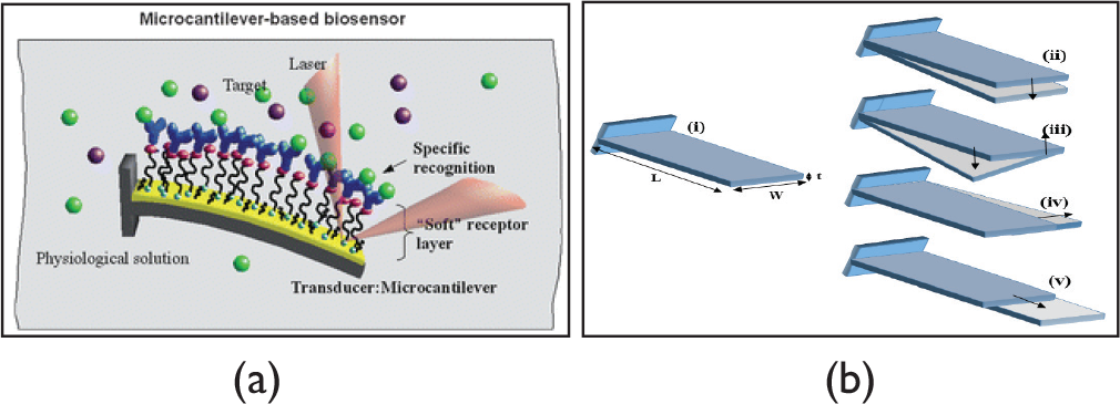
There are various entities associated with biosensors, the analyte which has to be detected, the substrate with a recognition element on which analyte sensing is performed, the way that the recognition element is immobilized to the sensor surface, the detection signal which comes out of the system, and the detection scheme through which the detection signal is quantized and interpreted. When an analyte has to be detected, the receptor element detects it and produces some kind of a signal (chemical/physical/optical) which is converted to some recognizable/quantized form that is detectable in nature.
Detection scheme in biosensors.
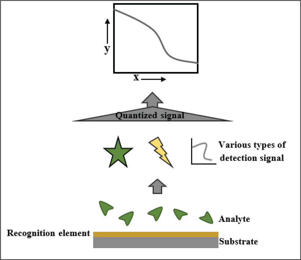
Figure 1 shows the detection scheme in biosensors in a step-by-step manner. The various analytes which can be detected through biosensors are either purely biological in nature, namely DNA, proteins, or cells, or non-biological in nature, namely ions (positive/negative, metallic/non-metallic), drugs, and several dissolved gases. 7 Majorly biosensors are divided into two categories, labeled detection or label-free detection. Labeled biosensors provide the advantage of being specific and highly sensitive in nature and work on the strategy of labeling some specific analyte with a selection and then reading out the quantity of the labeled analyte by a count of the labels. There can be two categories under this name, affinity biosensors, which utilize the affinity characteristics of the bio-recognition element (which can be antibody/antigens, DNA probes, aptamers, etc.) and the analyte (viz., antigen/antibodies, complementary DNA/RNA sequences, proteins, cells, ions, drugs, gases, etc.), and catalytic biosensors, which can detect the presence of the third chemical constituent generated from the binding of the recognition layer to the analyte of interest. While label-free biosensors deal with direct sensing and interpretation from the sensing data of various analytes on the detection platform, it does not need any secondary binding of a label through which detection will be carried out. These sensors are easy to use and serve as a rapid detection system but have a shortcoming of lower sensitivity or specificity of detection. These can effectively measure the difference in the chemical concentration of different analytes and other related aspects. In case of biosensors, the fabrication scheme utilized and the associated microfluidics deployed emerge as integral components of the sensing platforms, more so as sample handling is one of the basic elements in biosensor device planning and designing. Further, there are various detection schemes such as colorimetric (detection via observing color change through analyte addition on the recognition layer), optical (detection via observing change in optical characteristics), electrochemical (detection via observing change in electrical parameters) and mechanical detection (detection via change in mechanical parameters like mass) schemes which have to be integrated to incorporate the necessary transduction, making the measurement acquisition feasible. Different types of detection schemes require different types of measurement set-ups and measurement techniques. Their detailed study will be outlaid in forthcoming sections.
Biosensor fabrication techniques
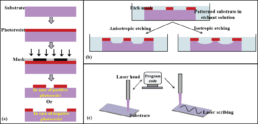
In the past 30 years, microsystem fabrication techniques have evolved as one of the most explored research areas due to the numerous applications that these systems find in fields such as pharmaceuticals, biomedical sensors, environment, defense needs, civilian applications, the energy domain, etc. More specifically, microsystem technologies for the biomedical sensing domain have attracted a lot of attention due to the evolution of point-of-care diagnostics for quick screening and investigation conducted at the bedside for patients. These devices necessitate specific characteristics in terms of high system integration efforts, efficient performance metrics, higher sensitivity values, shorter detection times, increased safety, lesser volume requirements, increased automation, reduced human intervention and handling, low overall costs, etc. 8 Manz et al. fabricated the first miniaturized total analysis system (μTAS) device which was able to perform sampling, transportation, separation, and detection. 9 There are various techniques which can be employed for fabricating biosensors. Among these, some processes are dedicated to the fabrication of the sensor architecture while others are used for devising sample handling and dispensing strategies for the platform. The fabrication techniques have evolved from traditional microelectronic processing techniques such as micromachining (laser micromachining), lithography and lift-off processes, chemical etching, electroplating, gas/plasma etching (more specifically deep reactive ion etching, DRIE), doping and implantation, thermal oxidation, wafer dicing, wire bonding, etc.
In the 1990s, micromachining and photolithography were the two most popular techniques for integrated circuit designing and microelectromechanical system (MEMS) fabrication. These processes were adapted to fabricate microstructures on glass and silica 10 for biomedical applications. As glass and silicon are quite expensive and sophisticated materials for substrates, alternate low-cost polymers like polymethylmethacrylate (PMMA), polyethylene, polydimethylsiloxane (PDMS), and SU-8 have also been heavily explored over the course of time. 11 The process of photolithography starts with a mask preparation step that may help in exposing the substrate to UV light selectively (as guided by the mask) and produces parts matching the end-user design requirements. Initially, the substrate is spin coated with the photoresist of various thicknesses using different grades of resists and is also controllable by spin speeds. After this, depending on the various-attained thicknesses of the photoresist film the substrate is pre-baked for various time durations following which the coated substrate is exposed to UV with a photo-mask for set time durations. After exposure, the baking can be done depending upon the type of photoresist used and subsequently the development process is done for 10–15 s in a developer solution. There are two different types of photoresist materials, one is positive-tone photoresist, in which the portions which are exposed to UV light dissolve away, and the other is negative-tone photoresist, where the portions which are exposed to UV light cross-bond and remain after the development process. 12 There are various enhancements to the existing photolithography technique which increases the process’s capability and also some other parallel techniques which make the MEMS process robust. Additionally, it’s pertinent to mention that most processes associated with photolithography are cleanroom processes which are carried out in a certain controlled manner for the processes to work well and deliver fully.
Apart from photolithography, micromachining is the other micro-fabrication technique which is the add-on used to subtractively or additively contribute to the wafer-level thickness to build MEMS architectures. The micromachining processes are comparatively robust in nature and do not require cleanroom facilities necessarily. Among the variety of micromachining processes, chemical etching (wet or dry) or laser micromachining processes is the one which is used in micro-fabrication.13–15 Chemical etching is the process of material removal through a chemical reaction. The substrate is dipped in an etchant solution and the portions which are directly exposed to the etchant solution get chemically modified and get dissolved/removed. Chemical etching can be isotropic or anisotropic in nature depending upon the substrate/etchant combination which is used for carrying out micromachining. Isotropic etching is homogenous or uniform etching throughout the substrate over areas exposed to the etchant solution with some of the portions under-etched below the masking zones, while in anisotropic etching, etching stops at the specified slowest-etching crystal plane. Dry etching of the substrate can be carried out by Light Amplification by Stimulated Emission of Radiation (LASER) micromachining which is a material removal technique where the material is evaporated through direct interaction of high-speed laser with the substrate material.15, 16 The laser is fed with a coded program through a software-driven control with which a substrate is scribed. Figure 2 represents the basic schematic of the micro-fabrication processes.
(a) Photolithography steps, (b) wet etching on the photo-lithographically patterned substrate, and (c) laser micromachining.
Apart from the well-established conventional processes, there are many additional processes which have come up based on requirements of high volume production, etc. In this context, there exists a whole class of processes known by the name of soft lithography, 17 which is purely based on printing, molding, and replication of soft materials (e.g., elastomers like PDMS). This class of processes which also includes selective molecular printing and deposition is very routinely used for fabricating microfluidic and sensory microchips. 10 Soft lithography relies on a master mold which is normally realized by patterns of channels, etc., that are already fabricated through patterning, etching, bonding, and subsequent integration. 18 Along with 2-D micro-features, 3-D features are also realized using soft lithography techniques. For this purpose, 3-D soft lithography is realized through the generation of multilayers and 3-D microstructures. 19 Micro-milling is also being used in addition to 3-D soft lithography for generating a variety of complex 3-D structures with curved cross-sections. 20 The other non-lithography techniques which are emerging these days for micro-fabrication involve various low-cost instrumentation such as printers (inkjet/laserjet/solid ink),21, 22 cutting plotters, adhesive vinyl films (xurography), 23 micro-milling, 24 microwire moulding, 25 etc.
The various combinations of these techniques can be used to fabricate microchips, help build microfluidics on microchips and the related microchannel design, and other strategies devised for the micro-fabricated chips, etc. The following sections respectively discuss the stated techniques and their applications in the fabrication of various modules of the sensing platforms.
Microchip fabrication
Microchips are the fundamental blocks or units in the current paradigm of science and technology which are used heavily for biosensing. As stated in the earlier sections there are many techniques which can be deployed to develop microchips ranging from lithography to printing, milling, molding, and so on so forth. A variety of shapes like interdigitated electrodes (IDEs), 26 2-electrode 27 /3-electrode configurations, 28 or insulators 29 can be carved over specific substrates and these carved shapes are meant to serve some specific purpose in sensing and detection. The most common substrate which is used to devise electrodes for the MEMS application is the silicon substrate. 26 Silicon substrate, in general, is very expensive and its handling is sophisticated. Therefore the idea of carrying out sensing using silicon substrates is being slowly replaced by other alternates such as paper, 30 polymer, 14 glass, 31 composites, 32 etc. These alternates offer more robustness to the detection and they are extremely cost effective. The patterning carried out on these substrates can be through machine guidance (as in paper), soft lithography (as in polymers), etching, etc. (as in glasses and composites), and some other processes.
In addition, the microchips are not compulsorily required to be some micro-patterned structures devised on specific silicon or glass substrates with the capability to carry out detection; rather, it can also in some embodiments be a micro-level assembly of various chemical/biological entities 33 in solutions as well as on substrates to detect a particular analyte of interest, which can be observed through some external means. These assemblies then offer the desired level of sensitivity of detection with various means. In these cases, it is the direct detection of the analyte that is carried out.
Microfluidics
Microfluidics is an inherently integrated domain to biosensors. As all biosensors need a flow of the analyte in some manner at the microscopic length scale, the study of this area is highly relevant. Microfluidics is majorly classified into three different streams, that is, channel-based microfluidics, digital microfluidics, and paper-based microfluidics. Channel-based microfluidics comprises an enclosed channel in which the liquid moves under the effects of pneumatic pressure. This system comprises pumps, valves, and mixers connected to a microchannel in some way. Channel-based microfluidics can dispense the fluid in a continuous manner or in the form of droplets depending upon specific application requirements. Continuous flow is persisted for applications such as continuous particle capture, 14 separation, 34 micro-total analysis chip, 26 etc., where the complete sensing/detection is accomplished on the same platform. Micropumps are the most important fluid-dispensing devices for fluid-handling purposes. They are used for dispensing the fluid at a specified rate. There lie various mechanisms for actuation within micropumps, namely electrostatic, 35 piezoelectric, 15 pneumatic, 36 thermo-pneumatic, electromagnetic, 37 electro-hydrodynamic, magneto-hydrodynamic, etc. Out of these mechanisms, piezoelectric actuation has been considered to be the most widely applied actuation scheme for pumping of fluids at the microscopic length scale. 38 Various materials such as polymers, polymer-metal composites, silicon, etc., are used for the fabrication of micropumps. A variety of fabrication techniques such as laser-based micromachining, lithography, lamination techniques, chemical etching, etc., are used for devising the platform architecture for micropumps. Olsson et al. fabricated the first valve-less micropump using silicon-etching techniques. 39 Carrozza et al. fabricated piezoelectrically driven micropumps on polymer-brass composites using stereolithography. 40 Kant et al. fabricated a digitally controlled micropump on the PMMA substrate using laser micromachining 15 and wet chemical-etching processes. The design of the chamber and the type of fluid motions are also very important aspects in the designing of micropumps. Circular chambers have been the most common shapes which have been fabricated within micropumping architectures. Truong et al. have reported piezoelectric micropumps (with the circular chamber) using a polymeric lamination technique. 41 But there remains a cross-contamination issue in the circular chambers as the complete fluid cannot ooze out of a circular chamber and there is a pertinent problem of residuals within such architectures. Kant et al. have shown a design optimization technique to achieve a modified chamber shape 42 which ensures the complete oozing out of the fluid from the chambers, thus avoiding cross-contamination issues to a large extent. The switching action of the flows within microchannels are controlled through microvalves. 43 There can be active as well as passive microvalves. For active microvalves, the actuation mode can be integrated (electrostatic, 44 piezoelectric, 45 thermal, 46 magnetic, or smart materials capable of changing phases like shape memory alloys, etc. 47 ) or external (pneumatic source 48 or external magnetic field, etc.).
Droplet microfluidics is used for generating discrete droplets of the analyte as suspended in an immiscible solution. 49 This provides high-throughput analysis especially as it is able to produce nanoliter-size droplets for single-cell sensitive detection/analysis, etc. Prior to this several groups have utilized droplet microfluidics for generating microbubbles, 50 irregular particles, 51 and hollow microcapsules 52 and further utilized them in a wide range of applications such as controlled delivery of drugs, dyes, enzymes, 53 diagnostics, imaging, artificial biomolecule synthesis, etc. 54 The application of droplet microfluidics is also extended to provide a platform that is able to perform multiple parallel isolated reactions in one go (each microbubble consists of a different reaction mixture). 50
Paper microfluidics, on the other hand, is a cost-effective technology which is majorly utilized in colorimetric detection schemes due to its white background. Passive fluid transport due to capillary action is a major mechanism of fluid flow on paper. 55 Paper microfluidics removes the need for expensive external devices for facilitating fluid flow. Additionally, usage of paper reduces the need for several physical or chemical immobilization techniques for the various chemicals mostly because the pores of the paper itself serve as the immobilization platform for most of the chemical/biological entities. 56 Additionally, multiple components such as separation domain, 57 reaction domain, 58 and mixing domain 59 can also be patterned on the paper easily to make it portable, disposable, and cost effective in nature. Patterning on the paper can be done by adding or removing agents which may change the surface energy thus making it hydrophobic. Selective removal of hydrophobic agents can also be carried out sometimes by spraying toluene 60 or through oxygen plasma exposure, 61 etc. These days specialized inkjet or solid ink (wax) printers are also being utilized to pattern papers in order to develop simple and cost-effective diagnostic platforms.62, 63 Various efforts have been made for enhancing the efficiency of detection of paper microfluidic devices. One such improvement was conducted by Fobel et al. who reported the integration of paper microflows to digital microfluidics. 64 Digital microfluidics is a completely defined technology of individually manipulating nanoliter-to-microliter-sized liquid droplets on an array of electrodes using the electric field. 65 Hence it can be concluded very well that various techniques for handling small volumes of fluids have been explored and these techniques are used in several combinations or schemes for devising fluid-flow strategies for biosensing purposes.
Microchannel fabrication
After defining the microfluidic architecture for biosensor platforms, microchannel fabrication becomes the next most important entity. In this domain, the material PDMS has been enormously used for soft-matter fabrication and has been heavily deployed in microfluidics/microchips. 10 This is a very apt material which is capable of irreversibly bonding to various solid substrates including itself under suitable conditions. For creating microchannels, a mold is devised on glass/silicon substrate through photolithography using a negative-tone photoresist (SU8). 66 This normally forms the master mold through which patterning is carried out over microchannels. PDMS is normally a liquid material which is commercially available in two parts, an elastomer and a curing agent. It is mixed in a particular ratio, normally 10:1, to develop a solid elastomer. After mixing, both the parts of the bubbles formed therein due to mixing are removed from the mixture using a desiccator and the mixture is poured onto a master mold and cured for some time to solidify. The solidified PDMS layer can be easily peeled off from the master mold to get the microchannels. Depending upon the various types of applications, various types of geometries/channels have been devised using 3-D soft lithography, 67 such as internal complex 3-D parts, 19 L-shaped serpentine microchannels (for improved mixing), 68 3-D microstructures as pumping valves (for biomicrofluidic applications), etc. The other non-lithographic techniques such as laser micromachining, 69 etching, rapid prototyping, 66 printing, plotting, and direct structure embedding are also used for fabricating microchannels. These techniques provide an advantage of being quite low cost as compared to the lithography-driven processes. Low-cost print-and-peel methods are enormously being utilized for creating a master mold for devising microfluidic components using laser jet, inkjet printers, and cutting plotters but the fabricated molds are not very much stable in nature. 21 Xurography has also been used for devising a master mold for making microchannels. 23 Xurography utilizes a cutting plotter machine and adhesive vinyl films for fabrication of microscopic features. Hence the master mold for replication processes or even direct writing of micro-features like microchannels, etc., can be fabricated using this technique. Apart from these machine-based fabrication schemes, different kinds of geometries such as nylon threads, 25 metal microwires, 70 and glass rods 71 are also utilized for fabricating circular microchannels.
Classification of biosensors
Biosensors can be broadly classified into two categories—depending on the type of the molecular recognition layer which is present, usually immobilized, over the sensory surface and depending on the type of utilized detection scheme. Based on the type of recognition layer, various direct or indirect methods can be deployed for sensing/detection of biological agents. The various recognition layers can include cell
Depending on the type of recognition elements
Cell based
These types of sensors are based on the biological composition and stimuli response characteristics of the cells. The response provided to external stimuli (maybe voltage, current, etc.) may depend on the intracellular/extracellular composition of live/dead cells. There are various non-invasive techniques available to detect such effects, namely colorimetric,
72
optical/fluorescence,
73
electrical,
74
or mechanical detection.
75
Some other phenomena which are frequently deployed in biosensing are dielectrophoresis (DEP),
76
electrophoresis,
77
polymerase chain reaction (PCR),
73
and live cell imaging,
78
which directly or indirectly assist cell
DEP
79
is a technique to manipulate the particles/cells through a non-uniform electric field as generated by the applied external voltage. The DEP force is given by the following equation
76
:
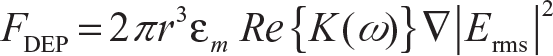
where r is the particle radius, εm is the electrical permittivity of the medium, K(ω) is the Clausius-Mossotti factor which further depends on electrical permittivity and the conductivity of the particle and the medium, and Erms is the root mean square value of the electric field. DEP can be positive (when particles move toward the increasing electric field) or negative (when particles move away from the increasing electric field) in nature depending upon the nature of the suspended particles, the medium in which particles are suspended, electrode geometry, and parameters of the applied electric field. DEP can be further categorized depending upon the type of electrodes used to carry out particle manipulation. There can be electrode-based DEP, electrode-less DEP, 29 light-induced DEP, 80 liquid electrode DEP, 81 carbon electrode DEP, 82 etc. Micro-fabricated metal electrodes can be 2-D (planar electrodes) or 3-D (extruded metal electrodes or doped silicon electrodes 83 ) in nature, while there can be an insulation scheme provided through silicon/glass/polymer matrices 84 between the two extremities of the metal electrodes, which can be used for electrode-less DEP. In comparison to the metal electrode-based DEP, the electrode-less one requires higher voltage. In liquid electrode DEP, conducting liquids are made to behave as electrodes which help in creating vertical equipotential surfaces and consequently DEP effects. On the other hand, light-induced DEP utilizes a photoconductive layer which is excited through light in order to create an electric field gradient. DEP is utilized in a variety of applications such as concentration, 14 manipulation, 85 separation, 86 etc., of cells. Apart from the sophisticated silicon platforms, 26 these days, DEP is also being carried out on the robust PCB platform which offers the benefit of both kinds of DEP, that is, electrode-based DEP and electrode-less DEP. 14
Another cell/particle manipulation technique is electrophoresis in which the particles move under an effect of uniform electric field contrary to that of the DEP in which a non-uniform field is needed. In electrophoresis, charged particles (mostly DNA in biosensors) move toward the opposite polarity at a certain speed and hence particle separation can be efficiently carried out using this technique. Particles are separated according to their sizes as smaller molecules move faster as compared to larger ones and hence get separated. Electrophoresis can be categorized into three sub-categories, namely gel electrophoresis, capillary electrophoresis, and surface electrophoresis. In gel electrophoresis, 87 porous gel matrice (usually made up of agarose, hydroxyl cellulose, etc.) is the medium through which particles move and separate. This is an internationally acclaimed technique to confirm the amplification of DNA samples through PCR, 87 while in capillary electrophoresis 88 the particles are made to move through single capillaries. Long strands of DNA molecules are also separated through yet another variant of electrophoresis which is surface electrophoresis 77 (the name given for electrophoresis on a flat surface).
DNA based
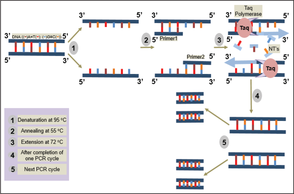
In this method, detection is carried out by looking at the relevant genetic make-up of different biological entities. The most widely used method here is the PCR. 89 PCR is a standardized process of multiplying/replicating DNA which was invented in 1985. DNA amplification is mainly carried out in three steps, denaturation, annealing, and extension. Denaturation is the separation of two single-stranded DNA from a double helical structure which takes place through heating the reactants at 95°C, annealing is the process of primer attachment at 3′ end of single-stranded DNA which takes place at around 55°C, and extension is the process of completing the DNA strand and the DNA backbone which takes place at around 72°C with the help of an enzyme called Taq polymerase. The schematic of the PCR is shown in Figure 3 which represents various PCR steps involved in PCR. The number of copies produced in PCR can be calculated by the formula 2 n , where n is the number of PCR cycles.
DNA replication can be conducted in a conventional thermal cycler instrument with repeated cycling of the temperature of the reactants so that the amplification process can happen. The same cycling can be carried over miniaturized sample volumes on micro-fabricated platforms. 73 The foremost benefit the on-chip PCR technology offers is that it completely removes human intervention that may otherwise be experienced in a DNA lab and makes the diagnostics possible as a point of care. A variant of microchip PCR, the quantitative PCR (qPCR), also known as real-time PCR (RT-PCR) completely eliminates the need for gel electrophoresis. In this variant, the amplified target DNA is visualized through an active fluorescence label which may either intercalate into the DNA structure (in case of SYBR green PCR) or act as a binding label with every cycle with every new strand, thus executing a change in emission frequency due to fluorescence resonance energy transfer (FRET) (in case of molecular beacon or TaqMan assay). 91 qPCR is a very beneficial technique in micro-total analysis systems which is carried on whole cell samples such as in the case of bacteria, etc. (also known as colony PCR). 26 In such cases, a single chip is utilized for performing multiple operations like pre-concentration through DEP and the subsequent PCR process. PCR can be majorly categorized into two types—one is liquid PCR where the primers lie in the liquid phase and the other is solid PCR where primers are immobilized on the substrates in order to carry out sample amplification. 92
Figure 3. PCR steps.
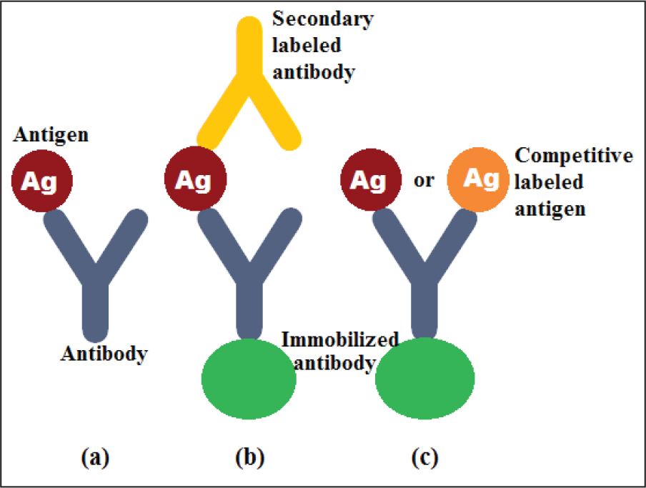
Antigen/antibody interaction mode. (a) Direct mode, (b) sandwich mode, and (c) competitive mode.
Nucleic acid probe based
In nucleic acid-based sensors, a hybridization scheme is utilized. A complementary nucleic acid to that of the target sequence is immobilized on a substrate through some adhesion mechanism. 93 The probe sequence can be a DNA oligomer 94 or its modified form like peptide DNA 95 and locked DNA. 96 Along with the complementary nucleic acid probes, hairpin-loop structure probes and molecular beacons are the potential probes which undergo structural change to produce a detection signal in labeled 97 as well as label-free 98 approaches.
Antigen/antibody based
Antigen/antibody-based sensors utilize the binding kinetics taking place between an antibody and an antigen as the recognition mechanism of the sensor. The kinetics work in several modes (Figure 4), namely direct mode (binding between non-labeled antibody and non-labeled antigen), sandwich mode (immobilized antibody binds to an antigen which further binds to secondary-labeled antibody), competitive mode (binding of limited binding site of immobilized antibody to competitive labeled and non-labeled antigen) and binding inhibition mode (inhibition of binding to immobilized non-labeled antibody and secondary-labeled antibody). 99
Aptamer based
Aptamers are oligonucleotide or peptide molecules which are capable of binding to a specific target molecule. These molecules are either available naturally or can be selected from a large random pool of genomic sequences. Aptamer-based biosensing can be compared with probe-type sensing with the aptamer sequences serving as the bio-recognition layer on the sensory surface and can be used to select a variety of analytes such as nucleic acids, drugs, amino acids, metal ions, organic dyes, etc., 100 and they can be easily modified depending on the requirement with fluorescent tags, metal nanoparticles, enzymes, etc. 101
The various applications of the discussed recognition element-based biosensors have been discussed in detail in the forthcoming sections under several detection schemes.
Depending on the type of detection technique
Colorimetric detection
Various applications of colorimetric detection techniques.
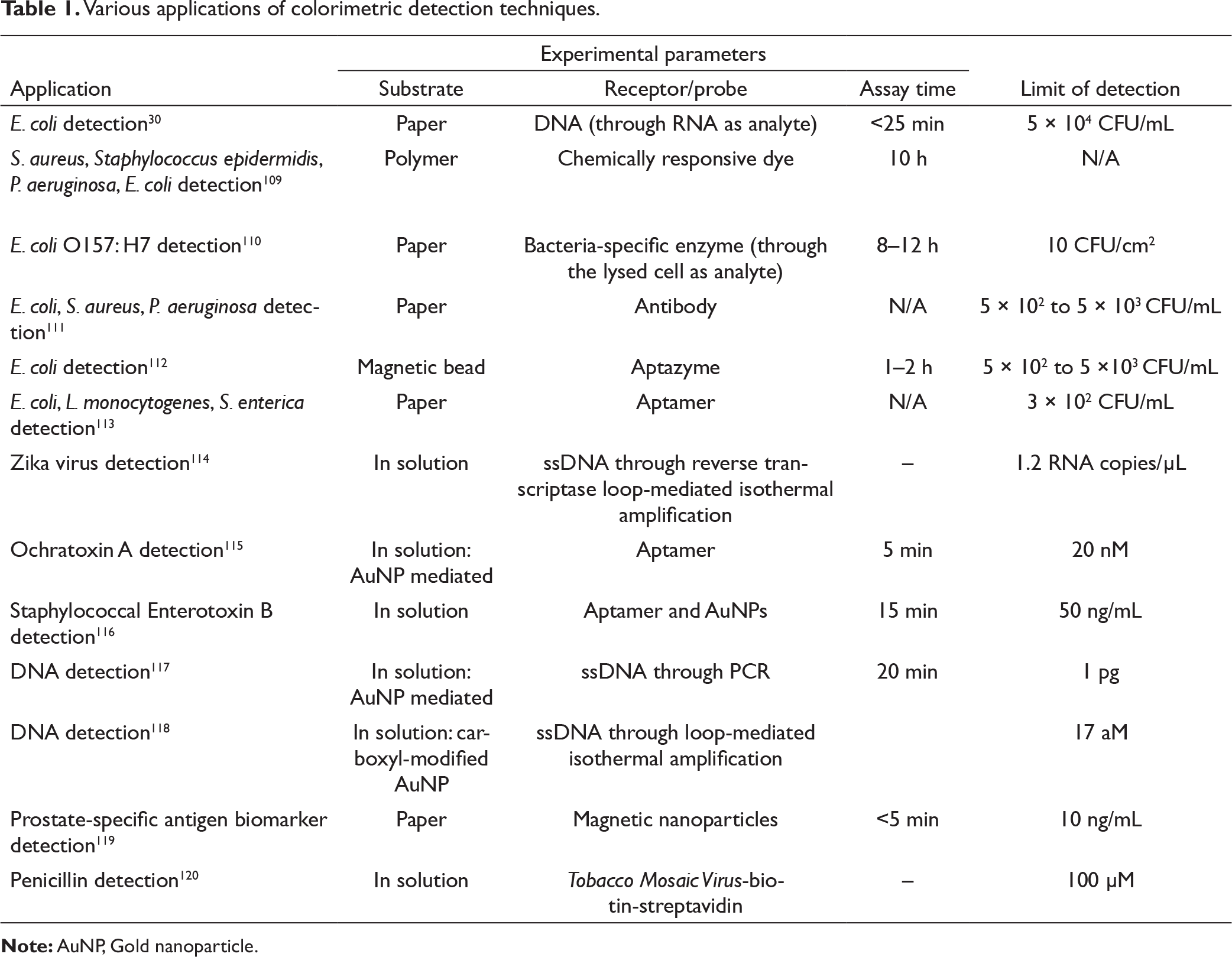
Figure 5. Color change as observed in the colorimetric sensing due to aggregation. (a) Schematic representation. (b) Surface plasmon absorbance band and solution color for 13-nm AuNPs in dispersed and aggregated condition.
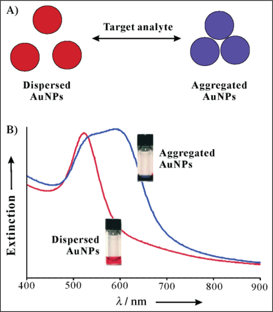
Table 1 represents various applications of colorimetric detection. Colorimetric detection has encompassed applications in detection of various pathogens/cells such as Escherichia coli, Staphylococcus aureus, Staphylococcus epidermidis, Pseudomonas aeruginosa, Listeria monocytogenes, Salmonella enterica, Zika virus, etc., or toxins like ochratoxin A and staphylococcal enterotoxin B; other biological entities such as DNA, biomarkers, and also drugs like penicillin, etc., have also been detected colorimetrically. The detection can be carried out on a variety of substrates such as papers, polymers, and magnetic beads or in solution form following specific reaction chemistry based on different recognition layers (as discussed in the earlier section) along with some combination of various nanoparticles. The detection time as observed through this technique can be observed to vary from a few minutes to several hours. The various reaction assays have shown different limits of detection as stated in Table 1.
Colorimetric detection is quite simple and is performed over a flat substrate/directly in solution, which provides a portable and cost-effective detection modality. But the major disadvantage that lies with this technique is its low-sensitivity detection with limited multiplexing and quantification capabilities. Several strategies have been used to enhance the detection sensitivity of colorimetric detection such as usage of magnetic beads, 121 metal nanoparticles, quantum dots, 113 chemiluminescent, substrates, etc.
Optical detection
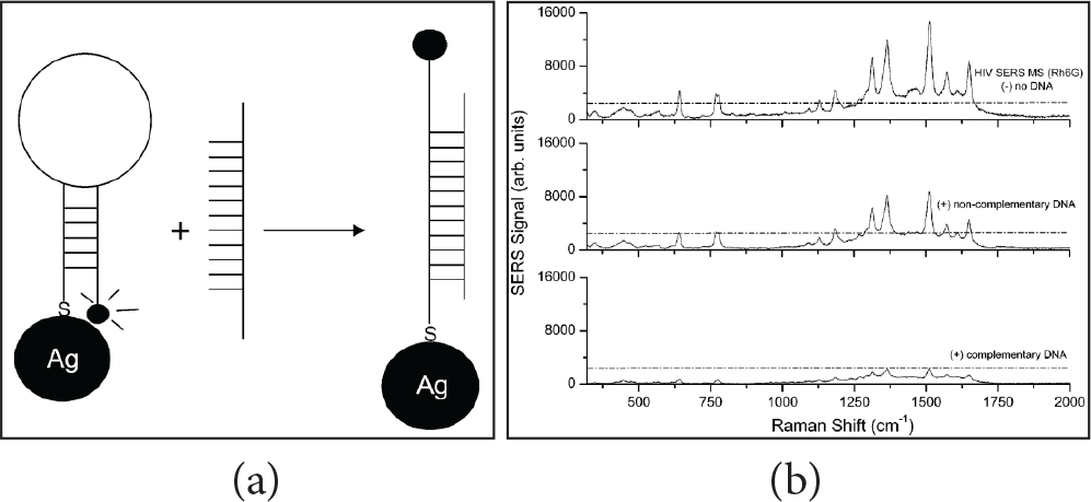
Optical detection is dependent on the change in the optical properties of the observed surface. This change can be gauged as an absorption, adsorption, transmittance, reflection, emission, scattering, and refraction change due to the occurrence of the sensing event that takes place. This event can be observed with or without the presence of a label (fluorophore). There can be various ways to carry out optical detection. Fluorescence intensity count in a solution can be directly monitored through a fluorescence microscope. This is a direct observation technique which can be utilized to quantitate DEP 14 and qPCR 26 processes. FRET is another optical detection scheme in which light-quenching phenomena are studied for the detection that takes place as a result of energy transfer from the donor to acceptor fluorophore molecule due to a change in the proximity between them. 122 The analyte quantification is done by an amount of quenching that occurs due to the interaction between the molecules and their surroundings which is indicative of the regular functioning of cells and also diseased states. Several fluorescent probes have been defined to fluoresce in a specific spectrum by enabling energy transfer when the molecules are in close proximity to one another. FRET is used intensively for studying molecular interactions between genetic expressions in the cells. FRET has evolved from in vitro to in vivo environments over a period of time for intra-molecular and inter-molecular applications. 123 Along with the detection of live cell proteins, 124 FRET is often efficiently used for single-molecule detection through the deployment of quantum dots, showing the FRET effect. 125 Raman spectroscopy utilizes a nonionizing laser as the excitation source. Photons are incident on the molecules and depending upon the energy of the incident photon and the energy gap in the molecule the molecules either absorb energy or scatter it or in some events let it pass through itself without any significant interaction. If this energy is the same as the difference of the energy levels (between both ground and excited states), they absorb the energy and get to the excited state and a fluorescence emission event occurs when these molecules get back to the ground state. When the electron cloud of the molecule is distorted, scattering starts. When only the electron cloud is distorted, an elastic distortion takes place and Rayleigh scattering starts to occur. As the vibrational state of the molecule changes, an inelastic distortion of the molecule occurs, and Raman scattering starts to occur. 126 Raman scattering has shown potential applications in biological aspects 127 and diagnostics. 128 Raman signals being weak can be compromised up to some extent as the samples are in solution form due to associated quenching effects. Water is very detrimental to such signals and is known as a quencher. Therefore an alternate known as surface-enhanced Raman spectroscopy (SERS) has been used which shows immense enhancement in Raman signals by utilizing the interaction of the molecule and the nanoscale metal surfaces over which they are placed. An enhancement in signals by a factor of 103–1014 has been routinely reported using substrates contributing to the SERS phenomena.129, 130 SERS has been extensively used for biological detection. Figure 6 elaborates the detection scheme of HIV Type-I DNA detected through SERS. 131
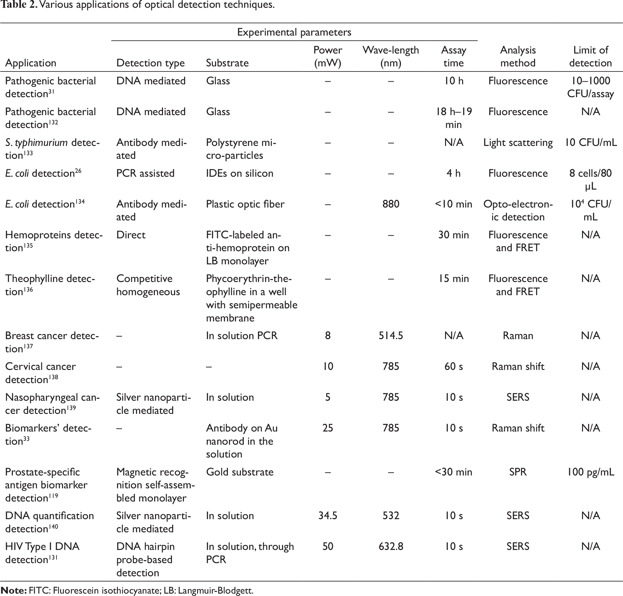
Table 2. Various applications of optical detection techniques.
Along with all these conventional optical detection schemes, photonic crystals are also being explored heavily for biosensing. These crystals consist of periodic dielectric materials arranged in a spatial manner which interact with light in a unique manner and provide high-efficiency reflection at particular wavelengths. 141 These crystals are well capable of detecting various pathogens, 142 DNA, 143 enzymes, 144 toxins, 145 etc. Different materials such as silicon, 146 glass, 147 polymer, 148 silk, 149 and colloids 150 can be utilized for fabricating a variety of photonic crystal structures, namely 1-D-, 2-D-, 3-D-orientated microcavities, 147 slabs, 151 porous geometries, 146 multilayered thin films, 152 and waveguides. 153 Different fabrication schemes utilized for fabricating these crystals are self-assembly 152 or lithography. 147
As illustrated, optical detection techniques are also very well represented in regard to the applicability in biosensing. They offer good sensitivity but are sophisticated and use complex instrumentation to observe the exact change in the optical signal. A properly calibrated system is a must condition for carrying out optical detection and systems may not be very amenable to field deployment.
Electrochemical detection
Electrochemical detection exploits both redox activity of the solution generating the signal or a reporter species and its biocatalytic reaction generating electrochemical signals. The reporter label is attached to the target molecule or there may also be a direct label-free electrochemical response of the analyte itself which is recorded as electrical activity. 154 There are various sensors which lie in this category. To name a few there are voltammetric sensors, amperometric sensors, impedimetric sensors, etc. Voltammetric sensors exploit the amount of current to quantify the analyte levels as obtained on current peaks due to redox activity across a range of the electric potentials. Hence the position and the limit of peaks define the analyte composition and concentration, respectively. The shift in the current peak is representative of the blockage of the sensing surface, which leads to slow electron transfer across the redox couple. Various sensors have been designed using voltammetric sensing schemes for detection of diseases such as cancer, 155 DNA, proteins, viruses, and cells. The real-time wearable sensors have also been devised for tracking heart rate using tattoo-based pH sensors. 156 Paper microfluidics strategies and carbon nanotubes have also been used for devising voltammetric sensors. 157 Amperometric sensors utilize redox activity of the catalytic products as obtained in the course of bio-recognition events on the sensing surface. 158 The current generated during consumption/generation of the electrochemically active constituents directly relates to the analyte. Various cells, 159 organic contents like glucose and, 160 gases 158 are measured through various electrode configurations. Various metal electrodes, carbon electrodes, and conducting polymer electrodes can be utilized for amperometric sensor applications.
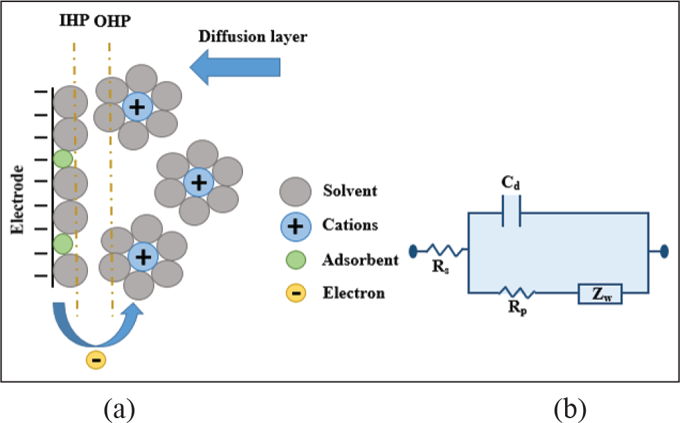
Electrochemical impedance sensing (EIS) utilizes a direct response of the analyte in terms of impedance (resistance/capacitance) change on the sensing surface. Its sensitive, easier, and a label-free detection scheme which monitors the change of charge transfer resistance and capacitance over a range of frequencies, and the corresponding characterization is done accordingly. The change in impedance is a key characteristic of double-layer capacitance which is formulated in the case when the electrodes are dipped in the analyte solution. The impedance formulation is given by Randle as
161
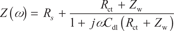
where Rs is substrate resistance, Rct is charge transfer resistance, Cdl is double-layer capacitance, and Zw is Warburg coefficient. Figure 7 shows the mechanism of double-layer formation and respective electrical circuit parameters.
Various applications of electrochemical detection techniques.
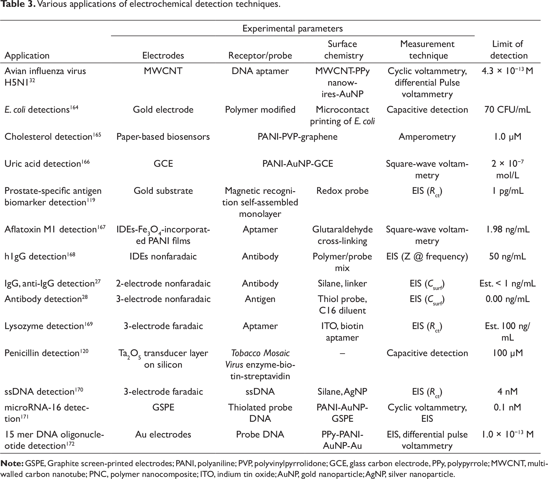
This type of detection is easy in nature but is very sensitive to the environment in which the detection is carried out. Different nanoparticles are also used enormously for enhancing the sensitivity of detection. There is again an integrated detection scheme which is gaining importance these days, known as electrochemical PCR. It deals with simultaneous DNA amplification and electrochemical detection of the amplified sample. This type of detection is carried out in various modalities, using several electroactive indicators such as redox couple, 173 dyes, 174 nanoparticles, 175 etc., for solution PCR as well as solid PCR. 176
Mechanical detection
Various applications of mechanical detection techniques.
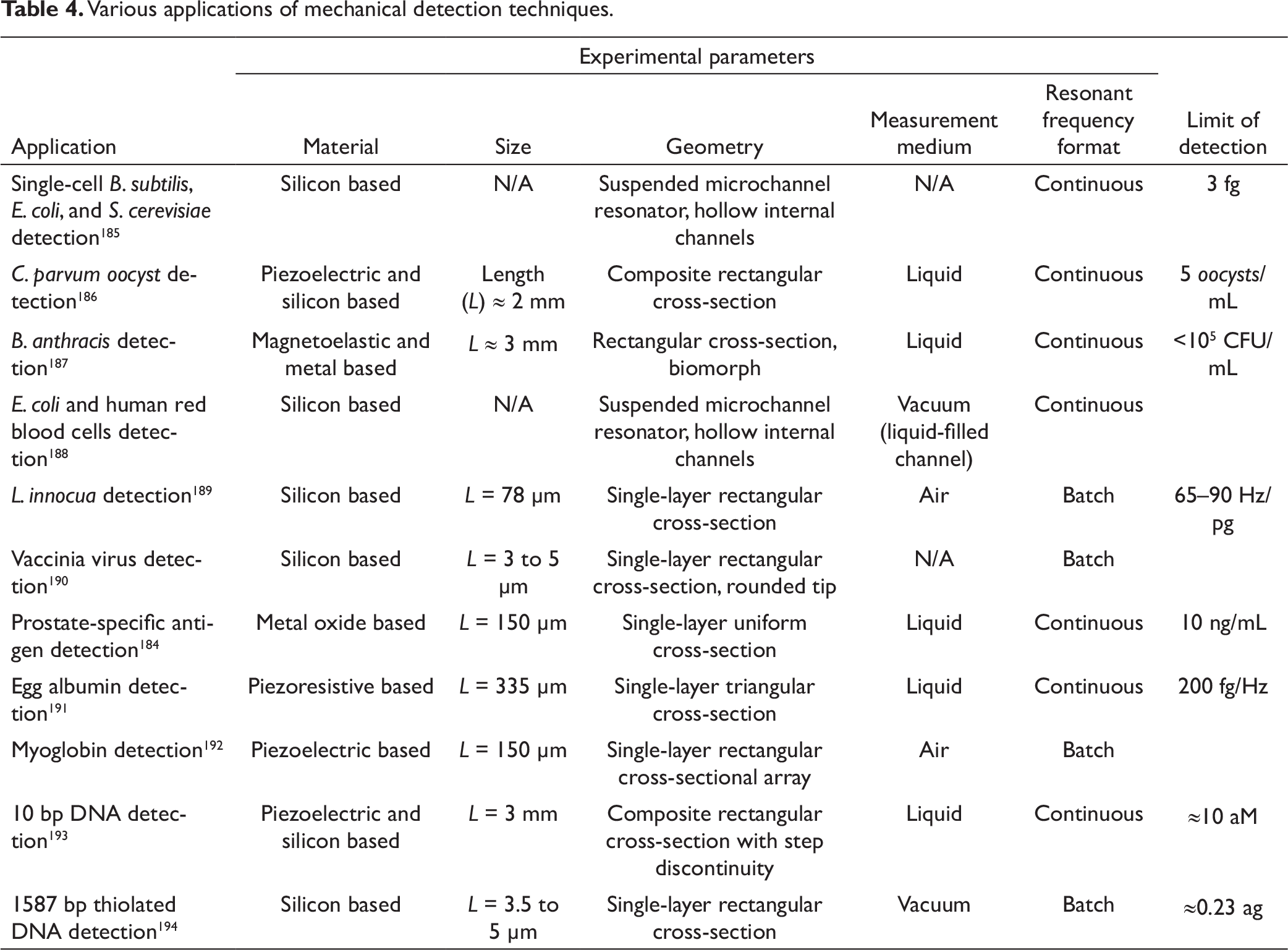
Cantilever-based biosensors are very sensitive to a variety of analyte concentrations, change in temperatures, changes in flow rate around the cantilever sensor, etc. The atomic force microscopy (AFM) technology is an example where these small mass sensors are heavily deployed. The tip of an AFM is such a cantilever and the microscope records bending and deflection of this small cantilever tip as it rasters over a surface to reconstruct the topology based on such bending signals. 181 The tip is further used to measure the force (between tip and analyte) through tip deflection or cantilever’s resonant frequency change. There are various factors which significantly affect the limit of detection in cantilever-based detection and these are properties of the cantilever (size and mass as smaller the size or lower the mass, better the sensitivity), the stiffness of the cantilever, and the sensitivity of the instrument which is deployed to monitor the deflection or vibrations. Depending upon such requirements, various fabrication strategies and several fabrication materials have been explored from time to time for cantilever fabrication. 179 Silicon-based materials such as silicon, silicon dioxide, and silicon nitride, piezoelectric, magnetoelastic, some metals, and polymers like SU-8 materials, etc., are among the most commonly used materials for fabricating cantilevers in different geometries and sizes. A similar geometry with different materials offers different resonant frequencies and hence various combinations in terms of material, geometry, and size can be optimized for a specific application. Silicon-based cantilevers have been extensively used, even for single-cell analysis. 182 Campbell and Mutharasan have used piezoelectric material-based cantilevers for detecting much-diluted pathogens. 183 Metal oxide-based cantilevers are used for detecting prostate cancer markers. 184 Table 4 summarizes the usage of different materials for cantilever-based sensing in various analytes such as microbes/cells/viruses (single-cell Bacillus subtilis, E. coli, and S. cerevisiae, Cryptosporidium parvum oocyst, Bacillus anthracis, Listeria innocua, vaccinia virus, red blood cells), proteins/antigens (egg albumin, myoglobin, prostate-specific antigen), DNA, etc. Different limits of detection have been achieved using different experimental parameters such as measurement methods, the medium in which the detection is carried out, cantilever shapes and geometries, etc. It is important to mention that the geometry and size of the cantilever are important in detection through cantilevers.
As stated in the table, various analytes have been detected through mechanical detection schemes on a variety of substrates/electrodes. The most common types of geometries which are realized in cantilever fabrication are rectangular, triangular, trapezoidal, etc. Different mediums like liquid, air, and vacuum are also used for carrying out mechanical detection. The major challenge that lies in this detection scheme is the fabrication of sensitive cantilevers.
Conclusions
This article averages out the various aspects of biosensors on the chip. It comprises various domains connected together to realize biosensors on the chip. These domains are substrate-related aspects, microfluidics associated with it and the channel scheme combined to realize the detection chip. This article further elaborates various classifications of biosensors depending mainly on the type of the bio-recognition layer and the types of detection methods. Various application areas of biosensors are also discussed in different measurement environments.
Future directions of biosensing research
In developing countries, the current business trends in biosensors are focused on sensitive bed side detection through card reading using various colorimetry schemes. The idea is to deploy a technology otherwise known as lateral flow assay with additional capabilities like higher sensitivity values and lower costs. There are some very obvious technology gaps which exist in the market and need to be bridged for those acute diseases which are life threatening as a function of their progression time such as dengue, malaria, chikungunya, etc. Incidentally the strategies used by the conventional marker of card-based detectors sometimes overlook the specific angles though diagnostic products may fit. For example, if we look at the dengue problem in Asian countries, the secondary infection due to repeated bites of a mosquito to a dengue affected patient may completely change the antigen profile in the blood. The cards developed conventionally may not work well in these cases due to higher limits of detection in the existing technologies which may be replaced by better innovative strategies to produce repeatability and reliability. In other words although physics-based sensing and new phenomena still form the first line of research in biosensing, there is a great need for case-specific reliability while application to real fields. The focus of the research should really now shift to product development in sensing and reliability of technologies.
Declaration of conflicting interests
The authors declared no potential conflicts of interest with respect to the research, authorship, and/or publication of this article.
Funding
The authors received no financial support for the research, authorship, and/or publication of this article.