
Abstract
Growing insight into bioelectric regulation of cell behavior has driven extensive use of electrical stimulation in in vitro studies over the last decade, notably in the enhancement of stem cell maturation. This has prompted the development of electrical stimulation bioreactors to provide the stimulation parameters required for such studies. However, biological outcomes are highly sensitive to stimulation parameters, electrode geometry, and electrode-electrolyte interface. These are factors that current commercial and custom systems handle differently; commercial platforms are robust but often cost-prohibitive, whereas custom open-source systems are cheaper but typically lack validated, user-friendly hardware and standardization. In this perspective, we argue for openly documented electrical stimulation platforms that are not only published but also made available through open-source science manufacturers. Coupling transparent designs with distributed assembly and open validation data offers the potential to lower current barriers to entry, enabling wider adoption of pre-characterized, affordable systems that improve reproducibility and mechanistic clarity.
Introduction
Exogenous electrical stimulation is an increasingly important tool for in vitro research, enabling control and interrogation of bioelectrical signaling in both excitable and non-excitable cell systems. However, biological responses to electrical stimulation (ES) are highly sensitive to stimulation parameters, electrode configuration, and system architecture. Consequently, experimental outcomes are often inseparable from the design and performance of the stimulation platform itself.
This sensitivity presents a particular challenge in areas such as stem cell maturation, where achieving adult-like phenotypes remains a fundamental limitation in regenerative medicine and disease modeling. Cells differentiated in vitro frequently exhibit immature structural, electrophysiological, metabolic, and transcriptional phenotypes when compared with their in vivo counterparts. This immaturity compromises physiological relevance, reducing the predictive validity of disease models and pharmacological testing platforms. Such discrepancies are widely considered contributors to translational inefficiencies in drug development, with several compounds that appear promising in early stages failing in clinical trials. Cellular immaturity similarly limits the functional performance and transplantation potential of laboratory-grown tissues engineered for therapeutic use.
Among the diverse methods for enhancing maturation, exogenous ES platforms are widely employed, particularly in excitable cell lines. Electrical stimulation has been associated with measurable improvements in maturation-related metrics across multiple lineages, including cardiac, neural, and musculoskeletal cells.
However, the technical and financial barriers to entry for in vitro research using these exogenous ES platforms remain considerable and are often prohibitive. Commercially available off-the-shelf systems offer user-friendly interfaces and validated performance; however, prices ranging from £15,000 to over £100,000 place them beyond the reach of many researchers. Consequently, bespoke, custom-built alternatives are prevalent in academic research. While they are more affordable, they require engineering expertise to design, construct, and validate these hardware systems while remaining compatible for use in cell culture. Moreover, the idiosyncrasies and inadequate characterization of each system, often tailored for a specific set of experimental results, result in significant inter-laboratory variability. The development and broader availability of affordable, standardized platforms that integrate seamlessly into established workflows are therefore essential to reducing inter-laboratory variability and enabling reproducible, comparable electrical stimulation studies.
Probing and Modulating Fundamental Cellular Behavior
Addressing these accessibility and reproducibility barriers is not merely a logistical concern; it is critical to advancing our understanding of how electric fields drive fundamental cellular processes. The exact biophysical mechanisms underlying the enhanced maturation and modulated differentiation across various cellular models remain open research questions. All cells maintain a transmembrane potential generated by their lipid bilayer, which is sustained by ion channels, pumps, and transporters. The membrane potential is sensitive to changes in ion transport and to external electric fields, potentially influencing voltage-gated signaling pathways, calcium dynamics, and downstream changes in gene expression. Regardless, there is still no consensus on the precise mechanisms by which specific stimulation parameters elicit these effects. ES bioreactors are particularly valuable in these systems, as they enable not only the application of exogenous electric fields but also the sensing and probing of the internal states of both excitable and non-excitable cells.
Some of the most mature examples of ES bioreactors are found in cardiac and neuronal research due to the inherent excitability of these tissues. In cardiomyocytes, ES (through cardiac pacing) is frequently used to study calcium handling alongside maturation protocols. This is particularly relevant in induced pluripotent stem cells (iPSCs) systems where standard differentiation protocols yield cells with immature phenotypic expression. 1 Therefore, electrical pacing protocols have become a commonly utilized strategy to enhance cell maturation and, consequently, to relate to physiologically equivalent tissue. Nevertheless, the intracellular signaling cascades that drive maturation remain incompletely defined, and the underlying biophysical mechanisms are still under active investigation. 2
In neural cells, ES bioreactors are similarly employed for effective maturation and differentiation of stem cells, including iPSC-derived models.3,4 The use of ES systems for neural systems extends beyond maturation, in particular to investigate the effects of exogenous electric fields on stem cell activation, neurite outgrowth, and regeneration. 5 Although the role of ES is often shown to be essential for neural cells to differentiate and activate, the reported outcomes vary substantially, suggesting that a comprehensive understanding of parameter-dependent effects and mechanisms has not yet been fully established. 6 Thus, to rigorously decouple and interpret the biological responses observed in electrically stimulated systems, the stimulation environment must be precisely defined and controlled through the characterization of field strengths, waveforms, frequencies, and electrode configurations. Even modest differences in electrode geometry, media conductivity, or boundary conditions can alter local current density and field gradients, such that nominally similar stimulation protocols activate distinct biophysical pathways and yield contradictory biological outcomes across laboratories. The sensitivity of these systems to stimulation parameters means that biological interpretation is inseparable from the design and performance of the stimulation platform itself. Without standardized control of electric field distribution and waveform fidelity, it is therefore difficult to determine whether observed phenotypic changes arise from defined bioelectric modulation or uncontrolled electrochemical artefacts.
Electrical Stimulation Bioreactors
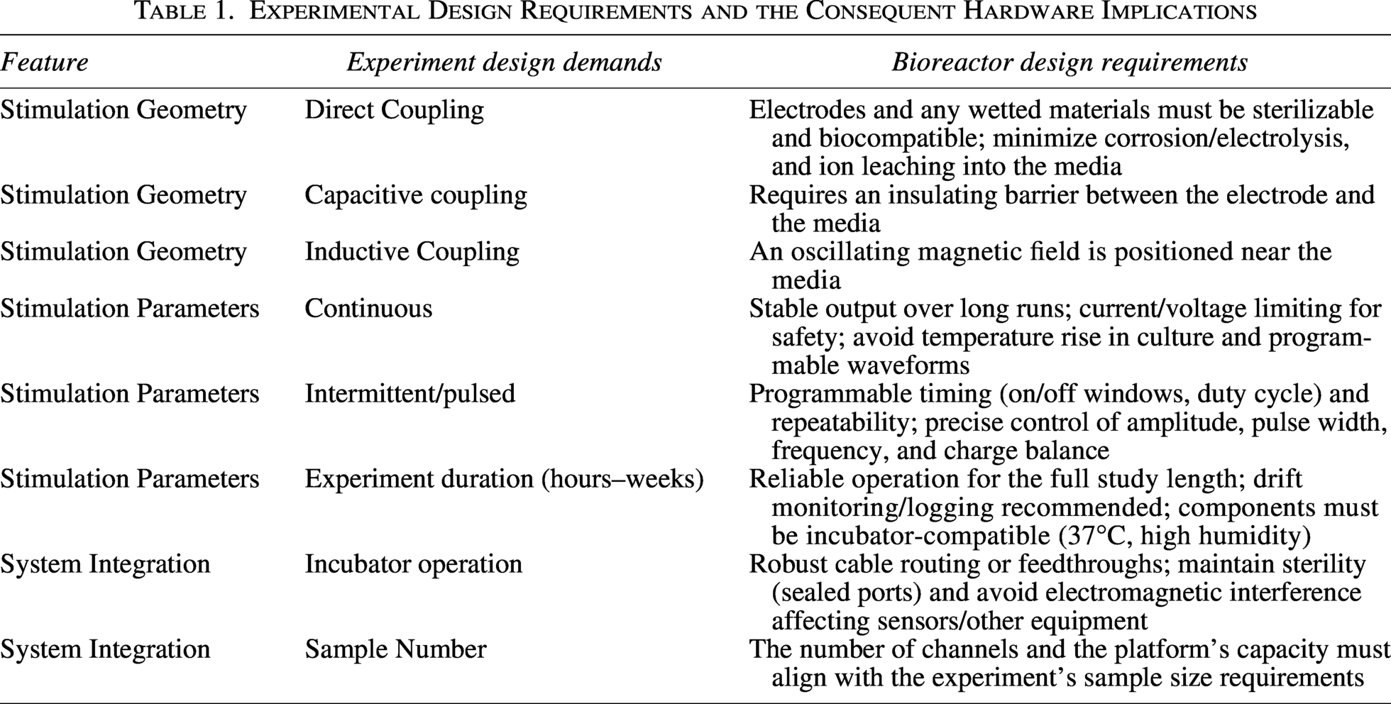
The electrical system architecture is inherently experiment-dependent, as different protocols impose distinct functional and performance constraints. Table 1 summarizes how specific experimental design requirements translate into corresponding hardware specifications. In practice, researchers must choose between commercially available systems and custom-built platforms. Consequently, balancing costs, flexibility, reproducibility, and technical support while ensuring that experimental objectives are met is a central challenge in this field.
Experimental Design Requirements and the Consequent Hardware Implications
Commercial systems
Several options for electrical bioreactors are commercially available, with the main players including Ion Optix, Curi Bio, and Axion Biosystems. Ion Optix’s culture pacing system is a well-established device that has been used in research laboratories for over 25 years to study cardiac and skeletal muscle cells. 7 Ion Optix’s system provides a programmable stimulation regime for up to eight plates (with 6-, 12-, and 24-well options) and real-time temperature monitoring. Additionally, mechanical stimulation capabilities can be incorporated by purchasing add-on components. A significant limitation of this device, however, is the inability to operate these systems within a standard incubator, as the electronics are not rated for high-humidity environments, and external cabling prevents proper sealing of the incubator doors when closed. Therefore, chronic pacing under physiological conditions is infeasible with the standard systems available.
Curi Bio is a relatively recent entrant to the market, established in 2015. Their ES products enable chronic pacing with the stimulation systems (StringrayTM and MantarrayTM), which are designed specifically to accommodate operation within incubators. 8 Curi Bio systems include a user-friendly UI that enables unique programme stimulation regimes for each well, with real-time data monitoring, including temperature as a key differentiator. Furthermore, mechanical stimulation for 3D culture is available on specific products. However, Curi Bio’s platforms can only stimulate one well plate at a time, with compatibility limited to their proprietary well plates.
Axion Biosystems provides a multi-microelectrode electrode array (MEA) capable of chronic pacing by creating a controlled microenvironment around the MEA plate. Unlike the large, direct-coupling electrode used in Curi Bio and Ion Optix systems, Axion Biosystem’s microelectrode arrays enable more localized control of the electric field. However, the power that can be supplied by the electrodes is limited by the microelectrodes’ diameters, which can lead to heating effects. A further advantage of the MEA includes the real-time data acquisition of temperature and label-free monitoring of cell death over the stimulation regime. 9
Collectively, these three commercial providers offer an insight into the user-friendly systems that are available to enable researchers to investigate the effects of ES without requiring substantial engineering expertise. They offer validated hardware, structured interfaces, and documentation that facilitate compliance with emerging reporting standards for a broad range of experimental demands. However, the cost of these systems ranges from approximately £15,000 to more than £100,000 for the more advanced systems, with additional recurring expenses for the purchase of ongoing proprietary consumables in some systems (e.g., Curi Bio and Axion Biosystems). For many laboratories, these financial constraints remain a decisive factor, prompting the development of custom in-house systems despite the advantages of commercial platforms.
Custom systems
When commercial systems are financially inaccessible, a custom-built system is often the only viable pathway for conducting ES experiments. Several research groups have successfully developed and published designs for low-cost ES bioreactors.10–12 An exemplar area of focus for these devices involves the differentiation and maturation of cardiomyocytes. Work by Licata, Gerstenhaber, and Lelkes 13 focused on developing a low-cost device, fabricated via 3D printing and custom electronics, to provide chronic pacing for 5 days. To validate the device Licata, Gerstenhaber, and Lelkes cultured cardiomyocytes differentiated from iPSCs, which exhibited greater synchronized contraction than controls. This device supported a programmable stimulation protocol for the cells and monitored the electrical signals. Additionally, a finite element modeling simulation was used to characterize the electric fields produced. However, the culture temperature was not monitored during stimulation, nor was the simulation experimentally validated.
Despite such examples of substantially lower-cost ES bioreactors, custom systems frequently face barriers to broader adoption. In many cases, expertise in electronics, programming, simulation, and device fabrication is required to build the device described in the report, even when CAD files, code-bases, and instructions are publicly available. Secondly, these systems are commonly designed to meet specific experimental requirements of the researchers developing the device. This results in the devices that are not optimized for general usability and scalability. In particular, UIs and calibration protocols may be limited, and systematic validation and formal fail-safe mechanisms are rarely implemented. Unlike commercial platforms, there is no official technical support, standardized documentation or warranty coverage for these systems.
In addition to geometric variability, insufficient characterization of electrode–electrolyte interfaces may introduce electrochemical artifacts, including pH shifts, interfacial gas evolution, and the generation of reactive species. Without systematic validation of charge balance and thermal stability, such effects may confound the interpretation of bioelectrical signaling responses. Standardized platforms with validated electrochemical performance are therefore essential not only for reproducibility but also for mechanistic clarity.
Despite their affordability, published evidence of independent replication and uptake of open-sourced designs remains limited. Open-source dissemination has facilitated knowledge transfer, as demonstrated through incremental and iterative refinement of some designs. Unlike mature open-source software ecosystems, ES bioreactors have yet to reach a critical mass of users to sustain a community that provides ongoing discourse and development of these open-source models. Consequently, while bespoke systems can approximate the functionality of commercial platforms at reduced cost, they generally lack the standardization, validation infrastructure, and scalability necessary to ensure consistent reproducibility across laboratories.
Minimum standard reporting
The diversity in platform architectures and implementation strategies described above underscores a broader issue: without consistent reporting of stimulation parameters and system characteristics, comparing results across studies becomes inherently difficult. Minimum standard reporting emerged in the early 2000s to improve reproducibility by providing a checklist of essential information that authors should report for each experiment. The Minimum Information About a Cellular Assay (MIACA), introduced in 2006, focused on cell-based assay experimentation and is therefore particularly relevant to in vitro ES studies; more detailed frameworks have since emerged, for instance, the additional reporting requirements for the MIACA guidelines proposed by Budde et al. 14
In 2019, Budde et al. proposed that reports using commercial systems should outline the manufacturer, model, and version of the software, whereas those employing custom-built platforms should describe the geometric dimensions, materials, and manufacturing processes used. For both platforms, the stimulation parameters outlined must include: circuitry configurations, applied current or voltage, waveform, frequency, duty cycle, stimulation duration, and rest period duration.
The adoption of such standards has substantively enhanced transparency.15,16 However, minimum reporting alone does not eliminate the structural barriers associated with implementing ES platforms. Even with comprehensive documentation, researchers must still navigate engineering design, fabrication, validation, and operational integration. Thus, while reporting standards improve interpretability, they do not address accessibility.
Open-Science Manufacture for Open-Source Designs
The development of a standardized, openly documented platform that can be purchased directly or assembled through open-science manufacturing networks presents a potential solution. Such an approach would enable researchers to easily reproduce the same circuitry, electrode geometries, and validated stimulation parameter ranges without having to design or manufacture the systems independently. Organizations such as LABmaker demonstrate the feasibility of such an approach by providing not-for-profit construction and assembly services for open-science instrumentation. Examples of similar systems include the StimJim, 17 which can deliver dual-channel arbitrary waveforms for ES experiments and provide charge balancing. In the specific case of the StimJim, this system comprises only the electrical circuitry; users would still need design and validate the hardware that interfaces with the cells. Therefore, the development of a fully integrated, open-source ES system that includes validated circuitry, electrode assemblies, and cell-culture-compatible hardware could overcome additional barriers to entry that minimum-standard reporting alone cannot, if made available for direct purchase or distributed assembly.
Importantly, the adoption of such a model does not require a centralized regulatory framework. Existing open-science manufacturing networks already assemble and distribute validated hardware for applications such as microbial culture and environmental sensing. 18 A similar approach could be adopted for ES platforms, whereby design files, calibration protocols, and validation data are openly published alongside distributed assembly services. Transparent documentation of field homogeneity, charge balance, and thermal stability alongside electrode material specifications and electrochemical characterization reports would allow users to independently verify system performance while maintaining the flexibility inherent to open hardware ecosystems.
Conclusion
The technologies available to research groups for exploring key questions in the ES field are both diverse and sophisticated. Commercially available options offer highly optimized, well-characterized systems that integrate smoothly into existing laboratory workflows while supporting reproducible experimentation. Where these tools are inaccessible or insufficiently flexible to address novel research aims, innovative, custom-built systems have enabled significant advances in our understanding of biophysical mechanisms, particularly when developed and documented alongside emerging minimum reporting standards.
Nevertheless, the barrier to entry for both custom and commercial systems is prohibitive for many research laboratories. Commercial systems can entail both high acquisition and operating costs, whereas custom systems are time-consuming to build and often require expertise in programming, electronics, and hardware fabrication. This dichotomy reveals an underserved segment within the ecosystem: an affordable, standardized, and readily deployable ES bioreactor.
A potentially transformative path forward may lie in the development of fully documented open-source ES systems that are not only published but also made available through open-source science manufacturers. By coupling transparent design with professional assembly and distribution, such platforms could provide pre-characterized, standardized hardware at substantially lower cost while preserving flexibility and reproducibility. Lowering these practical barriers would strengthen cross-laboratory comparability and reproducible integration of ES into in vitro studies, accelerating mechanistic clarity of how exogenous electrical cues modulate bioelectricity and cell behavior.
Data Availability Statement
For the purpose of Open Access, the authors have applied a CC BY public copyright licence to any Author Accepted Article (AAM) version arising from this submission. Data sharing is not applicable to this article as no new data were created or analysed in this work.
Footnotes
Acknowledgments
M.N. acknowledges support from the EPSRC BIONIC Hearts New Investigator Award (EP/Y004434/1). R.T.M. was funded by a studentship from the Department of Engineering Science, University of Oxford.
Author Disclosure Statement
The authors declare that they have no relevant financial or non-financial interests to report.
Funding Information
UKRI—Engineering and Physical Sciences Research Council (EPSRC) Grant No. EP/Y004434/1.
.