






Abstract
Barrett’s esophagus (BE) refers to the mucosal transition from normal squamous mucosa to a genetically unstable specialized columnar-type intestinal metaplasia in the esophagus. Recent sophisticated studies using orthogonal techniques of molecular and computational biology strongly support that BE is the predominant pathway to esophageal adenocarcinoma (EAC). In this treatise, we review the risk of BE transforming to EAC and the latest developments in risk assessment of BE progression to EAC. The progression rate of BE to EAC can vary from one study to another depending on the region of origin, definition of BE, and variations in data capture. Overall, the published studies suggest that the risk of BE transforming to EAC is less than 0.5% per year and that to high-grade dysplasia (HGD) or EAC is less than 1% per year. Clinical tools to predict risk such as the Progression in Barrett’s (PIB) have been developed but will need further validation. p53 is the biomarker for risk stratification and has stood the test of time but is still not being widely used due to incomplete correlation between the mutational status and protein expression. Molecular studies using genomics and proteomics approaches are being evaluated but need to demonstrate good clinical accuracy before being translated into practice.
Introduction
Barrett’s esophagus (BE) refers to the mucosal transition from normal squamous mucosa to a genetically unstable specialized columnar-type intestinal metaplasia in the esophagus and is the only known precursor to esophageal adenocarcinoma (EAC).1,2 The incidence of EAC has been increasing in the western hemisphere.3,4 Currently, the standard practice in managing patients with BE is histopathologic evaluation of the BE epithelium which determines surveillance intervals and eradication therapy. In this treatise, we review the risk of BE transforming to esophageal cancer and the latest developments in risk assessment of BE progression to cancer.
Do All EAC Arise From BE?
Before we quantify the risk of progression in BE, it is important to understand whether the pathway to EAC is via BE. It is well-accepted that many EACs are diagnosed without detectable BE at the time of endoscopy. Observations in 2 large cohorts of patients with EAC showed a significant difference in prognosis of EAC patients with and without detectable BE. 5 These data raise the question whether all EACs arise from the well-accepted BE pathway. Now we have insights from the fields of molecular biology as well as computational biology to suggest that the majority of EACs follow the BE pathway. Using a multiomics approach that included single cell sequencing of specimens of normal esophagus, squamocolumnar junction (from controls and from BE), normal stomach, BE, and EAC, the investigators showed that the cell of origin of all BE and EAC came from gastric cardia, irrespective of whether visible BE was seen at the time of endoscopy 6 suggesting a common origin of BE and EAC. Another study used computational tools to model the prevalence of GERD and BE along with progression rates to EAC and showed a high degree of concordance between the predicted and observed number of EACs in the Surveillance, Epidemiology, and End Results (SEER) database. 7 Based on this computational model, the investigators concluded that 90% of all EACs arise from BE. The above combination of molecular biology and computational data strongly supports that BE is the predominant pathway to EAC. Therefore, we can be confident that an accurate description of BE progression risks will be an effective way to understand the risk of EAC in the general population.6,7
Population-Based Risk of BE Progression From National and Other Large Registries
The progression rate of BE to EAC can vary from one study to another depending on the region of origin, definition of BE, and variations in data capture. In these risk estimates, we report on the combined incidence of high-grade dysplasia (HGD) and EAC. We decided to group HGD with EAC because HGD is a high-risk lesion and an actionable diagnosis. 8 The risks of progression to HGD and EAC are described separately in Table 1. The overall risk of progression to HGD or EAC was estimated to be 0.26%/year among 11 028 BE patients (defined by the presence of intestinal metaplasia) over a median follow-up of 5.2 years in the Danish Cancer and Pathology Registry, 9 whereas it was estimated to be 0.22%/year among 8522 BE patients (with and without intestinal metaplasia) over a mean follow-up of 7 years in the Northern Ireland Cancer Registry. 10 Both of these registries were national registries and excluded prevalent cases (HGD or EAC diagnosed within 1 year of BE diagnosis) to provide a true estimate of incident risk. Nationwide registries in the United States are difficult to create but an analysis of the National Veterans Cohort that included 29 536 patients (97% male) with BE from 121 facilities found the incidence rate of combined HGD/EAC to be 0.48%/year after excluding prevalent cases. 11 Another US study that included high-quality data from the Rochester Epidemiology project from Olmsted County, MN reported a progression rate of 0.79%/year over a mean follow-up of 7 years. 12 An important reason for higher progression rates in the Olmsted County study was that censoring of prevalent cases was restricted to within 6 months of index endoscopy (unlike the Danish and Irish registries that excluded prevalent cases within 12 months of index endoscopy). A multi-institutional study that included both veteran and non-veteran patients found the risk of progression to HGD/EAC to be 0.63%/year. 13 To summarize, the risk of progression from BE to HGD/EAC is variable across different cohorts but is less than 1% per year.9,14,15
Risk of BE Transforming to HGD and EAC.
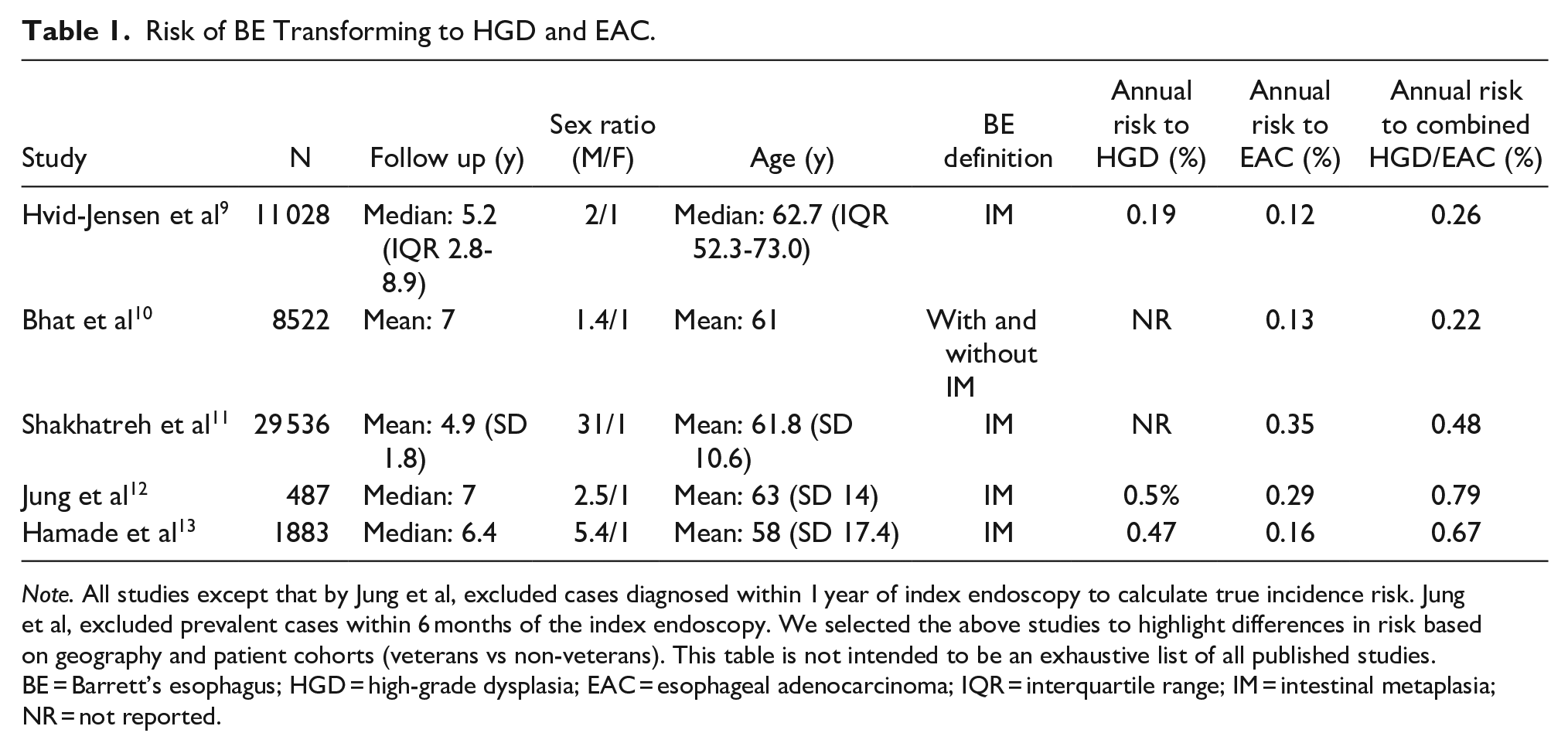
Note. All studies except that by Jung et al, excluded cases diagnosed within 1 year of index endoscopy to calculate true incidence risk. Jung et al, excluded prevalent cases within 6 months of the index endoscopy. We selected the above studies to highlight differences in risk based on geography and patient cohorts (veterans vs non-veterans). This table is not intended to be an exhaustive list of all published studies. BE = Barrett’s esophagus; HGD = high-grade dysplasia; EAC = esophageal adenocarcinoma; IQR = interquartile range; IM = intestinal metaplasia; NR = not reported.
How Does Exclusion of Prevalent Cases Affect Risk Estimates?
This topic was briefly discussed earlier but is important to rediscuss because it can affect the risk estimates of BE transformation. The precise natural history of BE is unknown. The duration of transformation from BE to EAC is unclear and difficult to calculate because the timing of BE initiation remains unclear. Multiple studies suggest that a significant majority of EACs may be prevalent rather than incident. In other words, a significant majority of EACs are diagnosed either on the index endoscopy or within 1 year of the index endoscopy. This can impact the risk calculations of incident EAC in BE. Revisiting the studies discussed above, by including EAC cases diagnosed within 1 year of the index endoscopy, the risk of EAC increased by approximately 50%, from 0.18% to 0.35%. 11 Similarly, in the Danish registry that included the entire Danish population of 5.4 million, the risk of EAC increased by approximately 60%, from 0.12% to 0.29%. 9 Similar observations were made in the Irish registry. 10 These differences in the risk of incident versus prevalent EAC in BE are relevant for counseling BE patients and also emphasize the need for careful examination of the BE segment at the time of endoscopy and perhaps, a short interval endoscopy within 1 year of BE diagnosis.
Risk of Progression in Patients Without Specialized Columnar Epithelium that is, Intestinal Metaplasia
The US guidelines require the presence of goblet cells in the columnar mucosa that is, intestinal metaplasia lining the tubular esophagus to diagnose BE, whereas the British and Asian guidelines exclude this requirement. 2 So does lack of intestinal metaplasia in the columnar mucosa affect the risk of progression to EAC? In the Northern Ireland Cancer Registry study, 10 lack of intestinal metaplasia on the index endoscopy was associated with a lower risk of progression to EAC when compared with its presence (0.07%/year vs 0.38%/year). However, other studies have suggested similar risk of progression with or without intestinal metaplasia.16,17 Additionally, the extent and frequency of DNA abnormalities were found to be similar in columnar mucosa with and without intestinal metaplasia. 18 Debate about the progression risks in BE with and without intestinal metaplasia rages on. Due to conflicting data regarding risk of progression without intestinal metaplasia and concern for sampling error with regards to detection of goblet cells in columnar esophageal mucosa, especially with short segments, many experts recommend to repeat endoscopy at 12 to 18 months with more sampling as that may increase the yield of detecting intestinal metaplasia by >20%. 19
Risk of Progression Based on BE Length
The risk of transformation to EAC in BE less than 1 cm is being discussed elsewhere but we briefly discuss the issue of BE length and risk of transformation. Historically, the relationship of BE length to risk of transformation has been in question 10 but more recent publications from various groups demonstrate that BE length is a strong predictor of EAC risk.20,21 In a large US multicenter study of ~2500 patients, the risk of BE progression increased by 12% for every 1 cm increase in length. 21 An analysis of the United Kingdom Barrett’s Oesophagus Registry controlled for multiple variables and showed BE length to be an independent predictor of progression risk over a mean duration of 5.7 years. 22 We performed a multi-center international study to show that the risk of progression in long-segment BE was higher than that in short-segment BE (0.91% vs 0.29%; P < .001). Taken together, these data suggest that the risk of BE transforming to cancer does depend on length. Based on these data, the British Society of Gastroenterology recommends more aggressive surveillance for BE lengths greater than 10 cm at a center of expertise. 23
Whether individuals with BE < 1 cm in length are at risk of progression to HGD/EAC has been difficult to evaluate because of frequent confusion with intestinal metaplasia at the gastroesophageal junction (ie, intestinal metaplasia of gastric cardia). In the earlier discussed study from Olmsted County, MN, 12 none of the 86 patients with intestinal metaplasia at the gastroesophageal junction (which could, in theory, be confused with ultrashort segment BE) developed HGD or EAC over a median 8 years of follow up. Similarly, none of the 167 patients with intestinal metaplasia at the gastroesophageal junction developed HGD or EAC in a multicenter cohort study with a median of 5.9 years of follow up. 24 The concepts around cancer risk in BE lengths <1 cm are being discussed elsewhere.
Risk of BE Progression Based on the Degree of Dysplasia
The earliest risk estimates that we reported were for all BE patients, irrespective of the degree of dysplasia. The histologic grade of BE is clearly an important predictor of risk. The risk of progression has been variable in BE with low-grade dysplasia (LGD) and this in part is due to high inter-observer variability even among expert pathologists in histologic diagnosis of LGD.25,26 The annual risk of progression from LGD to HGD/EAC in large epidemiological studies was ~1.5%,9,10 but this was likely an underestimation. In a randomized controlled trial comparing ablation to surveillance in patients with LGD at 9 European sites, the annual progression rate from LGD to HGD/EAC in the surveillance group was 26.5%. 27 A subsequent systematic review and meta-analysis, that included the above trial, showed a cumulative progression rate of 12.6% over follow up of 84 months. 28 The differences in the risk of LGD progression rates between epidemiological studies and clinical trials can be attributed to the histologic interpretation of LGD. The interobserver agreement of LGD among pathologists is poor thus raising doubts about the diagnosis of LGD, when read by a single pathologist. 29 In a remarkable study when LGD was confirmed by at least 2 pathologists, the risk of progression to HGD or EAC was 9.1% annually. 30 When LGD was confirmed by 3 pathologists, the annual rate of progression increased to up to 20%. 31
HGD is the penultimate precursor to EAC and is an actionable diagnosis with a high rate of progression to EAC. The annual progression rate from HGD to EAC has been variable in the literature as well, ranging from 16% to 60%. 32 but a recent pooled analysis estimated the annual progression rate from HGD to EAC to be 28.6%. 33
Based on above data, confirmed LGD and HGD (which is routinely confirmed by 2 pathologists) are important predictors of BE transforming to cancer.
Prediction Tools of Transformation Risk in Barrett’s Esophagus
In the era of precision medicine, applying general estimates of progression risk based on histopathologic evaluation to individual patients is fraught with inaccuracies since every patient is unique with different clinical and disease characteristics. Therefore, there is huge push that assessment of progression risk should be individualized. Furthermore, progression from NDBE to dysplasia and EAC is associated with a variety of genetical alterations and chromosomal abnormalities, even before dysplasia is visible endoscopically and detectable histologically (field effect). Moreover, most dysplastic areas are flat and patchy, rendering it difficult to recognize under white light endoscopy despite significant improvement toward high definitions lenses. Likewise, sampling error can occur as standard mucosal biopsies may miss dysplasia (false negative) and sampling is limited to small areas by following the Seattle protocol. The ideal risk assessment tool should include clinical, endoscopic, and histologic characteristics in addition to risk assessment from predictive tests to categorize individuals into risk groups and adjust therapy and surveillance periods accordingly.
Clinical Tools
A new tool that has been developed to predict risk of progression within BE is the Progression in Barrett’s (PIB) score. 21 PIB takes into consideration clinical (male sex: 9 points, smoking: 5 points), endoscopic (length of Barrett’s segment: 1 point/cm, to up to 10 cm maximum), and histologic (confirmed low grade dysplasia: 11 points) features from the index baseline endoscopic examination. It stratifies patients based on risk into 3 groups: low risk (0-10 points), intermediate (11-20 points), and high (>20 points). Based on previous analysis, annual progression rates in the 3 risk groups were 0.13%, 0.73%, and 2.1%, respectively. However, further external validation of this prediction model is crucial to establish the generalizability of the model before clinical use.
Molecular Tools
Barrett’s esophagus mucosa usually starts with molecular and cellular morphologic alterations before dysplasia can be visible or eventually detected histologically. 34 The only biomarker that has currently been recommended for risk stratification is p53, which has been recommended for clinical practice by the British Society of Gastroenterology. 23 p53 is the biomarker that has stood the test of time. 35 More recently, sophisticated studies using whole exome sequencing and whole genome sequencing again demonstrate that p53 is frequently mutated in progressors 36 and therefore should be able to risk stratify patients with BE. The problem with clinical application has been the technique of detection. p53 mutations do not directly correlate with protein expression by IHC staining. Therefore, routine IHC-based detection of p53 may not be as reliable and limits its use. A well-designed study evaluated the value of adding molecular data to risk prediction by performing IHC for multiple putative markers of cancer risk in BE. Remarkably, the investigators showed that superficial expression of Ki-67, which is done frequently for many different cancers, could not only improve the reproducibility of the diagnosis of dysplasia but also identified patients at higher risk for progression. 37 TissueCypher™ is another commercially available system that uses artificial intelligence and machine learning to provide multianalyte algorithmic assay which integrates data that identify and quantify 9 protein biomarkers in addition to changes in tissue structure.38,39 Although the TissueCypher™ assay was validated in well-established BE cohorts, 40 the sensitivity was only 29% and will need further improvement 41 before acquiring a definitive role in managing BE patients. With advancements in technology, proteomics analysis can be another tool for risk stratification in BE. The feasibility in formalin-fixed clinical specimens has been demonstrated 42 but needs further validation in larger cohorts before clinical application.
Conclusion
The risk of BE transforming to EAC is less than 0.5% per year and that to HGD or EAC is less than 1% per year. Despite huge advances in biomarker analysis, histologic grade remains the most clinically used predictor of risk to guide surveillance versus ablation. LGD is an important marker for progression and in particular, LGD confirmed by multiple pathologists carries an annual progression risk of up to 20% and approaches the high-risk profile of HGD. Clinical tools to predict risk such as the PIB have been developed but will need further validation. Although many different molecular assays have shown promise, more studies are needed before clinical application. Ultimately, a predictive model incorporating clinical, endoscopic, histologic, and biomarkers in addition to molecular and genetic analysis will be needed to optimally predict progression risk in each individual patient with BE in the era of precision medicine.
Footnotes
Declaration of Conflicting Interests
The author(s) declared the following potential conflicts of interest with respect to the research, authorship, and/or publication of this article: SS is on Advisory Board and Speaker Bureau-Castle Bioscience. AB received research support from Freenome Holdings and Stella Diagnostics and has an approved patent for a BE screening device.
Funding
The author(s) received no financial support for the research, authorship, and/or publication of this article.