
Abstract
Sex, racial, and ethnic disparities in the incidence and prevalence of Barrett’s esophagus and esophageal adenocarcinoma are well studied. Despite advances in surveillance techniques and multimodality treatment, disparities exist in outcomes with not all populations benefiting proportionately. A deeper understanding of the cause and nature of these disparities could help guide meaningful action toward mitigating these differences. This review focuses on the sex, racial and ethnic differences in the incidence, prevalence and outcomes of Barrett’s esophagus and esophageal adenocarcinoma, and delves into the literature on potential causes for the variation in presentation and outcomes.
Introduction
Barrett’s esophagus (BE) is a premalignant lesion for esophageal adenocarcinoma (EAC) that is characterized by the replacement of the normal esophageal squamous cell epithelium with mucus secreting columnar cell epithelium. BE is caused by the chronic exposure of the distal esophageal mucosa to refluxed gastric contents, which results in cellular injury, inflammation, and columnar metaplasia. The incidence of BE is strongly associated with gastro-esophageal reflux disease (GERD). 1 A recent meta-analysis reported a 7.2% prevalence of histologically confirmed BE in patients with GERD. 2 Other established risk factors for BE are older age, male sex and smoking. 1
Histopathologically, BE progresses from columnar metaplasia to dysplasia, and adenocarcinoma, if left untreated. The annual rate of progression of BE to EAC ranges between 0.06% and 0.31%, depending on the length of the Barrett segment. 3 A recent study reported a neoplasia detection rate of 3% at index endoscopy for patients with BE and chronic GERD. 4
Esophageal cancer remains a leading cause of cancer-related morbidity and mortality. Globally, esophageal cancer accounts for 3.1% of all new cancer diagnoses and 5.5% of all cancer-related deaths. There are notable geographic variations in the global burden of esophageal cancer; the incidence and mortality rates are highest in Eastern Asia and lowest in Central America. 5 Esophageal squamous cell cancer accounts for the vast majority of esophageal cancer diagnoses worldwide. However, in recent years, there has been a rise in the incidence of esophageal adenocarcinoma, particularly in western populations. 6 In the United States, it is estimated that there will be 20 640 new cases of esophageal cancer (16 510 in men and 4130 in women) and 16 410 deaths (13 250 in men and 3160 in women) attributable to esophageal cancer in 2022. 7 Esophageal adenocarcinoma is the predominant histology in the US, and is related to GERD, smoking, alcohol use, obesity, among other factors. 8
Numerous studies have highlighted sex, racial, and ethnic disparities in the incidence, prevalence and outcomes of BE and EAC.7,9-13 These disparities partly occur due to historic and contemporary social and economic inequalities. A better understanding of the cause and nature of these disparities could help guide meaningful action toward mitigating modifiable factors that drive these differences. This article reviews the most recent evidence on sex, racial and ethnic differences in BE and epidemiology of EAC.
Sex and Barrett’s Esophagus
BE is 2 to 4 times more common in men than women.2,9,14 In a recent meta-analysis of 28 136 patients with GERD, the pooled prevalence of histologically confirmed BE was 10.8% (95% CI: 6.6%-15.9%) in men and 4.8% (95% CI: 2.7%-7.5%) in women. 2 In another meta-analysis, the pooled male-to-female sex ratio for BE was 1.96 (95% CI: 1.77-2.17). 14 Irrespective of symptoms of GERD, men have a higher rate of histologically confirmed BE, 15 and are more likely to have long-segment Barrett’s (BE affecting greater than 3 cm of the esophagus) as compared to women, even in the setting of similar acid and bile exposure. 16
Men are also more likely than women to have a progression of their BE to EAC. In a recent multicenter prospective cohort study of 868 patients, the risk of neoplastic progression to high grade dysplasia and/or EAC was twice as high in men than women (HR: 2.3, 95% CI: 1.11-4.6). 12 Men also had a shorter time to neoplastic progression compared to women (2.1 year, IQR: 1.0-4.3 vs 4.6 year, IQR: 2.0-6.4). 12 In another meta-analysis, the rate of annual progression from BE to high grade dysplasia and/or EAC was 0.62%/year in women and 1.54%/year in men (OR: 0.44, CI: 0.30-0.65). 17 In addition to male sex, other variables that have been identified as predictors for the neoplastic progression of BE are older age, a history of smoking, low-grade dysplasia and longer length of the BE segment. 18
It remains unclear why men are more likely than women to progress from BE to EAC. Sex differences in the distribution of steroid hormones and body fat have emerged as possible explanations. Sex steroid hormones are involved in inflammatory processes and the expression of estrogen receptors in esophageal tissue has been linked with the development of EAC. 19 Studies have revealed a lower incidence of esophageal and gastric adenocarcinoma in men with prostate cancer who were likely on anti-androgenic therapy. 20 However, available evidence on this subject is conflicting as another large population-based study found no difference in the prevalence of EAC between men who were predominantly on anti-androgenic therapy for prostate cancer and the general male population. 21
In a meta-analysis that pooled data from 4 population-based case-control studies, breastfeeding was associated with a decreased risk of EAC in parous women (OR: 0.58, CI: 0.37-0.92). 22 Lower rates of EAC have also been reported in women on oral contraceptives and hormone replacement therapy. 23 A recent study found that higher concentrations of circulating dehydroepiandrosterone (DHEA), estradiol and free estradiol are associated with a reduced risk of esophageal and gastric adenocarcinoma. 24
There are sex differences in the distribution of body fat. Visceral abdominal obesity, which is more predominant in men, is more metabolically active than subcutaneous fat. 25 At high levels, visceral abdominal fat may result in hormonal and inflammatory changes, including alterations in the balance of insulin, leptin, and adiponectin. These metabolic changes have been linked with the development of BE and EAC. 26
Race, Ethnicity, and Barrett’s Esophagus
Of all the racial and ethnic groups, BE is most predominant in non-Hispanic whites.9-11 In a retrospective cross-sectional study of 2100 patients, the prevalence of BE was significantly higher in whites (6.1%) as compared to Hispanics (1.7%; P = .002) and blacks (1.6%; P = .004). On multivariable analysis, being Hispanic (OR: 0.38, CI: 0.18-0.84) or black (OR: 0.34, CI: 0.12-0.97) were independently associated with a decreased risk of BE. Similarly, in a large Veterans Affairs case-control study, the prevalence of BE was 4 times higher in non-Hispanic whites than blacks. 11
These racial and ethnic disparities do not seem to be related to differences in exposure to risk factors for BE.9,11 El-Serag et al, in a cross-sectional study that examined the association between race, GERD symptoms and endoscopic findings of erosive esophagitis, found no racial differences in the symptom severity of reflux (weekly heartburn and/or regurgitation: blacks—29%, whites—28%, other—25%; P = .80). Blacks, however, were less likely to have endoscopic findings of erosive esophagitis for the same frequency of GERD symptoms (adjusted odds ratio: 0.22-0.46; P < .001). 27 In their case-control study, Nguyen et al, 11 found blacks were protected against BE independent of all established risk factors (OR for blacks: 0.26: 95% CI: 0.17-0.38). Similarly, another study on patients with BE found no racial differences in the prevalence of hiatal hernia and long-segment Barrett’s among study subjects. 10
Genetics may partly account for the existing racial disparity in the prevalence of BE. There is some evidence to support an association between genetic factors and the incidence of GERD. A previous study reported a higher concordance for GERD in monozygotic as compared to dizygotic twins. 28 The recent meta-analysis of genome-wide association studies (GWAS) by Schröder et al 29 in over 16 000 patients has helped identify several novel risk loci for BE and EAC, and has provided some insight into potential strategies for prevention and intervention. The prevalence of BE in American Indian/Alaska Native (AI/AN) and the Asian American population are under-reported.
Some studies have evaluated race-based screening for esophageal cancer in patients with GERD symptoms. White race is an established risk factor for the development for BE and EAC, and Inadomi et al 30 showed that endoscopic screening of white men aged >50 year who have GERD symptoms was cost effective. However, it is also true that minority populations are diagnosed at an advanced stage, 31 and more recent guidelines have supported screening based on risk factors rather than race or ethnicity.32,33
Sex and Esophageal Adenocarcinoma
EAC is markedly more predominant in men.7,13 This trend has remained relatively stable over the last 4 decades. 34 Studies have shown that the male dominance is age-dependent. It increases with age until the age of 50 to 59 year, at which point it progressively decreases. 34 The sex-based difference in the prevalence of EAC is thought to be related to histology rather than anatomic location. 35 A population-based study conducted using the West of Scotland Cancer Registry noted a higher incidence of the intestinal-type of EAC in men. No sex-based difference in incidence was noted for the diffuse-type. 35 In this study, the male-to-female dominance was 3.4:1 for patients less than 50 year old, increased to 7.9:1 for patients aged 50 to 59 year old, and progressively decreased afterward. The age-specific incidence of the intestinal-type in women was delayed by about 17 year. 35
Rutegård et al 36 showed that men are more likely to be exposed to obesity and smoking. Men are also more likely to have erosive reflux disease. 14 There is evidence to suggest a stronger association between erosive reflux disease (rather than non-erosive reflux) and the incidence of EAC. 14
However, sex-related differences in the exposure to risk factors for EAC may not entirely account for the existing male predominance.13,36 For instance, while men are more likely to have abdominal obesity, a known risk factor for EAC, some data argue against abdominal obesity being the driver of the ongoing male dominance. In a population-based case control study from Sweden, the male predominance of EAC was similar in both overweight and lean study subjects on stratified analysis. The authors found no age-adjusted male predominance with higher BMI levels. 37 Studies have reported a higher prevalence of tobacco use in men than women. 36 However, in a large prospective cohort study by Freedman et al, 38 the incidence of EAC was higher in non-smokers (M/F ratio: 14.2, 95% CI: 5.1-39.5) than smokers (M/F ratio: 7.3, 95% CI: 4.6-11.7). Several other reports have shown a similar or stronger association between tobacco smoking and the risk of EAC in women.38,39 Helicobacter pylori infection and a plant-based diet are protective against the development of EAC.40,41 These variables have been extensively studied and their prevalence found to be similar in men and women.36,39 Aspirin and high dose proton pump inhibitors are also effective against the neoplastic progression of BE. 42 NSAID use is more prevalent in women. 43 The disproportionate use of NSAID in women alone is unlikely to account for the existing male dominance of EAC. Furthermore, the prevalence of long-term users of NSAID in the setting of GERD is likely low based on their potential to cause upper gastrointestinal disorders. 13
Differences related to the androgen-estrogen balance, pregnancy, lactation and the use of hormone replacement therapy in women may account for the existing male predominance of EAC.19,22-24,44-46 Compared to men, women seem to be relatively protected against the development of EAC till later life, which could explain the 17-year lag in incidence previously described by Derakhshan et al. 35 Further research is needed to fully understand the role of sex hormones and reproductive factors in the pathophysiology of EAC.
Race, Ethnicity, and Esophageal Adenocarcinoma
There are racial and ethnic disparities in the incidence of EAC, with non-Hispanic whites being disproportionally affected. The incidence of EAC is 4 to 5 times higher in whites than blacks, Asians/Pacific Islanders and native Americans, and is 2 times higher in whites than Hispanics.47,48 The prevalence of primary risk factors such as obesity, GERD, and smoking are similar between whites and blacks.27,47
Some proposed explanations for the existing racial disparity are differences in genetics, fat distribution, and H. pylori colonization. Obesity is more prevalent in blacks than whites. Yet blacks are less likely to have visceral abdominal fat than whites. 49 Visceral abdominal fat has been identified as a risk factor for EAC.25,26
A recent study that compared the esophageal squamous mucosa of African Americans to European Americans found significantly higher levels of glutathione S-transferase theta 2 (GSTT2) in the squamous mucosa of African Americans. 50 GSTT2 is a detoxifying enzyme that helps inactivate reactive oxygen species and prevent DNA damage from genotoxic stress. 51
H. pylori colonization which has an inverse relationship with the incidence of EAC is more prevalent in African Americans and Hispanics than non-Hispanic whites.52,53 Epplein et al 52 in a cohort study of 689 African American and white participants found that African Americans of low, medium, and high African ancestry had a 1.6-, 4.1- and 5.2-fold increased odds of seropositivity to H. pylori compared to whites.
Sex and Esophageal Cancer Survival
The 5-year overall survival of EAC is estimated to be 20%. 7 Multimodality therapy and surgery improve survival outcomes for esophageal cancer. There are sex disparities in treatment outcomes of EAC with women demonstrating better survival outcomes than men in several studies (Table 1).54,55 The European Cancer Registry data (EUROCARE) reported a significantly reduced risk of death in women with esophageal cancer compared to men. Using the Surveillance, Epidemiology and End Results Program data (SEER), Bohanes et al 54 examined the influence of sex on survival of esophageal cancer and found women to have a longer esophageal cancer-specific survival than men for both locoregional and metastatic esophageal cancer. However, when adjusted for age and histology, this survival advantage in women was lost in patients with adenocarcinoma histology. 54 A recent population-based cohort study that evaluated sex outcomes following curative surgery for esophageal cancer in Sweden also reported better survival outcomes in women. 56 The survival advantage for women was more pronounced for patients with early clinical stages, those treated with neoadjuvant therapy and those who suffered no postoperative complications. Notably, the survival benefit was greater for women with squamous cell histology than adenocarcinoma. 56 The reasons for the loss of survival advantage in women with EAC are unclear.
Studies Examining the Association Between Gender and Survival in Esophageal Cancer.
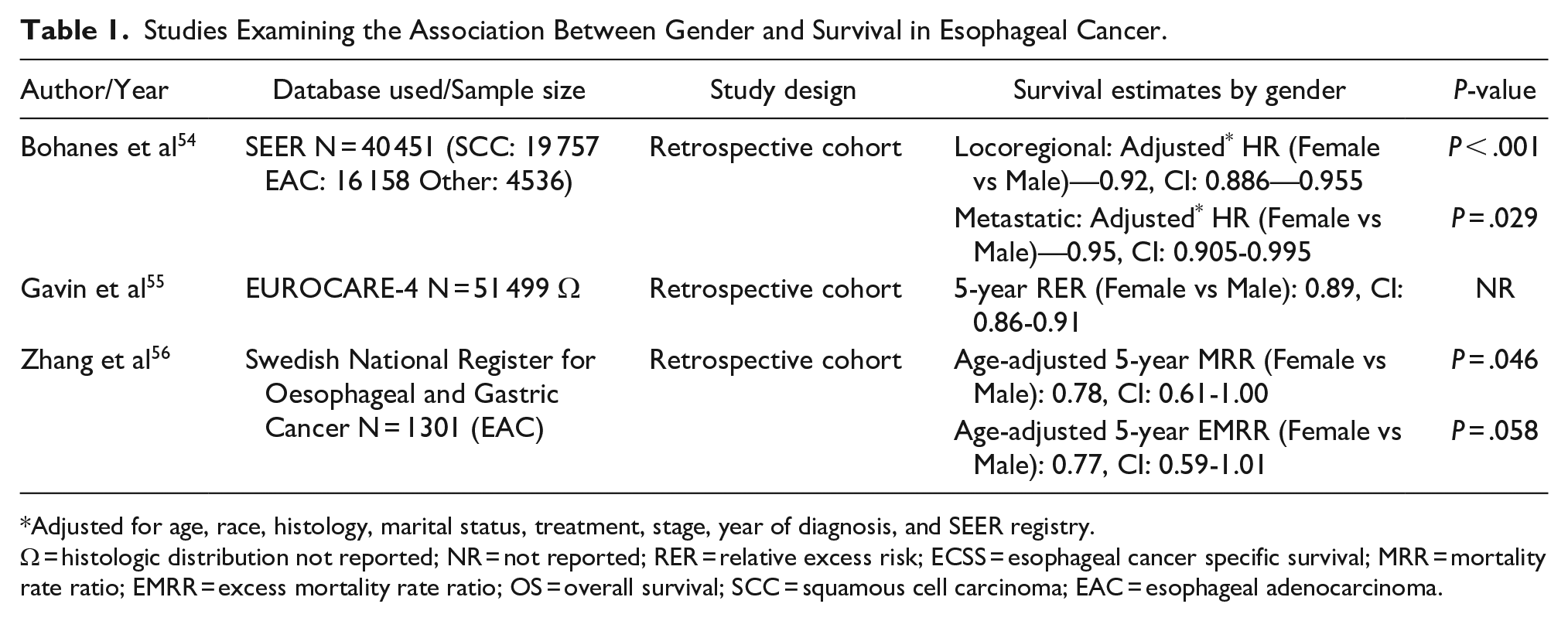
Adjusted for age, race, histology, marital status, treatment, stage, year of diagnosis, and SEER registry.
Ω = histologic distribution not reported; NR = not reported; RER = relative excess risk; ECSS = esophageal cancer specific survival; MRR = mortality rate ratio; EMRR = excess mortality rate ratio; OS = overall survival; SCC = squamous cell carcinoma; EAC = esophageal adenocarcinoma.
Race, Ethnicity, and Esophageal Cancer Survival
Several large studies on esophageal cancer have reported lower overall survival in racial minorities as compared to non-Hispanic whites (Table 2).31,57-59 Racial minorities are also more likely to be diagnosed at advanced stages and less likely to be undergo surgery for esophageal cancer.31,57,58 Using SEER data, Greenstein et al 57 reported a significantly lower esophageal cancer-specific survival in black than white patients (37%, CI: 28%-45% vs 60%, CI: 57%-63%; P < .001). Black patients were also significantly less likely to undergo resection than whites (44% vs 66%; P < .001) and more likely to receive only radiation therapy (43% vs 22%; P < .001). Dong et al 58 also reported a lower 5-year survival in demographically matched black than white patients with esophageal cancer (13.3% vs 18.4%). Using a 3-step tapered multivariable matching process, these authors sequentially showed a decrease in the 5-year survival difference between blacks and whites from 5.1% (P < .001) after matching for demographics alone, to 2.3% (P = .04) after matching for demographics and presentation. The survival difference subsequently disappeared after matching for treatment (P = .59), indicating that the underuse of treatment may account for disparities in survival between black and non-Hispanic whites. 58 Greenstein et al 57 reported similar findings in their analysis; after adjustment for T-status and treatment, black race was no longer associated with an increased hazard of esophageal cancer death.
Studies Examining the Association Between Race and Survival in Esophageal Cancer.
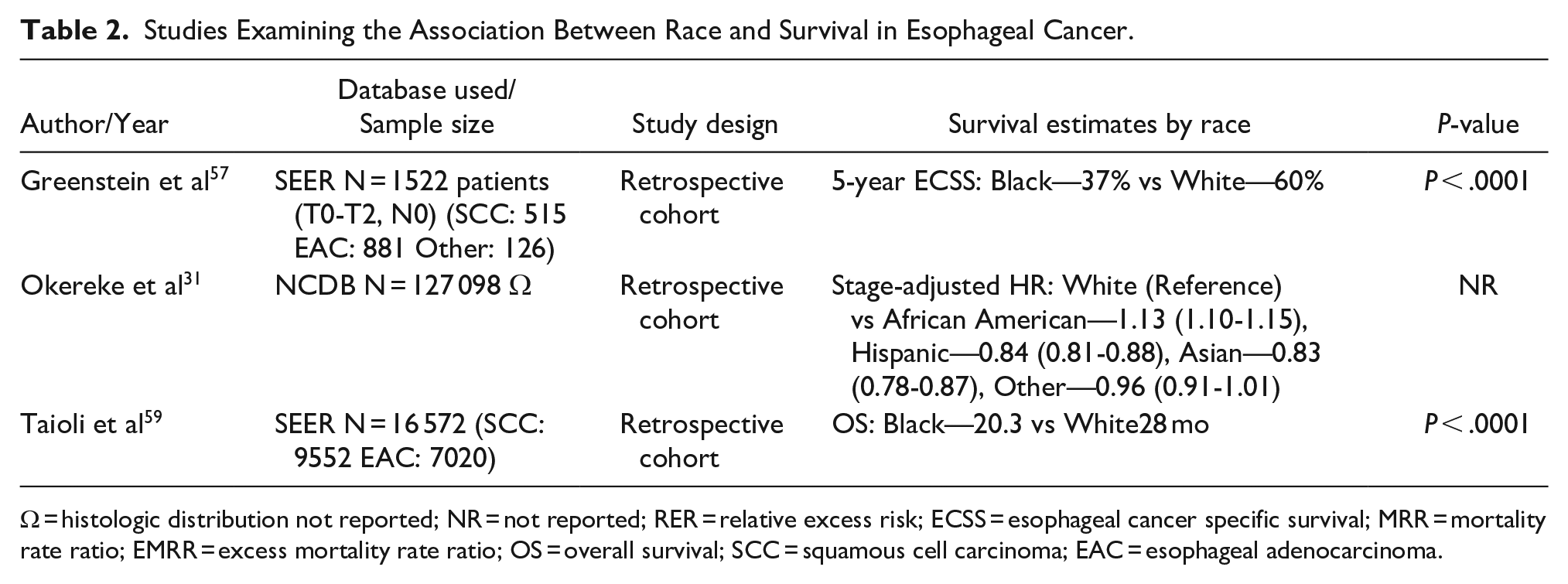
Ω = histologic distribution not reported; NR = not reported; RER = relative excess risk; ECSS = esophageal cancer specific survival; MRR = mortality rate ratio; EMRR = excess mortality rate ratio; OS = overall survival; SCC = squamous cell carcinoma; EAC = esophageal adenocarcinoma.
Despite being less likely than whites to receive surgery for esophageal cancer, a study described improved survival in Hispanics and Asians compared to whites. Blacks had the lowest survival of all racial groups. 31 This trend may be related to differences in tumor biology, lifestyle and epigenetics and has been termed the Hispanic paradox phenomenon. 60
The AI/AN population is an under-reported minority in several population-based studies. Geographic region appears to play a role in AI/AN cancer incidence and mortality, potentially due to varying social, cultural, and environmental factors. White et al 61 in their study across 6 geographic counties in the US, noted a higher mortality for AI/AN men with esophageal cancer in Alaska. Another recent study by the Alaska Native Tumor Registry compiling 50 year of cancer surveillance data showed that mortality from esophageal cancer was higher among AN compared to US whites. 62
The only large study addressing cancer risk in transgender patients noted that compared to cisgender, these patients may be diagnosed at advanced stages, be less likely to receive treatment and have worse survival for many cancer types, including non-Hodgkin lymphoma, bladder, and prostate cancer. 63 They quote that similar non-significant associations were found for other cancer sites, although they do not elaborate on esophageal cancer-specific data. This study underscores the need for more transgender focused research in the future.
The existing racial disparities in esophageal cancer survival that predominantly affect minorities may partly be related to patient- and provider-level factors. 64 Patient-level factors like sociocultural beliefs, risk aversion and treatment preferences may influence cancer prevention and health seeking behavior. 64 Patient-provider interaction may also be limited by biases, cultural differences, differences in values, experiences, and communication barriers. 57 Stiles et al 65 previously reported patient-level factors that are associated with a refusal of esophagectomy for cancer despite receiving a surgical treatment recommendation. These factors included non-white race, older age, female sex, and a lack of insurance. 65 Another study on patients with locoregional esophageal cancer noted a lower likelihood of any surgical consultation among black patients. 66 Among those who had a surgical evaluation, the likelihood of undergoing surgery was also significantly lower in black patients. 66 Regarding BE screening, primary care physicians who often represent the first point of contact for patients with chronic reflux experience difficulty identifying patients who should be screened and have a poor knowledge on screening guidelines. 67
Population-level analyses that specifically examine sex and ethnic disparities in EAC outcomes are scarce. Hence, definitive conclusions cannot be made regarding the association between these variables and EAC survival outcomes.
Conclusion
Sex, racial and ethnic disparities in the incidence and prevalence of BE and EAC are well established. It is important to note that although the incidence of BE and EAC is highest in non-Hispanic whites, the black population seems to have the worst survival. Ethnic minorities disproportionally suffer poor outcomes as they are often diagnosed at advanced stages and are less likely to receive surgical treatment. More data is needed on understudied populations such as AI/AN, Asian Americans, and transgender people. Available data should be used to guide surveillance and to improve access to care with a goal to mitigate modifiable factors affecting these disparities.