
Abstract
Background:
Advantages of using an intramedullary (IM) nail over a lateral plate for fixation of distal fibula fractures include the ability to allow for earlier weightbearing and return to sport, smaller incisions, and decreased hardware prominence. When comparing a fibular nail with an IM screw, the nail allows for syndesmosis fixation through the device and imparts rotational stability. Long-term studies support the viability and clinical outcomes of using an IM nail. In this technique video, a case is presented to illustrate the use of an IM nail for fixation of a distal fibula fracture.
Indications:
Distal fibula fractures should be treated operatively in the following cases: open fractures, bimalleolar fractures, or lateral malleolar fractures with medial clear space widening, displacement >3 mm, or decreased tib-fib overlap, increased talocrural angle, any talar displacement, fracture-dislocation, or nonunion. When treating these fractures operatively, it is crucial to get an anatomic reduction, as this will lead to a satisfactory outcome. By restoring the anatomy, the talar shift is decreased, and a normal tibiotalar contact area is approached.
Technique Description:
Percutaneous incisions are made around the fracture to place a clamp and hold anatomic reduction. Once reduced, a small incision is made 1cm distal to the fibular tip and in line with its axis. The start point is checked with the K-wire on anteroposterior and lateral views. An opening reamer is used, followed by a flexible guidewire and then a proximal reamer. Talons are deployed once the appropriate position is confirmed, and interlocking screws are placed through the guide. Syndesmosis fixation is added if necessary.
Results:
The success rate for achieving good outcomes is high at 90%. Patients should be counseled that their braking time while driving is normal at around 9 weeks and that overall recovery is about 2 years.
Discussion/Conclusion:
This technique should not be used if the 6.2-mm opening reamer will breach the cortex, and should not be used if a plate will provide superior fixation. Like with any nailing procedure, obtaining and maintaining the reduction is paramount. A proper start point is critical for optimal fixation.
Patient Consent Disclosure Statement:
The author(s) attests that consent has been obtained from any patient(s) appearing in this publication. If the individual may be identifiable, the author(s) has included a statement of release or other written form of approval from the patient(s) with this submission for publication.
This is a visual representation of the abstract.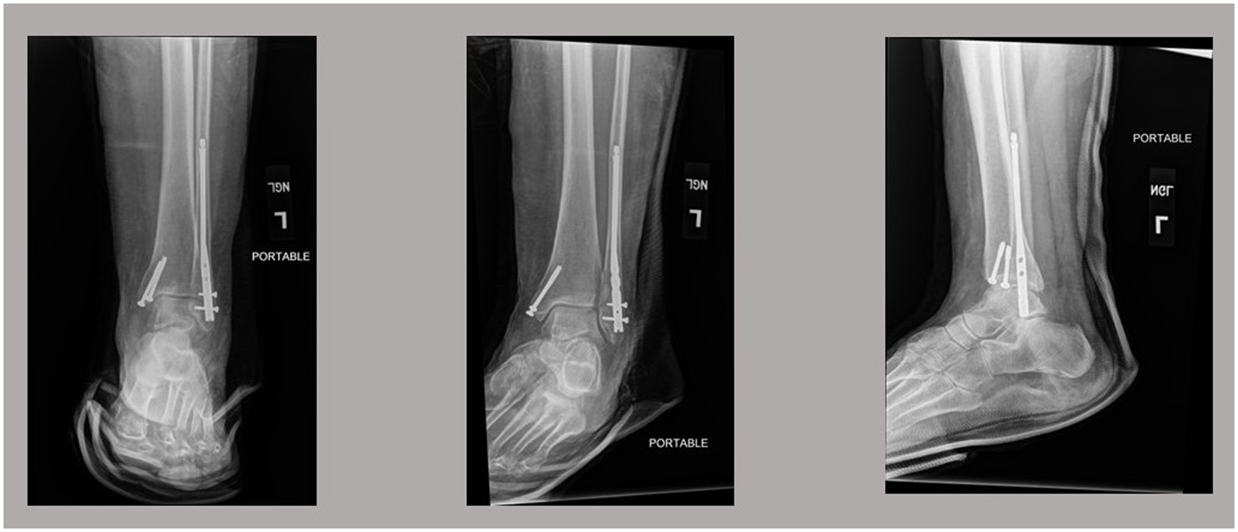
Video Transcript
This video demonstrates minimally invasive fixation of a distal fibula fracture with an intramedullary nail.
Background
Advantages of using an intramedullary (IM) nail over a lateral plate for fixation of distal fibula fractures include the ability to allow for earlier weightbearing and return to sport (RTS), smaller incisions, and decreased hardware prominence.1,2,3,5,6 Long-term studies support the viability and clinical outcomes after use of an IM nail.7,8,9,10,11 In this technique video, a case is presented to illustrate the use of an IM nail for distal fibula fracture fixation.
The patient is an 80-year-old female who was traveling to Turkey and had a syncopal episode on the airplane, resulting in a fall. She had an immediate onset of left ankle pain and inability to bear weight. Upon arrival in Turkey, she went to an urgent care, where she was splinted. She booked a flight back to San Jose and came to the emergency room locally, where she was admitted for surgical treatment. Physical examination was consistent with her injury, with moderate swelling and ecchymosis, and no neurovascular deficits.
Her radiographs showed this bimalleolar ankle fracture with moderate displacement of the medial malleolus and a transverse fracture of the distal fibula with slight lateral translation of the distal segment and shortening.
Indications
Given her radiograph findings, she met the indications for surgery. To review, distal fibula fractures should be treated operatively in the following cases. Open fractures, bimalleolar fractures, or lateral malleolar fractures with medial clear space widening, displacement >3 mm or decreased tib-fib overlap indicating syndesmosis injury, increased talocrural angle, which essentially means the fibula is short, any talar displacement, fracture-dislocation, or nonunion.6,7,11 When treating these fractures operatively, it is crucial to get an anatomic reduction, as this will lead to a satisfactory outcome. By restoring the anatomy, the talar shift is decreased, and a normal tibiotalar contact area is approached. The success rate6,7,11 for achieving good outcomes is high at 90%. Patients should be counseled that their braking time while driving is normal at around 9 weeks and that overall recovery is about 2 years.
When considering using a nail versus using a plate, the advantages of a nail include early weightbearing, greater freedom to operate despite moderate swelling, and decreased hardware prominence.1,2,3,4,5,6 With a nail versus a long screw, there is rotational control proximally and the option for syndesmosis fixation through the device.
Technique Description
For this case, a nail was selected for fixation of the fibula. The patient was positioned supine, at the end of the table, with a bump under the hip to put the extremity in neutral rotation. A foam ramp was placed under the leg. C-arm came in from the contralateral side, and a C-armor drape was used. A tourniquet was not necessary.
Steps of the Operative Plan
Fix the medial malleolus in standard fashion. Then, carry out fixation of the fibula. As with any fracture case, reduction must be obtained and maintained. Then, the nail is placed. The start point is critical.
After sterile prep and drape and before making incisions, the skin marker was used to mark out the level of the distal fibula fracture and the axis of the fibula. A freer elevator was used on the anteroposterior (AP) view to confirm the level of the fracture for planned small incisions and placement of a clamp, and the same elevator was used on the lateral view to confirm the fibular axis. The medial malleolus was fixed quickly with an open reduction and 2 partially threaded cannulated screws.
Then, attention was turned to fixation of the distal fibula. Two stab incisions were made, 1 posterosuperior to the oblique fracture line and 1 anteroinferior. One clamp was placed to anatomically reduce the fracture, and the reduction was checked on fluoroscopy. With the fracture reduced, a 1-cm long incision was made distally, 1 cm distal to the tip of the fibula and in line with the fibular axis.
The starting point was identified with the K-wire, checking both AP and lateral views to ensure it was at the very tip of the fibula. The K-wire was advanced along the fibular axis and was central on both views. Once the K-wire was advanced, the opening reamer was used, advancing until the flutes were buried.
Next, the flexible guidewire was used through the fracture finger tool to advance the guidewire up the fibula longer than the length of the nail.
The proximal reamer was used, and the larger of the 2 proximal reamer options was selected to better fill the canal.
With the canal prepared, the IM nail was placed in appropriate alignment and rotation.
The guide was used to ensure the nail was buried and aligned with the distal fibula, and the radiograph confirmed that the syndesmosis holes lined up at about 2 and 4 cm above the tibial plafond. Talons were deployed proximally by rotating the screwdriver, and rotational control was achieved. Distal interlock screws were placed through the guide.
The stress external rotation view showed that syndesmosis fixation was not necessary.
Final radiographic imaging demonstrated appropriate alignment and rotation.
How to Avoid Potential Complications With This Technique
Do not use an IM nail if the 6.2-mm opening reamer will breach the cortex, and do not use it if a plate will provide superior fixation. Like with any nailing procedure, obtaining and maintaining the reduction is paramount. A proper start point is critical for optimal fixation.
Results
The patient was permitted to weightbear as tolerated at 2 weeks and to start physical therapy. In general, patients should be counseled that RTS is 6 to 9 months.
Physical therapy should follow a stepwise protocol aimed at reducing swelling and improving range of motion in the early postoperative period, followed by strengthening, proprioception, and sport-specific activities.6,7,11
Outcomes in athletes depend primarily on the number of malleoli fractured. Patients with a single fractured malleolus have the best outcomes and the highest rates of RTS. Athletes with trimalleolar ankle fractures have longer recoveries and lower RTS rates.
Footnotes
One or more of the authors has declared the following potential conflict of interest or source of funding: J.M.L. is a paid consultant; receives honoraria for lectures, presentations, speakers bureau participation, manuscript writing, or educational events; and receives support for attending meetings and/or travel from Arthrex. AOSSM checks author disclosures against the Open Payments Database (OPD). AOSSM has not conducted an independent investigation on the OPD and disclaims any liability or responsibility relating thereto.