
Abstract
The increasing demand for alternative approaches to antibiotics has sparked renewed global interest in phage research worldwide. While numerous recent success stories are finding their way into the public eye, there are still some concerns related to phage treatments that need to be considered, particularly phage stability. However, phage stability-determining parameters have not been thoroughly evaluated across different phage-related applications. Differences in methodological approaches complicate comparisons between studies and hinder the establishment of standardized protocols for most applications. In the present work, a systematic review was performed using Web of Science and Scopus to identify studies on phage stability under varying environmental conditions, namely temperature, pH, salinity, and ultraviolet exposure between 2014 and 2024. Studies were considered eligible if conducted in buffers, media, or other suspensions and if phages were not manipulated to improve stability prior to evaluation. A total of 327 articles were analyzed and their limitations and implications for industrial and clinical applications evaluated. By addressing these gaps in current knowledge, we aim to provide a clearer understanding of the need for standardization in phage stability studies, ultimately enabling their effective application in diverse environments and enhancing their translation into practice.
Introduction
Bacteriophages (phages) are viruses that specifically infect bacteria and are increasingly considered to be a promising alternative to treat bacterial infections and tackle antibiotic resistance. 1
Since phage discovery in the early 20th century, phage therapy has become a common practice in the Soviet Union and continues in some of its former countries until today. 2 However, in other parts of the world, advances in the regulation framing phage therapy are still under development. Nonetheless, its use as an experimental therapy for the compassionate treatment of patients experiencing antibiotic failure has been increasing.3,4 Currently, Belgium, France, Sweden, the United Kingdom, the United States, Australia, and most recently Portugal have established phage therapy programs for compassionate use to treat human infectious diseases.5,6 After the first successful results of phage therapy in human medicine, knowledge was quickly translated to other areas, such as veterinary medicine, agriculture, aquaculture, and food industry, where this approach has been well received and commercial phage preparations have already been approved.4,7,8
While in human medicine the efficacy of phage therapy has proven to be an efficient alternative to fighting infections caused by drug-resistant bacteria, phages can be useful even when bacterial strains are still sensitive to antibiotics. As examples, one can refer to the treatment of infections caused by intracellular bacteria, infections caused by bacteria forming biofilms, bacterial chronic infections, infections in areas inaccessible to antibiotics (e.g., wounds and diabetic foot ulcers), which affect patients with poor circulation, and even in the treatment of bacterial systemic infections, as well as patients who have developed antibiotic allergies.9,10
In veterinary medicine, food industry, agriculture, and aquaculture, phages can also be used to fight antibiotic-resistant bacteria, as well as to mitigate bacterial outbreaks caused by emerging bacteria that can cause devastating losses and negatively impact sustainability, economy, food safety, and even food security. In aquaculture, the contamination of bivalve mollusks with harmful bacteria (often exacerbated by limited access to pristine water sources) poses a significant risk to consumer health. 11 Similarly, in agriculture, bacterial diseases, such as kiwi canker, can devastate orchards, 12 while animal husbandry faces ongoing challenges from pathogens such as Salmonella enterica. 13 Overall, the food industry is particularly vulnerable to threats posed by pathogenic bacteria, as these can contaminate food at any stage of the production pipeline, with bacteria being able to cause spoilage from fermentation, production of odors, and changes in texture (sliminess) and appearance (browning). 14
Phages offer a promising alternative treatment for clinical and industrial settings due to their specificity, a feature that allows them to target and eliminate specific pathogenic/spoilage bacteria without harming beneficial microbiota. Phage treatments can be applied directly to humans or animals, via injection, aerosols, topical application, or in feed.15–17 They are additionally used in agriculture through spraying or soil inoculation,18,19 in the food industry via surface disinfection by spraying or food package addition,20,21 and in aquaculture via feed or water addition to the fish tanks or during bivalve depuration.15,22,23 While the efficacy of phage therapy has proven to be an efficient alternative to conventional antibiotics, there are still some well-known concerns that need to be considered. The emergence of phage-resistant mutants, the narrow host range of phages, and the lack of relevant regulatory framework and phage stability are current bottlenecks that still need further research. Several approaches may control the development of bacterial phage resistance, such as the use of phage cocktails directly or sequentially, phage combined approaches (phage and antibiotics), the use of engineered phages, and the continuous search for novel phages.1,24,25 The problem of a narrow host range can be solved by using phage cocktails, 26 establishing phage libraries, 27 and expanding the host range of a single phage by direct in vitro evolution or using genetic engineering.25,28 On what concerns regulations, new dedicated regulatory frameworks for medicine use and also for decontamination/disinfection use in nonclinical applications have been improving and can pave the way for a more generalized application of phage therapy.4,29 Similarly, guidelines for phage isolation and preparation have also been developed for animal use (EMA/CVMP/NTWP/32862/2022), aiming to produce safer and more effective phage preparations.30,31 However, regardless of their purpose, all phage applications require virion stability, a feature affected by physicochemical conditions (Fig. 1).4,31 Unfortunately, specific guidelines for the evaluation of phage stability are yet to be clearly detailed, a caveat that certainly requires further attention by the scientific community.
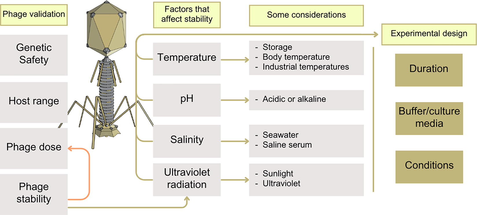
Process of bacteriophage characterization for application with emphasis on phage stability.
To successfully apply phage therapy, both in clinical and industrial settings, there are several stability-determining parameters that must be addressed prior to their application. 26 Phage dosing, the number of phages that are added for each available bacterial host, should be carefully studied beforehand, to safeguard that a high multiplicity of adsorption will take place in as many as possible available targets 32 without jeopardizing the infection through exposure to lower doses or by lysis from without. 33 While it should be relatively straightforward to calculate a dose for a suspension in a laboratory, transferring this application to a real-world clinical or industrial scenario can be challenging,32,34 particularly when pathogenic bacteria are located within animal tissues or biofilms. 35 Furthermore, environmental conditions, such as temperature and pH, in clinical and industrial settings, as well as salinity and ultraviolet (UV) radiation, particularly in industrial settings, can significantly affect phage viability.36,37 Most of these factors will experience more or less pronounced shifts throughout the year, particularly in outdoor applications. These factors can interfere with capsid stability and the preservation of genetic material, or even affect the genetic injection host recognition and adsorption, having a profound effect on phage infection outcome.38–40 This phage instability may cause a dosing failure and allow for the infected bacterial population to be replaced with resistant bacteria at a rate that will not promote recovery from the disease. 41 Understanding how phages will perform under different physiochemical conditions is, therefore, crucial for the development of effective phage therapy protocols. If variations in pH, temperature, or salinity foster a more pronounced phage decay, new phage doses or preservative agents/supports can be used to guarantee high phage numbers during treatment.34,42 In scenarios where solar UV radiation will cause phage decay (e.g., aquaculture or agriculture), phages can be applied at dusk to avoid periods of direct sun exposure.19,36 Nonetheless, since each phage will present a unique stability, it should be individually evaluated for a predetermined scenario prior to its application or combination in cocktail. However, unlike the other concerns associated with phage treatment, an in-depth understanding of the effect of environmental conditions on phage stability is still largely missing.
While research into phage stability has become a common property of the isolation and characterization process, most of the studies published to date solely focus on short-term experiments. These may not accurately reflect the procedures used in clinical and industrial settings since virions require time to diffuse through tissues or matrices to reach their targets. Moreover, the lack of standardization among phage stability studies, along with the lack of studies under environmental conditions that mimic potential therapeutic or preventive settings, means that the findings reported often lack direct applicability to different and/or real-world scenarios. The present systematic review summarizes the main findings on phage stability under varying environmental conditions (particularly temperature, pH, salinity, and UV exposure), while also examining their main limitations and implications for industrial use. By addressing these gaps in current knowledge, this review aims to provide a clearer understanding on how phages can be effectively applied in diverse environments, boosting their translation theory into practice.
Literature Search Methodology
A literature search was performed with Web of Science (WoS) and Scopus for studies that characterized phage stability with regard to temperature, pH, salinity, and UV radiation using the Preferred Reporting Items for Systematic reviews and Meta-Analyses (PRISMA) 2020 reporting guidelines. 43 PRISMA checklists for the systematic review are provided in the Supplementary Data (PRISMA 2020 checklist and PRISMA abstract checklist).
Eligibility criteria
Studies published in English language between 2014 and 2024 were considered eligible if they addressed any of the points aforementioned using suspensions of the bacteriophage in buffer solutions or media without any prior manipulation (genetic engineering of phage, lyophilization into dry powders, nebulization, fixation to surface, encapsulations or gel emulsions, etc.).
Search strategy and data extraction
Articles were extracted by J.D. from WoS and Scopus using the following keywords: bacteriophage stability OR phage stability AND pH AND salinity AND temperature AND Ultraviolet Radiation.
A total of 1267 potentially relevant studies were retrieved from the search and refined to 327 articles after removal of duplicates and by not fulfilling the inclusion criteria detailed above (Fig. 2 and Supplementary Tables S1, S2, S3 and S4). The abstracts of all articles were confirmed for potential interest in the review and in case of doubt, the articles would be analyzed for possible evaluation of phage stability under the criteria previously mentioned. The following data were accessed: year, phage name, suspensions used, temperature range, pH range, salinity range, UV conditions, and duration for each experiment.
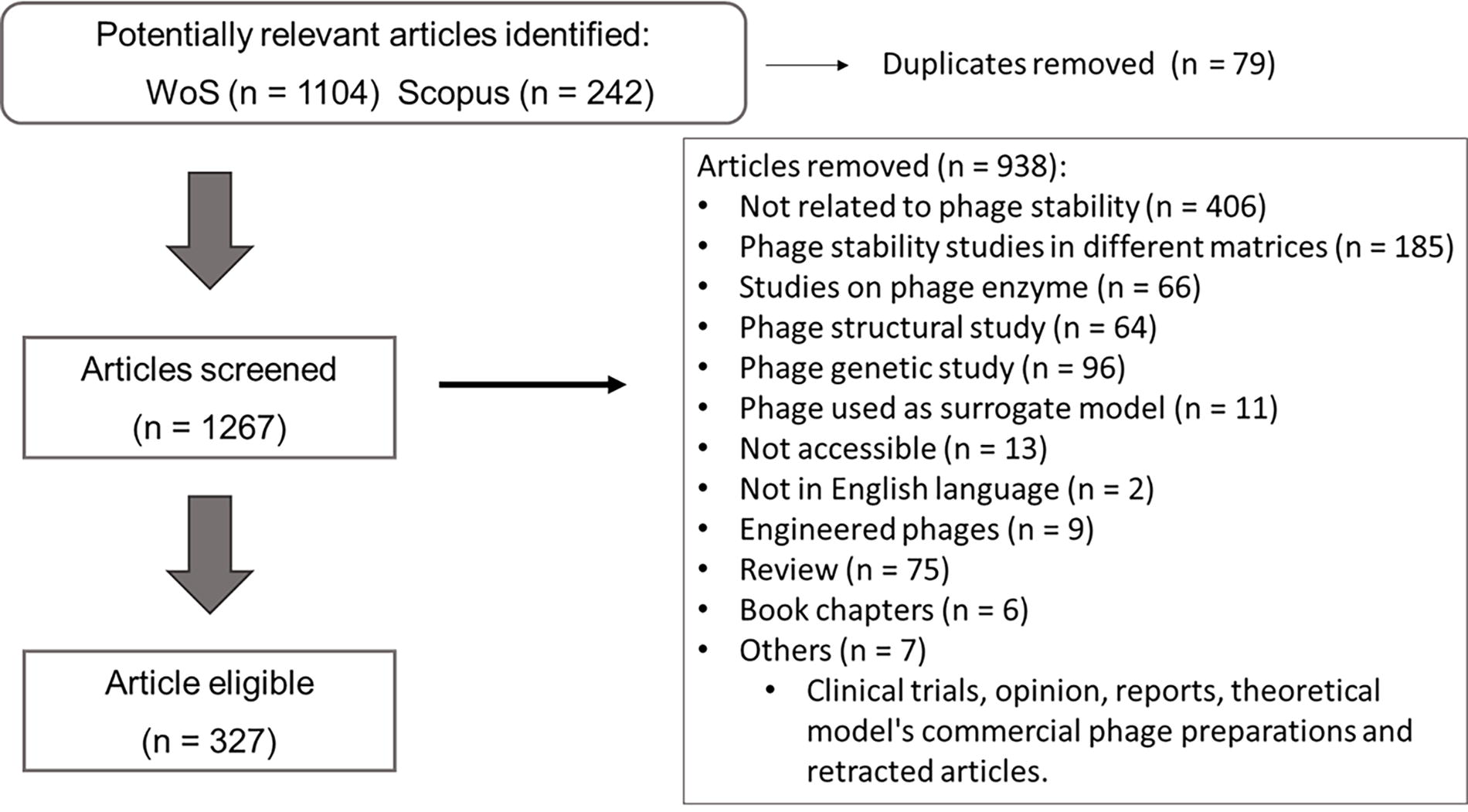
PRISMA flow diagram depicting the screening and selection process of peer-reviewed scientific publications on phage stability.
Data synthesis and analysis
The stability of each phage was described as reported by the authors of each study (stable, tolerant, detectable, etc.) (Supplementary Tables S1, S2, S3 and S4). Time of exposure was transferred to the same scale for comparison (minutes or hours). Concentrations of NaCl were transformed to salinity (% [w/v] NaCl) for comparison, but kept in the original description in the Supplementary Tables. If in a study more than one phage was isolated, several entries were added accordingly and comparisons were performed by phage entries and not by study. While the information of each study is described in the supporting information, not all information was comparable. Therefore, images presented in the article are only representative of all comparable phages. When possible, results were grouped by time intervals to assess trends. In these cases, a circle graph was added to represent the number of phages that were addressed.
Due to substantial methodological heterogeneity across studies, statistical synthesis or formal meta-analysis was not feasible. Instead, representative visualizations were used to summarize findings. Formal assessments of risk of bias, sensitivity analyses, certainty of evidence (e.g., GRADE), and effect measures were not conducted, as the review followed a narrative synthesis approach and was carried out by a single reviewer, limiting the feasibility of these methods. This introduces inherent limitations that may affect reproducibility. Data were processed using GraphPad Prism software version 8.4.3 (San Diego, CA, USA).
Environmental Factors Affecting Phage Stability
Temperature
Temperature can affect phage viability through different interactions with phage structure or its infection mechanism. 44 In general, the phage capsid is a thermoresistant structure that protects the nucleic acid of the phage, with protein denaturation being observed at temperatures of 87°C. 40 Furthermore, phage nucleic acid can be ejected from the capsid before reaching these higher temperatures, rendering the virion empty 38 and, as such, unable to cause infection. Phages can also carry enzymes that are crucial for cell infection and have varying thermostability. 45 If such enzymes exhibit low thermal stability, completely matured virions may still be present and not be detected through plating techniques, as these will lack infectivity. 46 Other phage constituents, such as tail tape measure protein (tail protein), the receptor binding protein (attachment to cell surface), spike proteins, and other structural molecules commonly present, feature different denaturation points and therefore detrimentally affect phage stability or, at least, cause a more deficient infectivity.39,40,46 One must highlight that ultimately, virion stability will most likely be determined by its most sensitive constituent.
In most studies, virion stability to higher temperatures was observed between 37°C and 45°C (Fig. 3A). Phage stability began to be negatively affected when temperatures were increased above 45°C, with some studies observing higher thermotolerance up to 60°C, with none or very slight reduction in phage titer when compared with phage groups exposed to lower temperatures (Supplementary Table S1). While phage incubation times varied among studies, most phages studied to date were incubated only for periods up to 1 h (Fig. 3B). This approach resulted in a considerable variation in results reported in the literature. In studies that incubated phage for only an hour, the most frequent maximum temperature at which the phage was considered stable was in average 45.9 ± 14.2°C (with a mean and mode temperature of 50°C). However, prolonged incubation periods fostered different conclusions, being the value reduced to 12.3 ± 12.6°C after more than a day of incubation (Fig. 3A). These findings suggest a possible overestimation of phage resistance to temperature under shorter incubation periods.
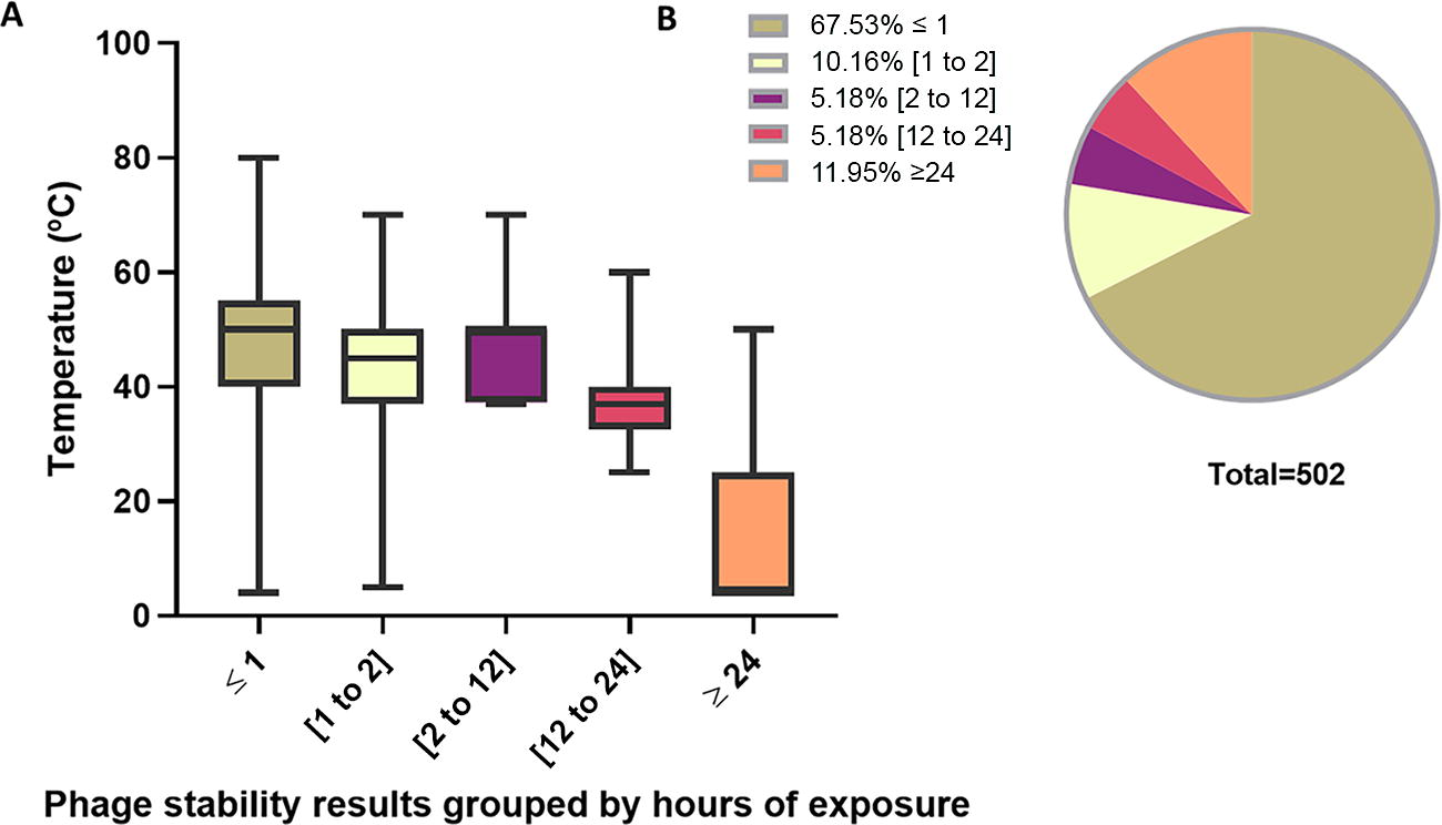
Phage’s maximum temperature stability by exposure interval. Only studies in which phage stability was determined were included. A total of 502 phages were used and the maximum temperature at which the phage was considered stable was grouped by period of exposure.
When phages were incubated for up to 2 h, a decrease in stability was observed, with an average maximum temperature resistance of 47.2 ± 10.1°C. These differences may be attributed to the time required for phage suspension to reach the desired temperature, as only a few studies describe a prewarming process of the medium to the selected temperature before phage inoculation (see the Experimental Period section).
Phage SaFB14 was considered stable up to 42°C for 6 h, 47 whereas phages vB_AbaS_TCUP2199 and CUB-EPI_14 were stable at 37°C for 24 h, but were affected by temperatures above 37°C.48,49 Contrarily, phage vB_VhaS-R18L showed a titer reduction when exposed to temperatures above 20°C for 48 h. 50 Interestingly, when phage PHA46 was incubated at temperatures ranging from 4°C to 37°C, a titer decrease was observed after only 12 h for all conditions and continued to decay during 2 weeks of exposure.
Similar variations in thermostability were observed when phages were exposed to lower temperatures. In general, most phage’s titer remained stable when kept at 4°C. However, differences in phage stability were observed when incubation temperature was lowered to −20°C or −80°C. Phage Hy01 and phage VVP001 showed stability for 12 h at −20°C, 51 while phage BF25/12 was stable for up to 1 year at −80°C. 52 However, phage vB_SmaM_PSM suffered inactivation after 1 h at −20°C. 53 Similar results were also observed for phage vB_EcoM-UFV13 at −20°C. 54 It is worth referring that phages Hy01 and VVP001 were inoculated in SM buffer, whereas phages vB_SmaM_PSM and vB_EcoM-UFV13 were both incubated in phosphate buffer saline (PBS) and Lysogeny broth (LB), respectively. As such, these differences may hinder a proper comparison and the differences recorded should be interpreted with caution. When phages LOCV2, LOCV5, and LOCARD were freeze–thawed in the same conditions, all phage titers were significantly reduced; however, a higher decrease was observed for phage LOCV2, whereas phage LOCARD titer remained high. 55 Contrarily, when phage ZCKP2 was incubated in SM buffer at −20°C, it did not present any significant level of inactivation. 56
Since each phage may present different susceptibility to freezing temperatures, preserving agents may be added during cold storage to improve stability for several months or years. 57 Similarly, for warmer temperatures, phages may have their stability improved by encapsulation. 42
pH
Phage stability is also known to be dependent on the acidity or alkalinity of the environment. 58 Shifts in pH can impact the infection outcome of viruses through modifications in their binding efficiency and/or the damaging of its enzymes. In addition, it can play an important role on the stability of the free virions, by disrupting the capsid or by inducing other structural changes.58,59 To best understand if a certain phage can be successfully applied, knowing its ability to withstand shifts in pH is paramount to guarantee that the goal of its use is successfully achieved. In marine aquacultures, phages should be expected to withstand slightly alkaline environments (pH ranging from 7.5 to 8.4), 36 whereas in other industries, such as their use in terrestrial livestock or human health, pH may vary in a more pronounced way, from very acidic to more alkaline environments.27,60,61
When the information available in scientific literature was organized by time interval, it was observed that prolonged incubations led to a narrower margin of pH tolerance (Fig. 4A). Between 12 and 24 h, or even more, pooled results suggest that the tolerance range and resistance to highly alkaline or highly acidic conditions decrease, along with a reduction in the number of reported phages being resistant to these conditions. In general, most isolated phages were stable in a pH ranging between 5 and 9 for 1 h and pH 6–8 after 24 h (Fig. 4A). However, there were considerable variations in results caused by methodological differences in the incubation temperature or buffer suspension used (further discussed in the Temperature section), features that in some studies were not thoroughly described (Supplementary Table S2). These differences may have caused additional variations that may have not been properly taken in the performed analysis. Despite this, several phages present strict pH tolerance. As an example, phage vB_Kpn_ZC2 presented stability only at pH 7, with a significant reduction occurring at pH equal or below 6 and equal or above 8 after an incubation of 4 h. 56 This strict tolerance was also observed for phages k2a, k2b, kp9, and k9coc (pH 7) and phage k2w6 (stable only at pH 6), despite a shorter incubation of only an hour. 62 Interestingly, when phage vB_VhaS-R18L was incubated in SM buffer with a pH range of 2–12, a decay in phage titer was observed in all test groups after 24 h of incubation. While the phage titer revealed a sharp decline in more extreme pH conditions namely equal or below 5 and equal or above 10, a decrease was also observed within the pH range of 6–9. 50 Similarly, when phage PHA46 was incubated in a pH range between 5 and 7, a reduction in titer of nearly 2 log PFU/mL was observed after 6 h. 63 Phage LOCV5 presented similar instability when incubated for 2 h in pH of 2.5, 6, and 10, whereas LOCV2 was not significantly reduced in pH of 6 and LOCARD was stable at pH 6 and 10. 55 However, certain phages seem to be able to withstand more acidic and alkaline environments. Phage Mac-1 was able to remain stable at a pH of 5 during 15 h. 64 In a more extended assay, 30 days, phages vB_AbaP_HB01 and vB_AbaM_HB02 remained viable at a pH of 4 for 2 days with a titer reduction of less than 1 log PFU/mL. Phage HB01 was inactivated after 30 days to the detection limit of the method, whereas phage HB02 titer remained above 50% of its initial value. 65
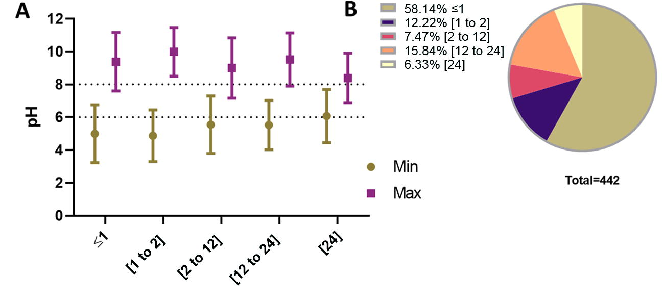
Phage pH stability grouped by incubation time interval.
UV radiation
UV radiation is one of the most relevant conditions for phage application.36,66 In numerous scenarios, phages will eventually be exposed to this type of radiation, either delivered from solar rays, for example, during application in orchards or aquacultures, or by UV-lamps, for example, at bivalve mollusk depuration units or food industry settings.67,68 While the UV spectrum is normally divided into UV-A (315–400 nm), UV-B (280–315 nm), and UV-C (100–280 nm), all these wavelengths are known to cause different types of damage to DNA, 69 thus negatively affecting phages in different ways. Studies of phage resistance to UV radiation are normally performed by placing phage suspensions under artificial lamps at variable distances. There are less studies performed under direct solar radiation or using artificial simulators of sunlight. As a multitude of exposure conditions were detailed in the different studies surveyed in the present work, the results reported also varied, from total phage decay in minutes to high titers maintained for hours (Supplementary Table S3). Furthermore, the different volumes used from as low as 200 µL 70 to larger volumes of 30 mL 36 (Supplementary Table S3) affected the thickness of the penetrated layer and possibly affected the outcomes of the studies.
In studies that evaluated the effect of specific wavelengths, UV-C was the most frequently used type of UV radiation and caused a higher decrease in phage viability, with all phages revealing decreases in titer and 52% being inactivated to the detection limit of the method after minutes up to 1–2 h).71–73 When phages were irradiated under UV-B, 72.7% had variations in titer and only 36.4% were totally inactivated to the detection limit of the method. Some phages were tolerant to this wavelength, with reductions of 0.1 log in 30 min 74 or 0.5 log in an hour. 75 However, others were more sensitive to this type of radiation, with reductions of 90% in 15 min 76 and inactivation to the detection limit of the method in 80 min. 77 While fewer studies analyzed the effects of UV-A radiation (Fig. 5), total inactivation was not recorded and only 45.5% of phages had variations in titer. However, a decrease of 2.13 log for phage PHB09 78 and significant differences in phage titer were still recorded after 20 and 30 min for phages PcaP1EGY and PcaP2EGY. 79 In less specific wavelengths (direct sunlight or solar simulators), phages revealed a higher tolerance (1–2.5 log reduction after more than 6 h).
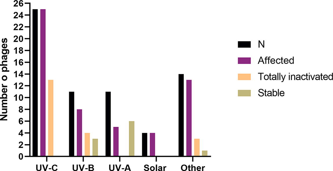
Studies on phage stability under different types of ultraviolet radiation (UV). From left to right—number of phages studied for each category (n); number of phages that displayed titer reduction (affected); number of phages that were inactivated to the detection limit of the method (totally inactivated), and number of phages considered stable during the study (stable). Experimental duration range by UV type: UV-A (30–60 min); UV-B (60–1440 min); UV-C (2–360 min); solar (360–720 min), and others (12–120 min). Detailed information is presented in Supplementary Table S3.
When exposed to UV-C radiation, phage TE consistently decreased from 16 log PFU/mL to 4.2 log PFU/mL after 120 min of exposure. 80 Similarly, when exposed to UV-C (250 nm), phage ZCSS1 decreased and, after 60 min, no phage particles were detectable. 37 When phages Psst1, Psst2, and Psst4 were exposed to UV-C (253.7 nm) for 140 min, both phages Psst1 and Psst2 were totally inactivated after 80 min, whereas phage Psst4 was only completely inactivated after 140 min. 71 In studies that used different types of UV radiation wavelength, phages were able to survive longer periods. Phages L541, W41, MLA23, and LBH01 were more affected by UV-B than by UV-A radiation during 1 h of exposure. 74 The titer of phage P421 slowly declined when exposed to UV-B for the first 30 min, at which point, it began to rapidly decline and no phage particles were detectable after 80 min. 77 Under UV-A, a decrease of 2.13 log PFU/mL was observed for phage PHB09 after 60 min. 78 Similarly, significant differences in titer were also recorded for phages PcaP1EGY and PcaP2EGY after 30 min. 79 When phage AS-D was exposed to UV-B radiation for 24 h, a decrease of less than 2 log PFU/mL was observed. 36 Contrarily, when the same experiment was performed for phage φ6, total inactivation was observed after 8 h of exposure. 81 In a different study, phages Fifi044 and Fifi318 were placed under a Class ABB Solar simulator for 6 h. While phage Fifi044 presented a decrease from 7.2 to 5.6 log PFU/mL, phage Fifi318 decreased only by 1 log PFU/mL 82 during the same period of exposure. Similarly, when phage AS-D was incubated under direct sunlight, a decrease of 2.3 log PFU/mL was recorded. 36
Salinity
Salinity is also known to be an environmental destabilizer for phage structure.58,66,83 It can affect phage thermotolerance, the susceptibility to pH variations, and its infectivity. 58 These issues may hinder the outcomes of phage application in scenarios where there is increased salinity, namely in saltwater, or during food production. Some phages may tolerate increases in salinity to 8% (w/v), 37 8.18% (w/v), 37 and 9% (w/v). 84 Contrarily, others display a very low stability at 0.5% (w/v), 85 1% (w/v), 66 or 2% (w/v) 86 (Supplementary Table S4). In general, most phages remained stable in salinity between 0% (w/v) and 6% (w/v) (Fig. 6), with exposure durations of 1 h (N = 11), less than 12 h (N = 6), and 1 day or more (N = 11). However, some of these variations can be the result of phage adaptation to new conditions, not only of salinity, but also of temperature, mainly if the used buffer or medium was not preheated, or interactions with pH if this parameter had not been previously adjusted. In extended experiments, phage titers remained high even after more than 30 days at a salinity of 3% (w/v)36,58,83 (Supplementary Table S4).
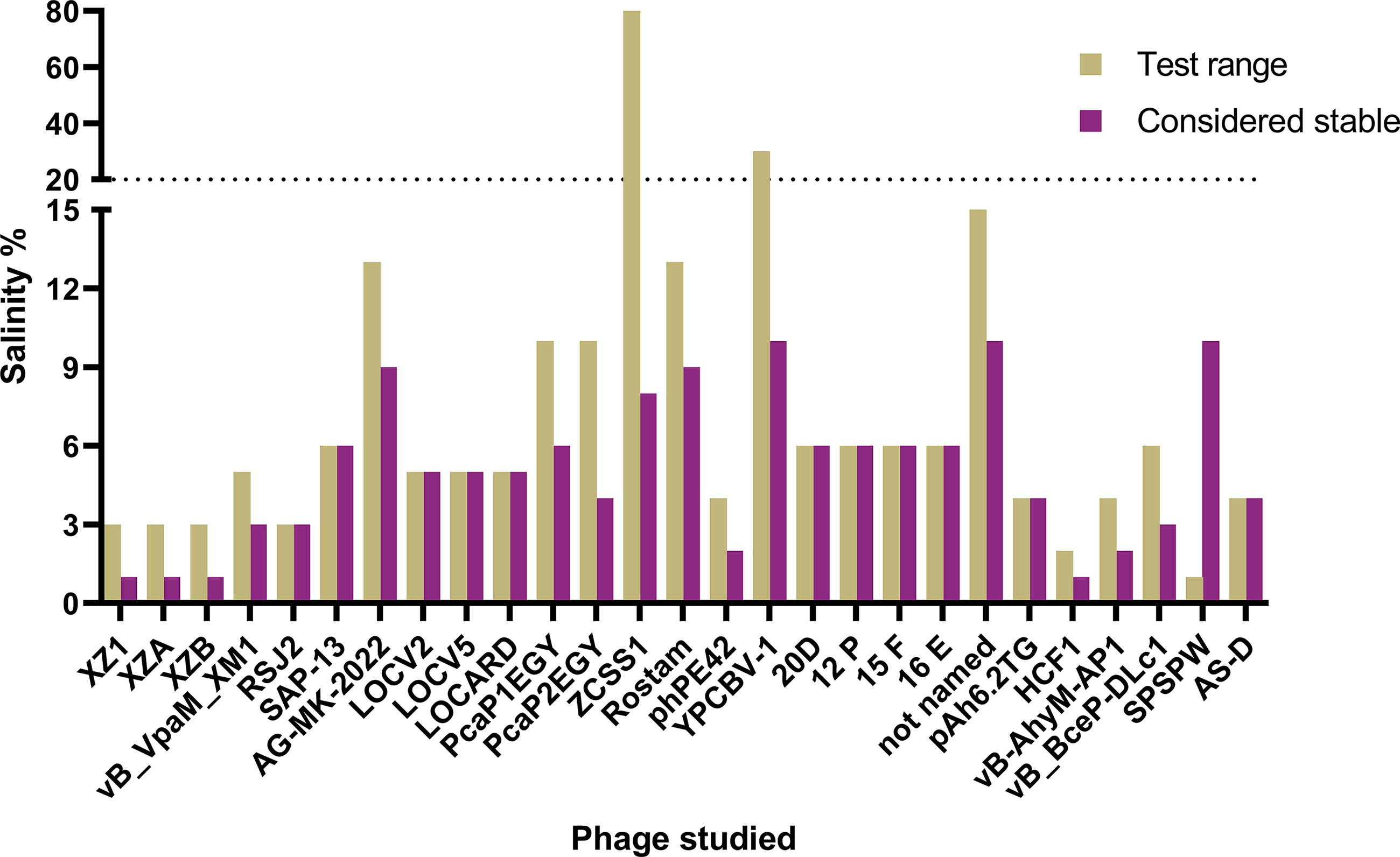
Studies on phage stability to different salinity by individual phage. Phages were inoculated in different solutions with varying concentrations of NaCl. Left column—the maximum salinity the phage was exposed to during the study (test range). Right—maximum salinity at which the phage was considered stable. The results of the studies were converted to the % (w/v) of NaCl for comparison. Detailed information is presented in Supplementary Table S4.
When phage AG-MK-2022 was incubated to a final titer of 109 PFU/mL in a wide range of NaCl concentrations (1–11% [w/v]) for an hour, no significant reduction was observed in concentrations ≤9% (w/v); however, further increasing NaCl concentration (to 11% and 13% [w/v]) resulted in a significant reduction of titer of 0.73 and 0.88 log PFU/mL, respectively. 84 Similar results were observed for phage Rostam (no significant differences between 1% and 9% (w/v) and a decrease in titer at 11% and 13% (w/v) NaCl of 0.7 and 0.87 log PFU/mL, respectively. 87 Phages PcaP1EGY and PcaP2EGY were also stable in NaCl concentrations of 2% and 4% (w/v) but decreased in concentrations of 6% (w/v). 79 Phage ZCSS1 was inoculated to a final titer of 109 PFU/mL and presented stability in NaCl concentration of 0.14M to 1.4M (≈0.7% to 6.8% [w/v]). 37 In a more extended incubation of 24 h in concentrations of 0–3.5% (w/v), phage phPE42 experienced a significant decrease of about 1.1 log PFU/mL at 3.5% (w/v) NaCl. 88 In another study, phages 15F, 20D, 12P, and 16E were incubated at a concentration of above 8 log PFU/mL for 24 h at different salinities (0–6% [w/v]). Despite phages 15F and 16E having presented a strong stability at up to 6% (w/v) NaCl, differences in phage titer were observed in salinity above 3% (w/v) for phage 20D and above 0% (w/v) for phage 12P. Nonetheless, all phage titers remained high (≥8 log PFU/mL) during the experimental period and were considered stable. 89 In a more extended experiment, phage YPCBV-1 was incubated for 28 days (108-109 PFU/mL) under different NaCl concentrations, ranging from 0% to 30% (w/v) NaCl. During that period, the phage titer remained higher at 30% (w/v) NaCl, with about 40% survival, than in all other concentrations tested. 83 Phage AS-D was inoculated to a final titer of 107 PFU/mL and remained viable for 107 days at a salinity of 15%, 20%, and 35% (w/v), with a final titer of about 4 log PFU/mL and with no significant differences between salinities. 36
Other Factors Affecting Phage Stability
Experimental period
The evaluation of phage stability during storage, months to years, has been routinely performed using different preservatives and temperatures. 90 However, in most recent studies, it has become common practice to evaluate phage stability under different conditions during short periods of time, most frequently an hour (Figs. 3B and 4B). Despite presenting reliable information for more extreme conditions (e.g., low and high values of pH, or high values of temperature), in general, a short exposure period may neither allow to adequately infer the stability of the phage, nor provide insights into its ecological impact. 91 Due to the short experimental period, most results retrieved from these assays can be affected by the initial adaptation of the phage to the new conditions, and not just by the variables under study. For example, when one aims to evaluate the effect of pH on phage stability, the temperature of the suspension being used must be at the optimal value for the phage to thrive; otherwise, the experimental results will be blurred by the negative effects promoted by the temperature. This aspect is very important in short-term experiments, as there is no time for the temperature to stabilize, unless the suspension is previously heated or cooled to its optimal value. In fact, when studies that determine the influence of temperature in phage stability were grouped (Fig. 3), it was observed that prolonged incubations led researchers to determine lower maximum temperature thresholds than short-exposure studies. Indeed, in the selected studies used to evaluate the effect of temperature in phage stability, only 13 of a total of 313 studies have indicated that the suspensions being used were prewarmed prior to phage inoculation, corresponding to solely 23 phages of the 483 total phages addressed in all the studies retrieved (Supplementary Table S1). In the case of pH experiments, short-term experiments did not even allow to reach any conclusive information (Fig. 4A). Nevertheless, a variation profile arises when incubation was extended for more than 6 h (Fig. 4A). Therefore, the experimental period should be considered an important aspect to be taken into account when evaluating phage stability. Understanding phage decay is paramount to design efficient protocols against pathogenic or spoilage bacteria and maintain the infection kinetics at optimal levels. 32
Furthermore, these short-term exposures do not mimic potential therapeutic or preventive applications in clinical and industrial settings, as effective bacterial inactivation by phages in vitro has been observed only after more than an hour of treatment.92–96 This means that the findings of these short-term experiments often lack direct applicability to real-world scenarios.
Buffer and culture media used in the experiments
Most stability assays are performed in SM buffer (see Supplementary Tables S1, S2, S3 and S4), a buffer that preserves phage stability, 97 thus allowing a more appropriate measurement of the impact of the tested variables. Nonetheless, some studies also use PBS and culture media, such as tryptic soy broth (TSB), LB, or brain heart infusion (BHI) (see Supplementary Tables S1, S2, S3 and S4). When addressing the stability of phages, the selection of the appropriate suspension to be used should be carefully considered. Phages YpYeO9 and YpEc11 revealed very similar stability at BHI broth, SM buffer, and PBS, with titer remaining unaffected over 6 months of storage at 4°C. 98 As such, any of these suspensions can apparently be used to successfully study the stability of these phages in relation to a given parameter, thus avoiding negative interferences from the type of suspension used during the study. Contrarily, differences in stability between PBS and LB broth were observed for phage PS3-1. 99 Temperature stability for phage DLc1 was also different when experiments were either performed in water or TSB. 100 In addition, some phages may present divalent cation requirements (e.g., calcium or magnesium) or NaCl.83,101 However, such requirements may come at a cost to phage stability, as observed for phage YPCBV-1, which became more efficient, with higher adsorption and progeny, at NaCl concentrations of 10%, but less stable at these salinities. 83 It is worth highlighting that if phages are to be used in certain scenarios, studying their stability in conditions that mimic such scenarios for future applications will most likely provide more valuable insights. As examples one may refer the use of artificial seawater or autoclaved seawater for marine aquaculture scenarios, milk derivates for food processes, or simulated gastric fluids for animal husbandry.21,36,61,102 The information resulting from this type of testing should allow protocols developed under laboratory conditions to be more reliably transposed into practice. As an example, when phages were incubated in physiological saline concentrations, LOCV2 and LOCV5 revealed to be less stable than LOCARD, 55 an important observation for the success of clinical applications of these phages.
Absence of threshold values
The divergence in the interpretation of results is also a factor that must be considered for the successful use of phages. While in some studies it is considered that phages are stable at a specific variable if the titer remains above 80%,103,104 in other studies, statistically significant differences are considered by directly comparing results with those of control groups. 82 In addition, some studies also consider phages to be stable despite an observable decrease in titers103,105–110 or relatively stable despite significant differences in titers. 111
Conclusion and Guidelines for Future Research
To successfully apply phages as therapeutical or decontamination agents in industrial applications, the impact of environmental conditions on phage stability needs to be routinely addressed in all studies. However, the absence of specific guidelines/standardization can lead to a misinterpretation of results concerning phage stability, which will difficult phage treatment translation from theory to practice.
Although it is already well established that environmental conditions, such as temperature (high stability between 4°C and 37°C for 24 h), pH (high stability between 6 and 8), salinity [high stability up to 6% (w/v) saline], and UV radiation (less affected by UV-A and UV-B), can significantly affect phage viability, results recorded to date suggest that long-term experiments, rather than short-term experiments (of an hour or less), should be considered to obtain more accurate results. As phage inactivation of bacteria takes more than an hour before being effective (some phages may even take more than 6 h), it is mandatory that phage stability is evaluated for longer periods, preferably never under 6 h. In studies focused on phage applications in industrial settings such as food, agriculture, or aquaculture, the experimental duration may need to be extended for weeks to months and conditions adapted to better reflect real-world application outcomes and assess potential ecological impacts. 91 In addition, to support practical implementation, researchers should consider prolonged experimental periods under conditions that simulate phage stability during storage, handling, and distribution, potentially over months or years and at appropriate temperatures. In cases where phages exhibit poor stability under conditions expected during application, the use of hydrogels or encapsulation techniques may enhance their protection and improve success.30,42
With regard to the buffer and medium used to characterize phage stability, the lack of specific standardization has resulted in several studies that are difficult to compare and ultimately lack a direct applicability to real-world scenarios. The absence of threshold values makes this comparison even more difficult. 26 To address this caveat, it is important to establish guidelines, standardize experimental periods, and define suspensions (buffers/culture media) to be used, and threshold values for a phage to be considered stable in relation to a given condition. In phages that are intended for human use, understanding the stability in suspensions such as physiological saline 55 or in other regulated suspensions may provide more practical information. 30 Simultaneously, one must also consider existing regulatory standards26,30 to safeguard the kinetics of infection 32 and facilitate the development of more effective dosing strategies. 26
Footnotes
Authors’ Contributions
J.D., C.P., R.C., and A.A. conceived the idea. J.D. performed the data curation and analysis. C.P., R.C., and A.A. supervised the work. J.D. wrote the first draft and C.P., R.C., and A.A. reviewed and edited.
Author Disclosure Statement
The authors declare no conflict of interest.
Funding Information
The authors acknowledge financial support to the UID Centro de Estudos do Ambiente e Mar (CESAM) + LA/P/0094/2020 through national funds. J.D. thanks the Portuguese Foundation for Science and Technology (FCT) for his doctoral grant (DOI:10.54499/2021.05519.BD). C.P. acknowledges the FCT for Junior Research contract (DOI:10.54499/CEECIND/03974/2017/CP1459/CT0022).