






Abstract
Introduction:
Increasing numbers of gender-affirming facial surgeries are being sought out and performed.1,2 Feminization of the upper third of the face often involves a frontal sinus setback to increase the nasofrontal angle. It is recommended to minimize disturbance of the frontal sinus mucosa and frontal outflow tracts, as this can lead to postoperative frontal sinus disease or mucoceles.3,4 These complications have been rarely reported in the literature.
Materials and Methods:
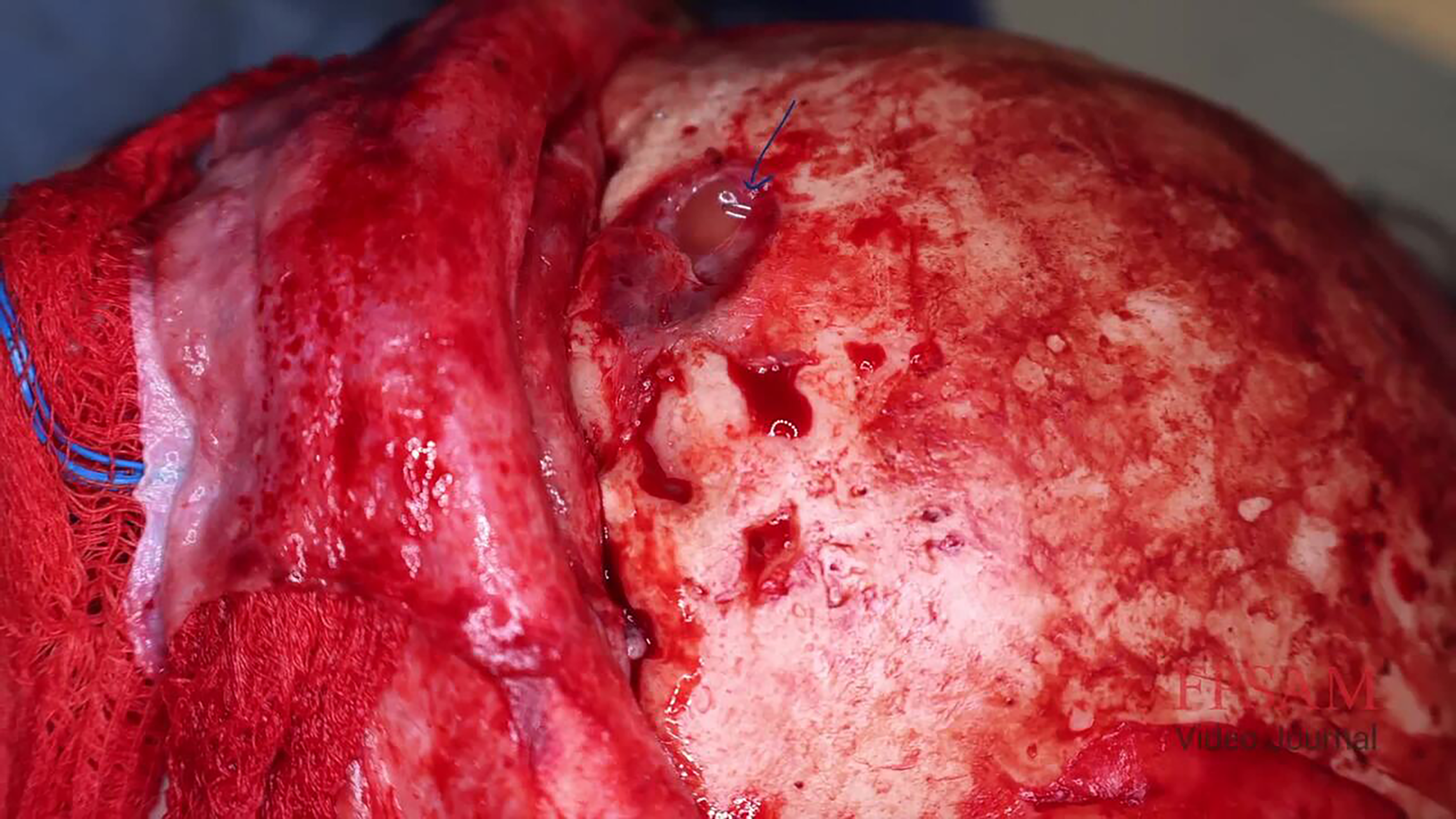
After obtaining institutional review board approval, we compiled radiologic imaging and surgical videos. We describe the case of a 36-year-old transgender female who developed a frontal sinus mucocele 13 years after their initial frontal sinus setback. The patient reported a 10-year history of worsening headaches and frontal sinus pressure that increased with physical exertion. On physical examination, the patient was noted to have a soft tissue fullness over the right glabella that was mobile and was not indurated or inflamed. Computed tomography imaging demonstrated a frontal sinus mucocele eroding through the anterior table. Recommendations were for a revision bicoronal approach through a prior hairline advancement incision to access the entire anterior table of the frontal sinus. The frontal sinus mucocele was noted to be eroding through the anterior table in multiple locations, corresponding to the site of the glabella swelling. Virtual surgical planning designed a cutting guide for the piezo to remove the entire anterior table. When the entire frontal sinus was exposed, it appeared that the wires the previous surgeon had used to fixate the anterior table had fallen into the right frontal sinus. This created a band of scar tissue that prevented the appropriate drainage of the right frontal sinus despite patent outflow tracts. This was the nidus for the mucocele formation. The frontal sinus mucocele was drained, and the outflow tracts were left undisturbed. A split calvarial bone graft was harvested from the left parietal bone using patient-specific cutting guides and was used to reconstruct the entire anterior table. The bone was fixated into place with two mini plates and screws. The wound was then closed up in typical fashion.
Results:
Patient recovered well postoperatively. The previously noted glabella swelling was completely resolved. The frontal sinus pressure and headaches were no longer present. Postoperative imaging was obtained 3 months later and demonstrated a patent frontal sinus and well-healed osteotomy sites.
Conclusions:
Frontal sinus mucocele formation after frontal sinus setback is a rare but potential complication. Routine imaging post frontal sinus setback is not recommended unless there are symptoms concerning for mucocele. A split calvarial bone graft is a viable option for reconstruction of a destroyed anterior table.
The authors involved in this video have no financial disclosures or conflicts of interest related to the materials.
The patient has signed both an authorization to use and disclose protected health information and a media release consent form.
Runtime of video: 3 mins 7 secs.
Keywords
Get full access to this article
View all access options for this article.