
Abstract
Development of the novel delivery provides optimal drug delivery via facilitated transcorneal penetration and ultimately improved retention time in eye. The present study was to fabricate and evaluate the performance of ocular inserts of Olopatadine hydrochloride by consuming polymers such as hydroxypropyl methylcellulose, gelatin, and chitosan at various concentrations with different plasticizers using solvent casting technique. Prepared formulations were evaluated for thickness, weight variations, drug content, surface pH, swelling studies, and in vitro drug release. Fourier transform infrared spectroscopy studies showed that interaction occurred between polymer and drug, and successful drug entrapment occurred in the matrix. All formulations showed excellent film-forming capacity with good thickness and weight uniformity. Folding endurance proved that the inserts were soft, elastic, and flexible. Chitosan-based ocular inserts showed greater swelling index as compared to gelatin and hydroxypropyl methylcellulose inserts. In vitro studies showed prolonged drug release and followed zero-order kinetic model and non-Fickian drug release.
Introduction
The ocular inserts show significant improvement in the treatment of eye ailments. 1 Ocular inserts are defined as sterile, thin, multilayered, drug-impregnated, solid, or semi-solid consistency devices placed into the cul-de-sac or conjunctiva sac, whose size and shape are especially designed for ophthalmic applications. 2 Ocular inserts are polymeric supports with or without entrapped drug. Drug entrapment is usually achieved by a dispersion or a solution within the polymeric support with enhanced ocular residence and sustained active moiety release into the eye. 3 The insert includes a body portion which is sized to the position within an eyelid lachrymal canaliculus. The inserts are classified according to their solubility as insoluble, soluble, or bioerodible inserts. 4 Olopatadine hydrochloride (HCl) is a dual-action selective histamine H1 receptor antagonist and mast cell stabilizer with anti-allergic activity which prevents histamine release from the mast cells. 5 In addition, it also blocks histamine H1 receptors, thereby preventing histamine from binding to these receptors. 6 Both actions prevent the effects of histamine on capillaries, thereby preventing histamine-induced pain and itching of mucous membranes.7,8 In this work, HCl was selected due to its therapeutic potential reported in literature.7,8 The developed formulation was characterized for various in vitro parameters such as swelling ratio, drug contents, and in vitro properties such as drug release and ocular irritation. The ocular irritation of the formulation was evaluated to assess its potential as an ophthalmic drug delivery system.
Experimental
Materials
Olopatadine HCl (Commercial name is Patanol, 99.98% pure) was donated by Innvotek Pharmaceuticals Islamabad, Pakistan. Hydroxypropyl methylcellulose (commonly known as hypromellose), chitosan (commonly known as deacetylchitin), and gelatin were all purchased from Sigma Aldrich Ltd, USA. Glycerin, acetic acid, potassium dihydrogen phosphate, sodium hydroxide, and sodium chloride were of pure analytical grade.
Film fabrication
Amount of drug loaded on a single insert
The theoretical Olopatadine HCl loaded on each insert was calculated by measuring the area of a single petri dish and the total inserts (radius of 0.3 cm) that can be prepared on a single petri dish by dividing the area of the petri dish by the area of single insert. 9
HPMC and gelatin film preparation
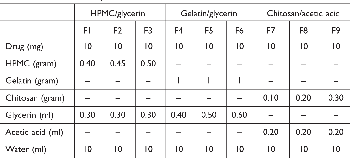
Films were prepared by adding polymer hydroxypropyl methylcellulose (HPMC) (0.40, 0.45, and 0.50 g, respectively, in formulations F1, F2, and F3), gelatin (1 g in each formulation F4, F5, and F6), and glycerin 0.30 ml in each formulation in 10 ml of water in case of HPMC-based formulations and 0.40, 0.50, and 0.60 ml of glycerin in 10 ml of water in gelatin-based formulations with constant stirring on magnetic stirrer. Bubbles made during stirring and adding Olopatadine HCl 10 mg in each formulation were allowed to set 20–30 min before sonication. After sonication was done for 30 min to ensure complete bubble removal, the homogenous mixture was poured into a petri dish. Solvent was evaporated at room temperature for 48 h. After complete drying, the film was separated from the petri dish, and inserts were made with a special cutter. Inserts were then stored separately in desiccators. 10
Preparation of films using chitosan
Films were prepared by dissolving measuring amount (0.10, 0.20, and 0.30 g in formulation F7, F8, and F9, respectively) of chitosan in 1% acidic water. After chitosan was completely dissolved, Olopatadine HCl 10 mg was added, mixed, and then stirred for almost 6 h, and the resultant solution was poured into a petri dish and was covered with inverted funnel and placed at room temperature for 24–48 h for drying. Layer was then separated from petri dish and cut with die into 6 mm diameter circles. Inserts were then placed in desiccator before further use. 11
Ocular inserts composition
Table 1 shows the inserts formulations.
Formulation compositions for the ocular inserts.
Pre-formulation studies
Compatibility studies
Compatibility studies were conducted in order to check any physical interaction between drug and polymers.
Fourier transform-infrared spectroscopy
Olopatadine HCl alone and as physical mixtures containing polymers were analyzed using FTIR spectrophotometry.
Differential scanning calorimetry
Olopatadine HCl alone and as physical mixture with polymer were analyzed using DSC technique. This provides information of thermal changes that do not involve a change in sample mass. The changes that take place in polymers upon heating were observed. A 5 mg sample was taken, and temperature was increased 10°C/min up to 300°C.
Thermogravimetric analysis
Thermal characterization of pure drug, polymer, and their physical mixtures was performed with calorimeter. The thermogravimetric analysis is a method of thermal analysis in which physical and chemical properties of materials are measured as a function of increasing temperature (with constant heating rate). It determines the volatile content and decomposition temperature of a substance.
Characterization of ocular inserts
Physical appearance
Inserts prepared by using different polymers were evaluated for their physical characteristics like color, texture, appearance, and translucency.
Scanning electron microscopy
Scanning electron microscopy was used to analyze the structural morphology of the formulated inserts.
Surface pH
Inserts were placed in petri dish, and 0.05 ml distilled water was poured on them, and after 10 min, pH was measured by using pH paper. The color that developed after 60 s was matched with the standard color chart.12,13
Thickness
Thickness of three randomly selected ocular inserts was measured using micrometer screw gauge. Mean thickness and standard deviation were calculated. 14
Weight uniformity
Digital balance of accuracy was used for weighing. For the determination of weight of inserts from each formulation based on three different polymers, in order to determine uniformity of film, procedure involved individual weighing of 10 inserts from each formulation. Average weight and standard deviation were then calculated. 15
Folding endurance
Folding endurance is used for measuring of flexibility of inserts in quantitative terms. In order to determine the folding endurance of prepared inserts, three inserts were randomly selected from each formulation and were folded at the same place for several times till the insert broke at that point. Folding endurance is the number of times of folding of inserts that went successfully without breaking. 16
Swelling studies

Drug content
Content uniformity was determined using three inserts from each films, and USP phosphate buffer of pH 7.4 was prepared and used. A 100 ml buffer was taken in flask, and inserts were placed in it after dissolving. One milliliter of this solution was then serially diluted after filtration and analyzed under UV spectrophotometer against blank for estimating drug content uniformity.19,20
In vitro release studies
In vitro release studies for ocular inserts were performed using USP dissolution apparatus, and phosphate buffer of pH 7.4 was prepared. Inserts were placed inside the cellophane membrane that acted as the donor compartment, were attached to the lower shaft of dissolution apparatus, and beaker was filled with phosphate buffer that acted as receptor compartment, maintained at 37°C with slight stirring. Release studies were performed by taking samples and replacing the same with buffer solution, at specified time intervals 15 min, 30 min, 1, 1.5, 2, 2.5, 3, 4, 5, 6, 7, and 8 h. The drug release then was determined in each sample insert with a UV–vis spectrophotometer at λ 298 nm.
Release kinetic models
In order to analyze the mechanism of release kinetics of the drug, various kinetic models like zero-order, first-order, Korsmeyer Peppas, and Higuchi model can be used for plotting the results from in vitro release profiles. Drug release mechanism can be easily elaborated and well understood using these mathematical modeling; in addition, these can cut off the number of experiments to be performed in order to determine the formulation to be optimized. Fickian or non-Fickian diffusion of drug can be seen by these for formulations having diffusion-based release. 21
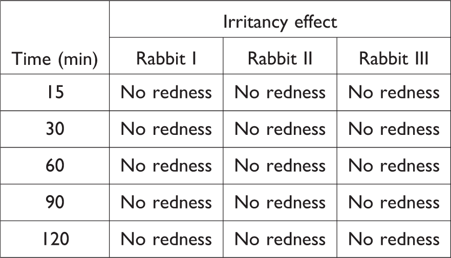
Ocular irritation studies
Irritation studies were performed on three albino rabbits for selected formulation from chitosan-based inserts in order to check any irritancy effect and bio adhesion. The rabbits were properly feed, cared during the study, and the animal ethics committee of the Faculty of Pharmacy, Baha Uddin Zakariya University approved the research protocol involving the animal study. F9 was selected because of its pH, swelling index, and in vitro release study. After placement of insert, irritancy was checked at certain intervals. 22
Stability studies of optimized formulation
The optimized formulation of the insert F9 was then studied for stability purpose. It was kept in a chamber at 40°C and 65% relative humidity for 3 months. 23
Results
This study proposed to develop ocular inserts using different polymers that were biodegradable, bio adhesive, and biocompatible in nature. Olopatadine HCl was used as anti-allergic agent to be incorporated into ocular inserts for treating allergic conjunctivitis. The visual aspects of the ocular inserts, results of FTIR of Olopatadine HCl, FTIR of physical mixture of Olopatadine HCl along with polymers, thermogravimetric analysis of physical mixture, DSC of physical mixture of drug and polymer, scanning electron microscopy of the ocular insert, swelling studies of ocular inserts, percentage release of drug from ocular inserts, and stability studies of optimized formulation F9 were shown in Figures 1 to 9, respectively. Table 2 tabulates the weight uniformity, folding endurance, surface pH, and thickness of inserts. Tables 3 and 4 tabulate the release kinetics and ocular irritancy.
Discussion
Film formation success depends on the prepared inserts being smooth in texture, translucent, and has uniform appearance without any imperfections according to the reported literature. In this regard, all prepared formulations were visually inspected. Each formulation was translucent, homogenous, and elastic. No cracks, imperfections, or phase separation between the drug and matrix was observed in any formulation which indicates that the components were uniformly distributed. Inserts of HPMC F1, F2, and F3 were transparent in color. Gelatin-based inserts F4, F5, and F6 were light yellow. Chitosan-based inserts F7, F8, and F9 were light brown (see Figure 1).
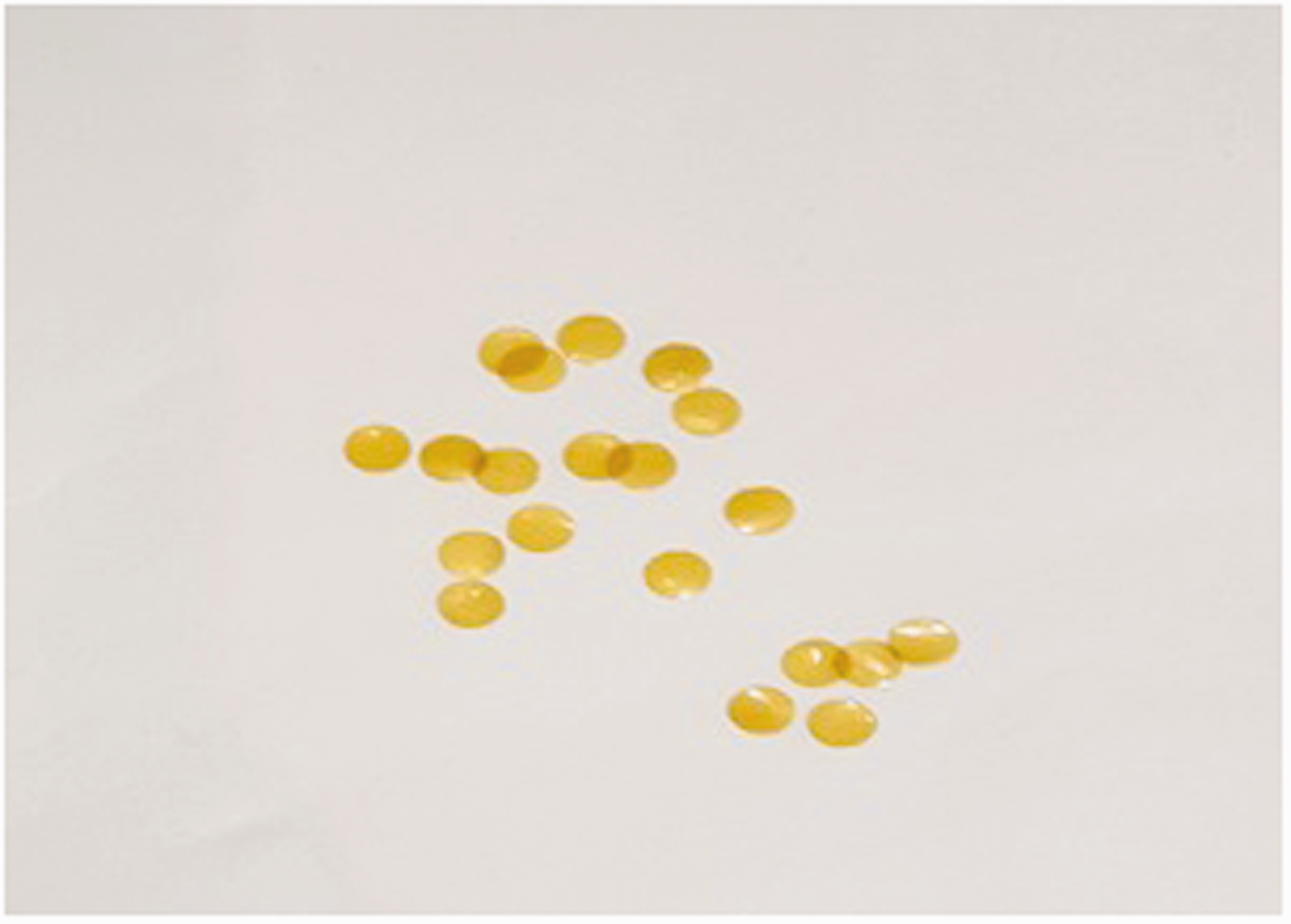
Visual aspect of the ocular insert.
All the polymers showed excellent film-forming capacity. Srivastava and Pathak 24 reported similar results.
Pre-formulation studies were conducted in order to check active ingredient purity and investigate if any physicochemical interaction among polymers and drug existed. These included melting point determination for analyzing drug purity and FTIR, thermogravimetric analysis, and DSC to check for interactions.
FTIR studies were conducted, and observance of functional group region showed that the interaction occurred between polymer and drug and successful drug entrapment occurred in matrix. Figure 2 shows the Olopatadine HCl absorption band at 2687 cm–1 (carboxylic acid OH stretch) and characteristic peak 1714 cm–1 (carboxylic acid C=O stretch) via FTIR.
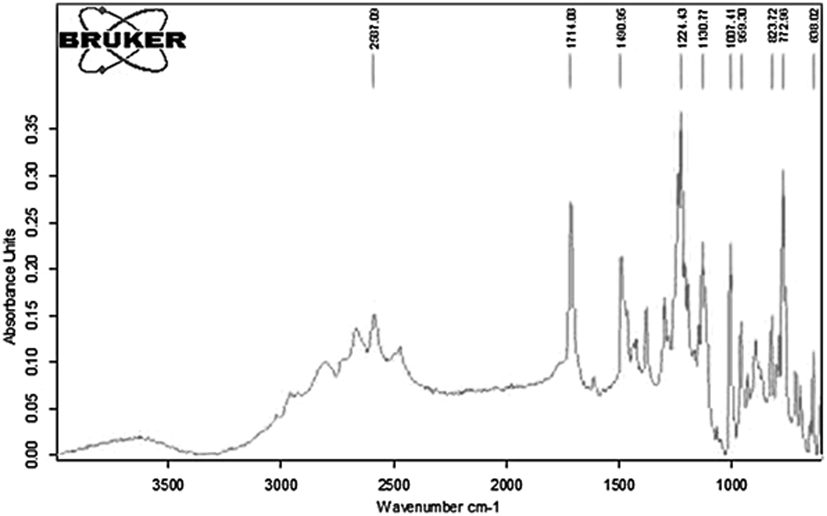
FTIR spectra of Olopatadine HCl.
FTIR spectra of HPMC, gelatin, and chitosan were also observed and recorded. Chitosan showed 3294 cm–1 (alcohol phenol pH stretch) and 1588 cm–1 (aromatic C=C bending) (Figure 3). Physical Olopatadine HCl and chitosan mixtures showed the stretch and bend at 2588 and 1714 cm–1 (Figure 3).
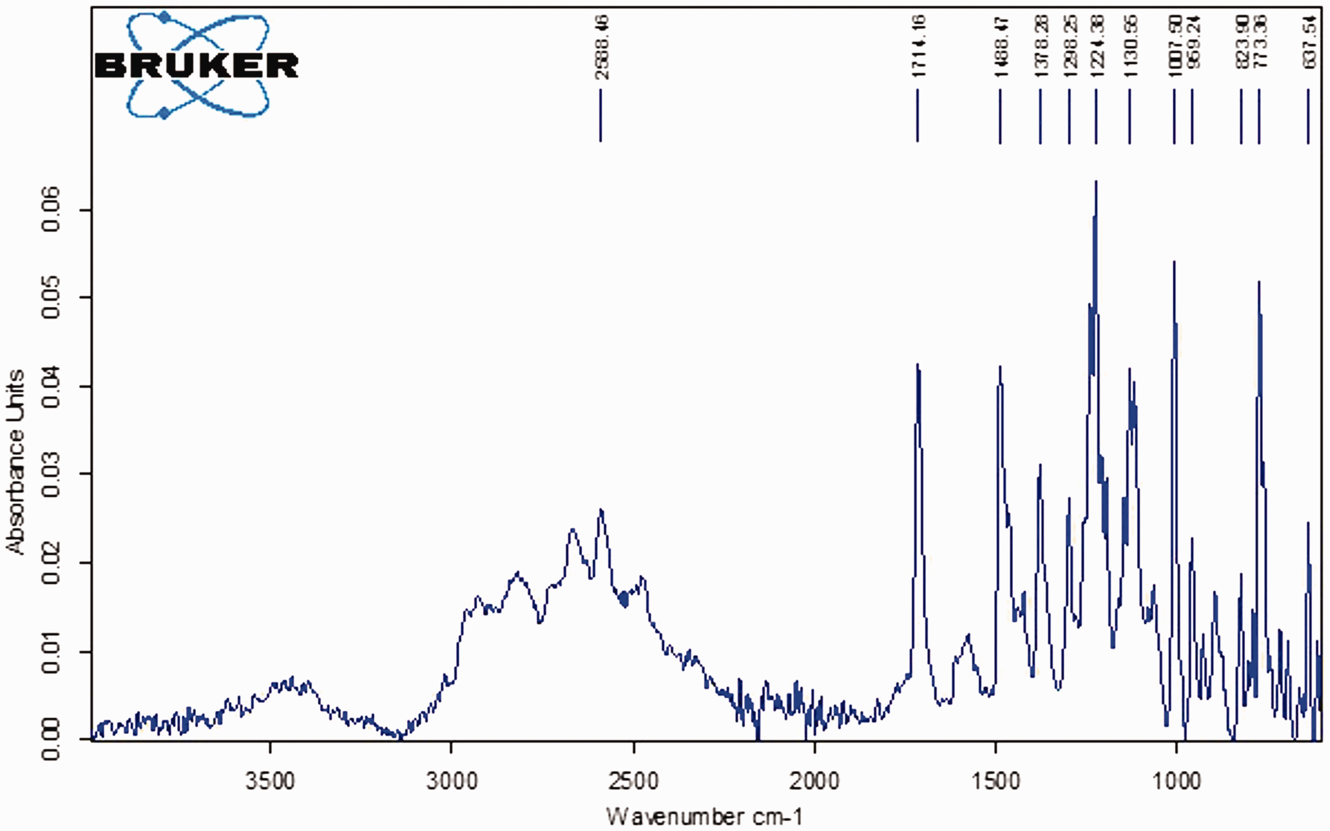
FTIR spectra for Olopatadine HCl and Chitosan mixture.
So, no interaction in physical mixtures was observed. Chavan and Lasonkar 25 reported similar results in 2013.
This analysis was performed prior to differential scanning calorimetric to determine the volatile content and decomposition temperature of active ingredient and physical mixture. Samples were heated to greater than 450°C. Weight loss is attributed to physical transitions or chemical reactions like decomposition, combustion, vaporization, evaporation, sublimation, or drying. Almost 99% was lost at 428°C. Chitosan TGA was calculated from 4.26 mg sample which showed a gradual weight loss as the temperature increased and 20% was lost at 100°C (see Figure 4).
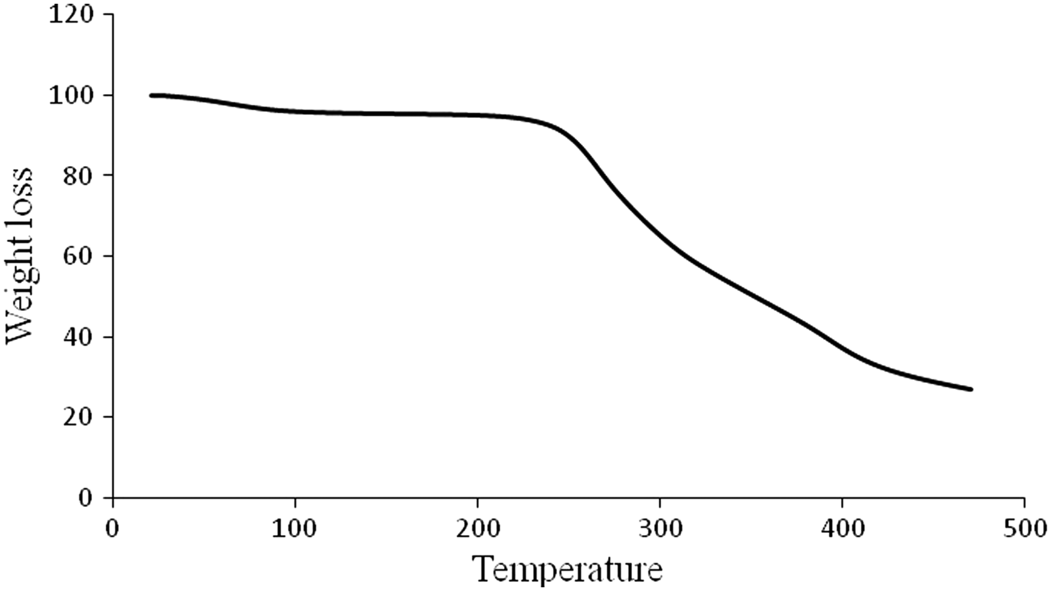
Weight loss versus temperature for Olopatadine HCl and Chitosan mixture.
Figure 4 shows the TGA for chitosan and Olopatadine HCl mixture (7.750 mg). There is a slight change in mass with increased temperature till 244°C and then a constant rate weight loss. However, there was still 20% left at 470°C. Similar results were reported by Łaszcz et al. 26 in 2016 while studying structural and physicochemical studies of Olopatadine HCl conformational polymorphs. 26
Differential scanning calorimetry was done for Olopatadine HCl, chitosan, and its physical mixture with chitosan, and both showed the endothermic reaction. Broad endothermic dip of Olopatadine HCl shows the dehydration reaction (physical change) in Figure 5.
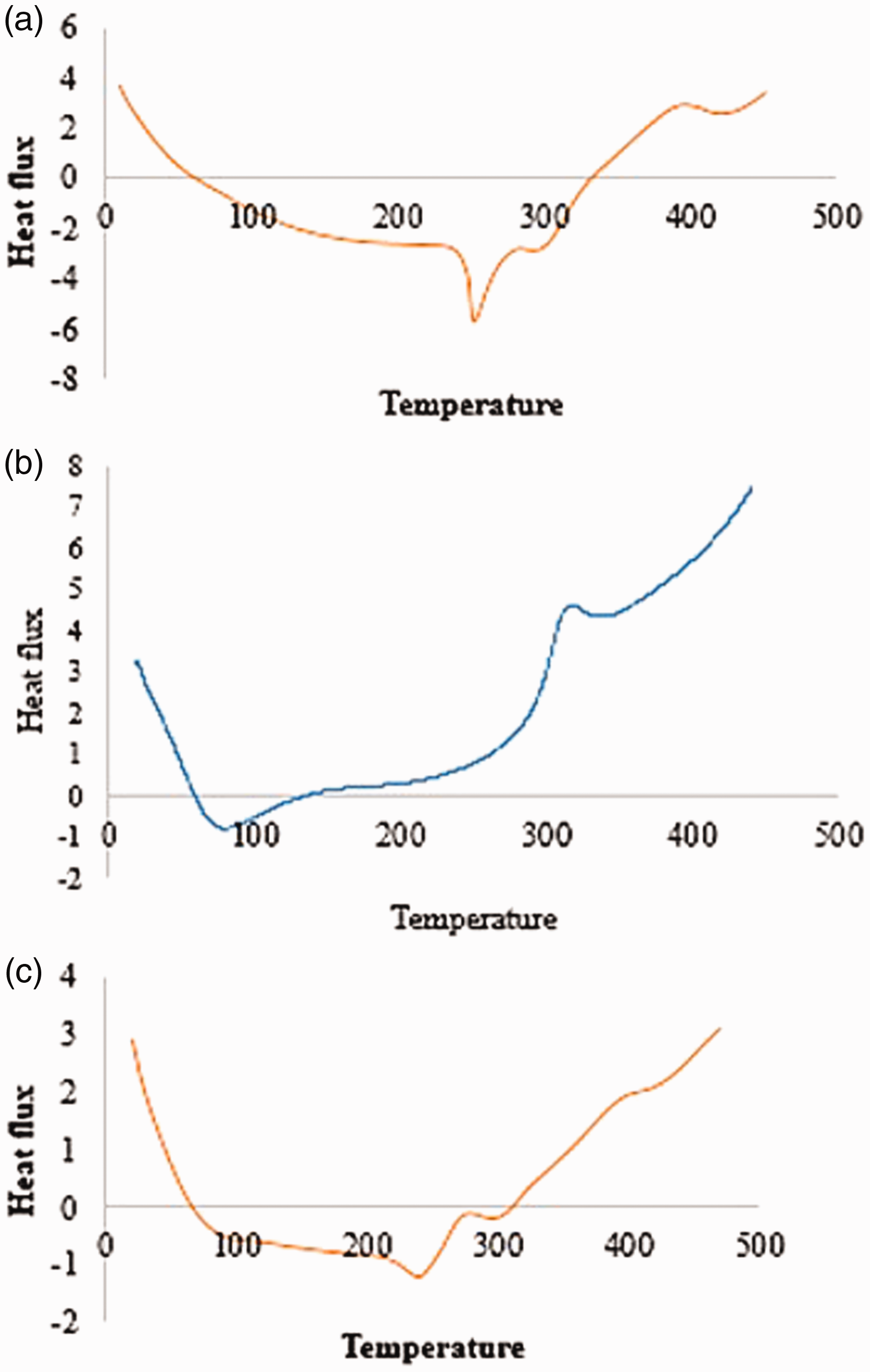
Heat flux versus temperature for Olopatadine HCl (A), Chitosan (B) and mixture (C).
Chitosan had a sudden short endothermic dip with increasing temperature which indicated that dehydration occurred as in Figure 5. Physical mixture showed the endothermic reaction. As no glass transition temperature or crystallization point was observed, it was concluded that the drug, chitosan, and its physical mixture are in amorphous forms and do not tend to align in crystalline structure. Scanning electron microscopy showed a rough porous structure (see in Figure 6).
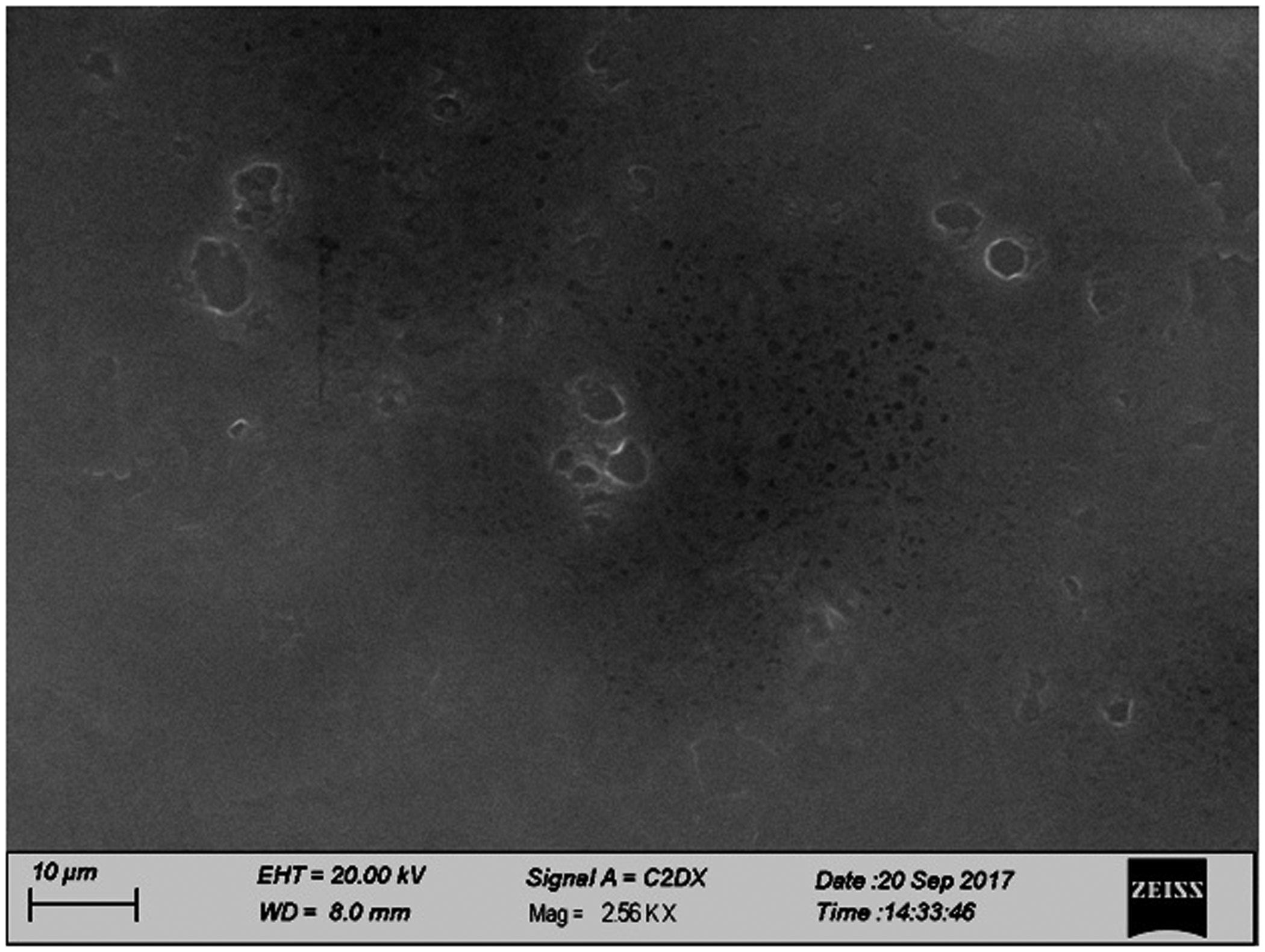
SEM of optimized formulation.
Surface pH varied from 6.0 to 6.5 for HPMC-based inserts F1, F2, and F3. Gelatin-based inserts F4, F5, and F6 pH varied from 6.5 to 7.0 and chitosan-based inserts F7, F8, and F9 the pH was between 7.0 and 7.5 (see Table 2). Thus, inserts would not cause irritation to eye when applied. The insert diameters were 6 mm, and thickness varied from 0.09 to 0.12 mm for HPMC-based inserts. Gelatin-based inserts F4, F5, and F6 varied from 0.15 to 0.21 mm. Chitosan-based inserts F7, F8, and F9 varied from 0.09 to 0.10 mm (see Table 2).
Surface pH, thickness, weight uniformity and folding endurance of ocular inserts.
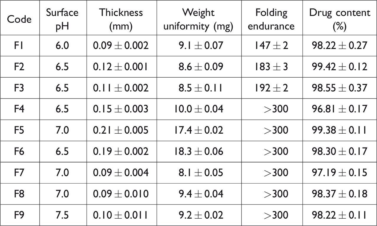
The weight measurements showed that ocular inserts are not uniform varying from 8.1 to 18.3 with chitosan-based formulation F7 having the minimum weight and gelatin-based formulation F6 having maximum weight (Table 2). Each formulation showed excellent folding endurance from 147 to more than 300-fold and no breaking, which showed that the inserts were soft, plastic, and flexible. Drug content uniformity test was performed. The results showed that the drug content was uniform. The percentage varied from 96.81 ± 0.17 for F4 to 99.42 ± 0.12 for F2 (see in Table 2).
Different degrees of swelling were likely to be observed while using hydrophilic polymers having different types and structures which depend upon the resistance to matrix network structure to the movement of molecules. Swelling of any polymer is essential as to initiate bio adhesiveness that begins just after the swelling begins. Polymer adhesion then increases due to more hydration of polymer; however, excessive hydration may lead to lower adhesion. Similar results were reported by Sultana et al., 27 while formulating ocular inserts for controlled delivery of pefloxacin mesylate. Rate and extent of drug release is also dependent on swelling and hydration. Therefore, this parameter is of prime importance for predicting bio adhesive strength and drug release. Figure 7 shows the swelling behavior for the investigated inserts.
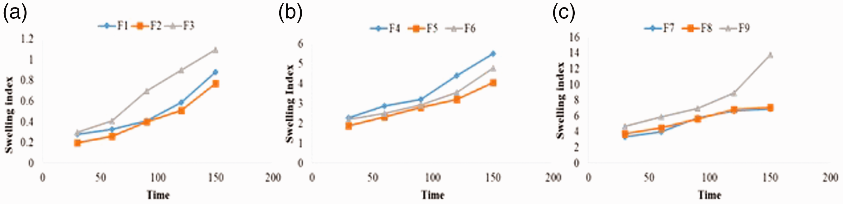
Swelling studies for ocular inserts from F1 to F9.
One sees that chitosan and gelatin inserts showed higher swelling index. HPMC-based inserts (F1–F3) showed expansion in size with the passage of time that can be attributed to its hydrophilic nature. Gelatin inserts (F4–F6) also showed higher swelling index, and the expansion in thickness was seen. Inserts of chitosan (F7–F9) showed radial swelling and expansion, and F7 was unable to maintain its integrity. When removed from swelling medium, it fragmented easily. However, chitosan inserts showed maximum swelling index with excellent swelling capacity, and they were handled very carefully as reported earlier. 28 Chitosan-based ocular inserts F7, F8, and F9 showed greater swelling index as compared to gelatin and HPMC inserts as shown in Figure 6. Each formulation showed good results on swelling analysis which was performed to correlate the release rate and polymer network formed.
In vitro release studies were performed on each formulation. Samples were taken from dissolution media at pre-defined intervals, and absorbance was measured using UV spectrophotometer. Absorbance was then converted into percent release. All inserts showed a gradual drug release. HPMC inserts F1, F2, and F3 were from 6 to 8 h while the maximum release from gelatin inserts F4, F5, and F6 was within 6 h. However, chitosan-based inserts F7, F8, and F9 drug release varied from 4 to 8 h (Figure 8).
Percent cumulative drug release profile versus time for F1 to F9.
Chitosan-based formulations F7 released 100% in 4 h, F8 released 100% in 6 h while F9 released 80% in 8 h. As this study was done to find a formulation that satisfied the objective where there is 80% drug release in 8 h, the F9 formulation meets the objective. Formulation F9 is considered to be better because its sustained drug release is 80% in 8 h. This was also reported earlier. 29
Release models were applied to all the formulation, and comparing the zero-order and first-order graphs showed that the formulations follow zero-order kinetics i.e. the drug release does not depend upon concentration. Korsmeyer Peppas model was applied to determine the mechanism by which drug releases from the polymer solid matrix. Formulations tended to follow non-Fickian diffusion (Table 3).
Release kinetics of Olopatadine HCl at pH 7.4.
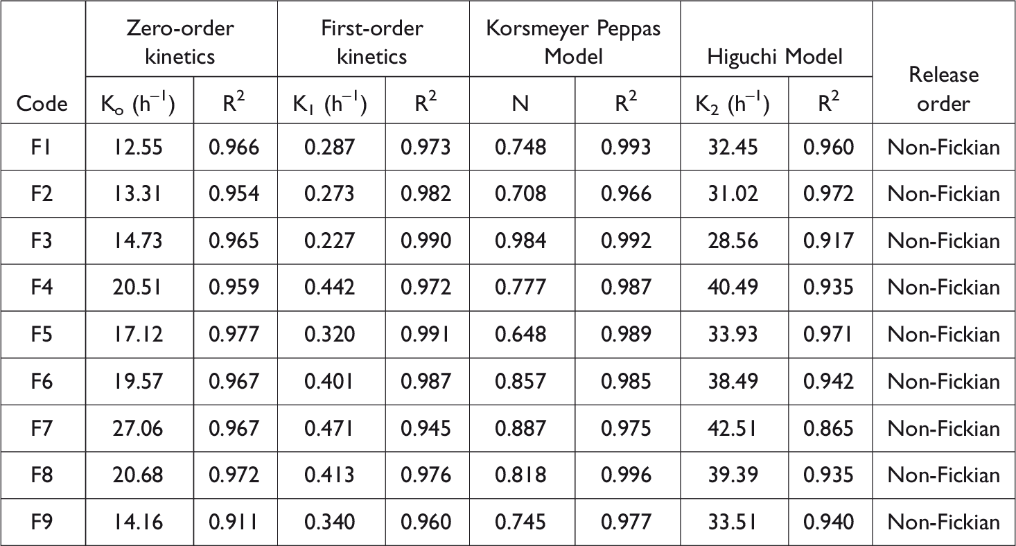
All formulations followed zero-order release kinetic model and best fitted in Korsmeyer Peppas model and showed non-Fickian drug release; similar results were also reported earlier. 30 Also, Di Colo et al. 29 studied the effect of chitosan on in vitro release and ocular delivery of Ofloxacin from erodible inserts based on poly(ethylene oxide). 29 Stability studies of optimized formulation of ocular insert F9 showed comprehensive and better release of drug during the investigation period (Figure 9). The criteria to optimize the formulation were the in vitro percent cumulative drug release profile.
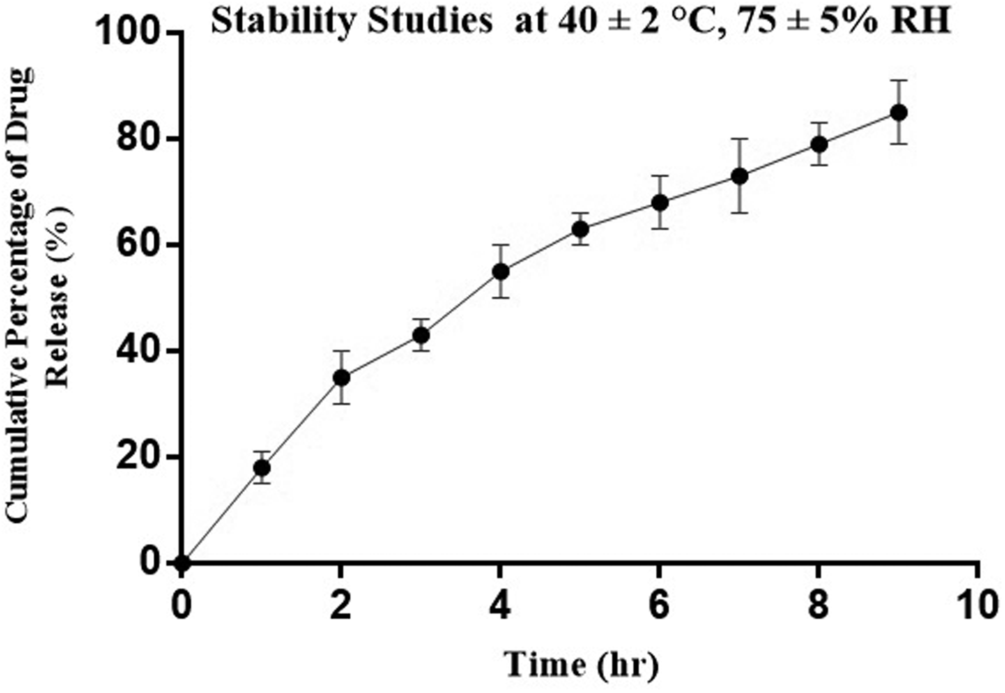
Stability studies of the optimized formulation.
Irritation studies were performed on an albino rabbit for optimized formulation F9 to check any irritancy effect and bio adhesion. No irritancy was found (Table 4), and chitosan-based formulation showed no irritancy as was reported earlier. 31
Ocular Irritation studies for F9.
Conclusion
Each formulation showed precise drug release, and no burst effect was seen with any formulation and chitosan-based inserts. F9 (0.30 g chitosan based) among all showed better results in swelling studies as well as in vitro testing. The study developed a dosage form for prolonged release to eye. Therefore, ocular inserts were a better alternative dosage form in terms of economic factor, better drug delivery without using any excipient.
Footnotes
Acknowledgements
The authors are thankful to Innvotek Pharmaceutical (Pvt) Ltd, Pakistan, for providing Olopatadine HCl for the research work.
Declaration of Conflicting Interests
The author(s) declared no potential conflicts of interest with respect to the research, authorship, and/or publication of this article.
Funding
The author(s) received no financial support for the research, authorship, and/or publication of this article.