
Abstract
Bone ingrowth into and through porous coatings on orthopaedic implants can substantially improve fixation. However, the introduction of pores increases surface roughness and also the risk of bacterial adherence, which can lead to infection (in extreme cases, to death) and complicate implant surgery due to the high risk of revision being required. Improving osseointegration without increasing infection risk is therefore a major challenge in implantology. Staphylococcal adhesion and biofilm formation on Ti surfaces of varying roughness and porosity have been investigated in vitro. Porous pure titanium coatings, obtained by a PM processing route based on the electrophoretic deposition of TiH2 followed by thermal treatment in vacuum, significantly reduced bacterial colonisation of the surface compared with a state-of-the-art vacuum plasma sprayed coating. Further reduction of biofilm formation could be obtained by additional surface modification.
An important indication for revision arthroplasty is infection.1,2 It has been observed that bacteria causing prosthetic joint infections grow in highly structured biofilms, sessile communities of microorganisms adhering to the biomaterial embedded in a matrix of extracellular polymeric substance (EPS) that they themselves produce.3,4 This protective environment enables bacteria to escape the host's defences and antibiotic attack. Moreover, it has been suggested that biofilm-embedded bacteria exhibit an increased resistance to antibiotics due to a horizontal transfer of resistance genes within the biofilm.4–6 Therefore, antibiotic treatment is often insufficient to eradicate biofilm-related implant infections, hence debridement (surgical removal of the infected tissue) and in some cases even complete removal of the implant is inevitable. 5
Titanium alloys are widely applied as implant materials because of their combination of outstanding properties such as excellent biocompatibility, high specific strength and good fatigue resistance. 7 Porous Ti structures or coatings are of special interest because they enable bone ingrowth into the porous structure and as such establish a biological anchorage of the implant in the host bone. However, porous structures intrinsically exhibit increased surface roughness, which is associated with an enlarged risk of bacterial adherence.8,9 So, when envisaging a porous coating for effective osseointegration, measures should be taken to minimise bacterial colonisation.
Recently, the present authors developed a novel processing route for porous Ti coatings capable of tailoring surface roughness and pore morphology.10–13 The resulting coatings allowed bone ingrowth in vivo.11,13,14 Although a general meta-analysis of these coatings revealed decreased biofilm formation and enhanced cell proliferation and gene expression in vitro, 15 further validation of the coatings for implant applications is required via an in-depth assessment of the bacterial response in combination with a thorough surface characterisation to enable in vitro–in vivo correlation. The goal of the present study was to investigate the effect of surface roughness, pore topology and surface modification of the porous Ti coatings on bacterial colonisation relative to state-of-the-art Ti implant surfaces: polished, machined, bead-blasted and vacuum plasma sprayed (VPS). Two bacterial strains that are most frequently isolated from infected prostheses, namely Staphylococcus aureus (S. aureus) and Staphylococcus epidermidis (S. epidermidis), were used.5,6
Materials and methods
Discs of machined Ti–6Al–4V alloy (grade 5, 15·5 mm diameter, 2 mm thickness, LIMA Lto) were used as substrate material and control. Additionally, three clinically relevant Ti surfaces were prepared as control groups: polished samples were prepared by grinding with SiC paper (4000 grit, Hermes) followed by polishing with an oxide polishing suspension of colloidal silica (OP-S, Struers); alternatively, bead-blasting (Helipro, d.o.o.) was carried out using high purity Al2O3 particles (d50 = 1·0–1·6 μm) and, as a porous reference surface, a state-of-the-art porous VPS Ti coating (Alhenia AG) was included. Electrophoretic deposition (EPD) of TiH2 powder suspensions or combinations of a suspension and an emulsion followed by vacuum dehydrogenation and sintering was used to produce experimental grades of porous pure Ti coatings, as described elsewhere.10–13 In short, different TiH2 starting powders grain sizes (∼10 μm for grade VM and ∼40 μm for grade P, Chemetall GmbH) allowed porous titanium coatings with varying surface roughness and pore morphology to be engineered.10,11 These coatings are denominated EPD Ti(Vm) and EPD Ti(P) respectively, referring to the starting powder grade. Additional spherical pores could be introduced into the coatings by EPD of the same Vm or P TiH2 powder based suspensions, which are used to engineer the interpore struts, in combination with a TiH2 (grade Vm) stabilised emulsion, the droplet size of which acts as a template for the final pores in the sintered coating.12,13 These emulsion template based grades are referred to as EPD Ti(Vm/Vm) and EPD Ti(P/Vm).
Additionally, a surface modification of the experimental porous coatings was included. Micro-arc oxidation (MAO), performed at 150 V in a 1 M H3PO4 solution with hydroxyapatite and CaCl2 additions at the University of Bayreuth, was used to apply a TiO2 layer containing Ca2+ and PO43− ions on the inner pore surface. 16 These samples are described as EPD Ti(P)+MAO.
Surface characterisation included three-dimensional roughness measurements using white light interferometry (WLI, Wyko NT 3300 optical profiler, Veeco Metrology Inc.), surface wettability analysis by the sessile drop method (CAM 200, KSV Instruments) and characterisation of the pore structure (porosity, pore size, interconnecting pore channel size (IPC)) by mercury intrusion porosimetry (MIP, AutoPore IV 9500, Micromeritics) in combination with image analysis (PPM2OOF software, NIST or CT-analyser, Skyscan) on SEM images of five representative metallographic cross-sections. MIP was performed to assess the open porosity and interconnecting pore channel sizes; image analysis was used to calculate the total porosity.
The bacteria strains used for this study were clinical isolates with an affinity for biofilm formation, 17 S. aureus ATCC25923 and S. epidermidis 1457. First, the bacteria were incubated overnight in 5 mL of Tryptic Soy Broth (TSB, Bacto, Becton Dickinson) in a shaking incubator (model G25, New Brunswick Scientific) at 300 rev min−1 at 37°C. After 25-fold dilution in TSB and re-incubation for 1 h under the same conditions, the resulting early exponential growth phase culture was adjusted to obtain an OD600 of 0·3 and further 10000-fold diluted in TSB, resulting in a bacterial suspension of (1–3)×104 cells/mL.
Autoclave sterilised discs were placed in a 12-well plate (Cellstar, Greiner Bio-One) in 2·5 mL of the bacterial suspension. To allow bacterial adhesion to the surface, the discs were first incubated under static conditions for 2 h at 37°C. Further incubation was performed on a 3D rotating platform (Belly Dancer, Stovall Live Sciences) at 60 rev min−1 at 30°C for 24, 48 or 72 h. Experiments were run in triplicate.
Qualitative visualisation of the biofilm was done by confocal laser scanning microscopy (CLSM, Leica TCS SP5, Leica Microsystems) after 24 h of incubation or by SEM after 48 h of incubation. For quantitative analysis of the biofilm formation, the discs were removed from the well plates after the test period and gently rinsed by dipping three times in an excess of saline to remove planktonic cells. Next, to detach and disrupt the biofilms to obtain a bacterial suspension, the discs were placed into 10 mL of saline and sonicated (Bransonic 2510E-MT, Branson) for 2 min at 40 kHz followed by 15 s of vortexing. This latter step was repeated twice. To quantify the bacterial cell concentration, 10-fold serial dilutions of the supernatant were plated on Tryptic Soy Agar (TSA, BBL, Becton Dickinson) followed by colony forming unit (cfu) counting after 20 h of incubation at 37°C. Second, flow cytometry (FCM, FACSCalibur, Becton Dickinson) in combination with a Live/Dead BacLight bacterial viability and counting kit (Molecular Probes) was applied.
As reference material, VPS Ti coated discs were tested in parallel with all other samples. By setting the number of viable bacteria recovered from the VPS discs to unity, an interexperimental quantitative comparison of the biofilm formation on different samples could be made.
Results and discussion
The introduction of open pores inherently alters the surface topography. The results summarised in Table 1 indicate that for the experimental EPD Ti coatings, a gradual increase in porosity and IPC size was accompanied by an increase in surface roughness. This is reflected by an increase of the amplitude parameters Sa and Sz describing the height variation of the surfaces, but also by a higher Sdr value, indicating a strong increase in surface area. Since surface topography/roughness is known to influence bacterial adherence strongly, it is likely porous coatings can enlarge the risk of implant infections.18–20
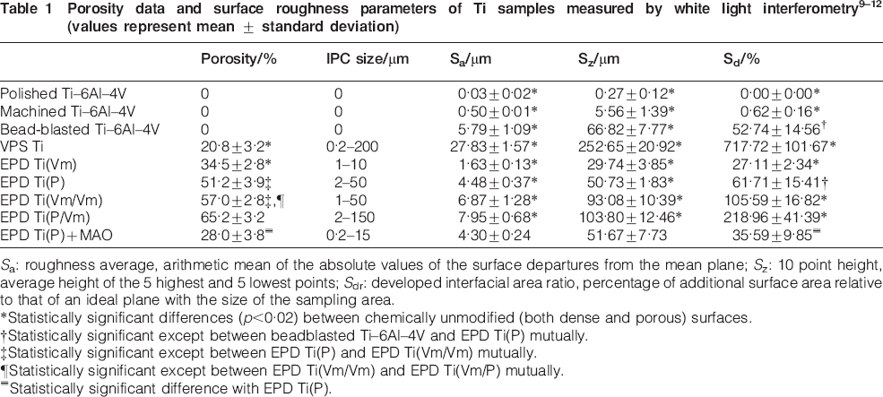
Sa: roughness average, arithmetic mean of the absolute values of the surface departures from the mean plane; Sz: 10 point height, average height of the 5 highest and 5 lowest points; Sdr: developed interfacial area ratio, percentage of additional surface area relative to that of an ideal plane with the size of the sampling area.
*Statistically significant differences (p<0·02) between chemically unmodified (both dense and porous) surfaces.
†Statistically significant except between beadblasted Ti–6Al–4V and EPD Ti(P) mutually.
‡Statistically significant except between EPD Ti(P) and EPD Ti(Vm/Vm) mutually.
¶Statistically significant except between EPD Ti(Vm/Vm) and EPD Ti(Vm/P) mutually.
≡Statistically significant difference with EPD Ti(P).
For this study, S. aureus and S. epidermidis were selected as clinically relevant test microorganisms; these species together account for almost two-thirds of implant-related infections. 5 The presence of EPS observed by both CLSM after 24 h (results not shown) and SEM after 48 h (Fig. 1) confirms the formation of a biofilm. At both time points, the EPS layer is most pronounced on VPS Ti surfaces and considerably more bacteria are detected on VPS Ti than on the other surfaces (Fig. 2). Furthermore, the qualitative analysis revealed that bacteria preferentially agglomerated in surface depressions. Microcolonies were formed in the grooves of the machined Ti–6Al–4V, in the open surface pores of EPD Ti surfaces and to a much larger extent in those of the VPS (Fig. 2). 21 This is consistent with previous findings that bacteria tend to accumulate in concave surface features, maximising the contact area with the material to be protected from environmental forces. 22
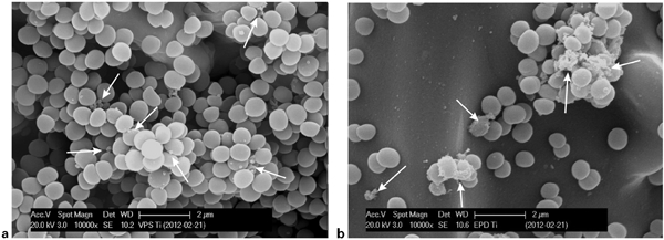
SEM micrographs of S. epidermidis colonisation after 48 h incubation on a VPS Ti and b EPD Ti(P): arrows indicate EPS formation
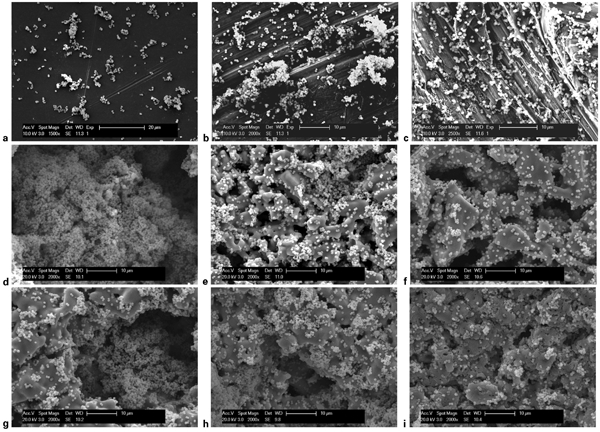
SEM micrographs of S. epidermidis colonisation after 48 h incubation on porous Ti surfaces: a polished Ti–6Al–4V; b machined Ti–6Al–4V; c bead-blasted Ti–6Al–4V; d VPS Ti; e EPD Ti(Vm); f EPD Ti(P); g EPD Ti(Vm/Vm); h EPD Ti(P/Vm); i EPD Ti(P)+MAO
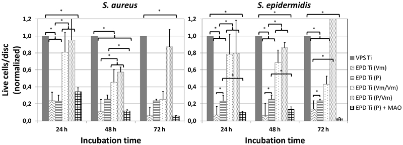
Quantitative analysis confirms the high susceptibility of porous coatings to biofilm formation, which can be correlated with increasing surface roughness (Fig. 3–5). As a result, the experimental porous Ti coating EPD Ti(P), which exhibits moderate roughness compared with VPS Ti, also shows strongly decreased biofilm formation (Fig. 3). Further reducing the surface roughness using smaller sized starting powders, as for EPD Ti(Vm), gives amounts of biofilm comparable even with polished and machined surfaces after 48 and 72 h of incubation. 21
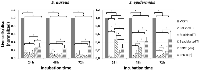
Biofilm formation after 24, 48 and 72 h of incubation on various dense and porous Ti surfaces for S. aureus and S. epidermidis: data presented as mean values ± standard deviations; *indicates statistically significant differences (p<0·1)
Biofilm formation after 24, 48 and 72 h of incubation on various dense and porous Ti surfaces for S. aureus and S. epidermidis: data presented as mean values ± standard deviations; *indicates statistically significant differences (p<0·1)
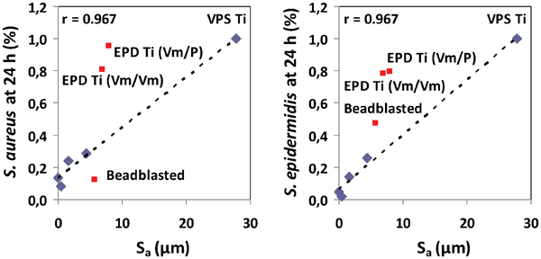
Correlation between surface roughness Sa and S. aureus and S. epidermidis biofilm formation after 24 h of incubation for various dense and porous Ti surfaces
The expanded porosity and IPC size of the EPD Ti(Vm/Vm) and Ti(P/Vm) coatings significantly increased the surface roughness, height variation and surface area (Table 1). However, the biofilm formation on these coating surfaces after 24 h (Fig. 4) was similar to the levels observed on VPS Ti, which has much higher roughness parameters (Table 1). As contact angles are comparable for all EPD Ti coatings (results not shown), this cannot be a result of additional hydrophobicity effects. The larger surface colonisation must be accounted for by the presence of additional spherical open surface pores, where bacteria can accumulate, as is confirmed by Fig. 2g and h .
The linear correlation at moderate roughnesses between the amount of adhered bacteria and the surface roughness does not apply for beadblasted Ti–6Al–4V (Fig. 5), confirming that it is not only topological surface features that govern biofilm formation, but that hydrophobicity also has a strong influence. While the hydrophilic nature of the beadblasted Ti–6Al–4V surface, resulting from the incorporation of remnants of the blasting medium into the surface, seems to decrease the S. aureus biofilm formation, it promotes the adhesion of S. epidermidis. For both bacterial strains, the amount of adhered bacteria was still considerably lower for beadblasted Ti–6Al–4V than for VPS Ti.
To prevent bacterial adhesion and/or subsequent biofilm formation, several surface modifications (both topographical and chemical) have already been explored for non-porous surfaces. 8 For porous coatings, however, to preserve the possibility of osseointegration, it is important that these surface modifications do not alter the pore structure. On flat substrates, a Ca incorporating anodic oxide was reported to decrease bacterial adhesion and enhance bone contact. 23
The Ca incorporating oxide coating (EPD Ti(P)+MAO) significantly decreased bacterial colonisation of the surface compared with the non-modified EPD Ti(P) (Fig. 4). It is suggested that this is due to the strong hydrophilic nature of the surface. It is well established that hydrophilic materials are more resistant to bacterial adhesion.18,19 However, as these surfaces also exhibit a significant decrease in Sdr due to filling of the pores by the oxide layer (Table 1), the contribution of a reduced surface area cannot be entirely ruled out. Nevertheless, if tuning of the processing parameters makes it possible to preserve the original pore characteristics of the Ti coating, MAO could be a promising surface modification technology to reduce bacterial adhesion. 21
Conclusion
Several experimental porous pure Ti coatings for orthopaedic implant applications, with varying surface roughness and pore morphology, have been compared with four clinically relevant implant surfaces for short term in vitro surface colonisation by S. aureus and S. epidermidis bacteria strains. It was observed that bacterial colonisation is linearly correlated with surface roughness for pure Ti or Ti alloy surfaces, whereas chemically modified Ti surfaces such as bead-blasted surfaces may show strong deviations from this relationship, depending on the bacterial strain. The newly developed porous titanium coatings obtained by EPD of TiH2 powder suspensions exhibit a significantly reduced bacterial colonisation relative to a state-of-the-art porous VPS Ti coating. Moreover, for appropriate starting powder particle size, S. aureus and S. epidermidis biofilm formation was found to be as low as that on polished or machined Ti–6Al–4V surfaces. The introduction of additional spherical pores into the coating by EPD of TiH2 powder stabilised emulsions, although only slightly increasing the surface roughness, had a significant effect on biofilm formation, as bacteria tend to accumulate in the open pores. These results indicate the importance of surface functionalisation to reduce bacterial adhesion for porous coatings, if the envisioned enhancement of the osseointegration capacity of the porous coatings is to be realised.
Footnotes
Acknowledgements
The research leading to these results has received funding from the European Commission's Sixth Framework Programme under project contract NMP3-CT-2006-026501 (Meddelcoat) and the Seventh Framework Programme (FP7/2007-2013) under the grant agreement Coatim (project 278425) and the Research Fund of KU Leuven under project IDO/06/013 and GOA/08/007. This is an edited version of a presentation at Euro PM 2012, organised by EPMA in Basel, Switzerland on 10–12 October 2012.