
Abstract
Current orthopaedic implant technology focuses on fixation by osseointegration to maximise the implant longevity and reduce the need for burdensome and expensive revision surgery. In this respect, porous Ti coated implants, which enable bone ingrowth into the porous structure and the establishment of biological anchoring of the implant, are of interest. In previous work a new powder metallurgical processing route was reported for the application of porous Ti coatings on Ti alloy substrates by electrophoretic deposition (EPD) of TiH2 powder suspensions. To validate the function of these coatings for potential clinical applications, the early peri-implant bone response was evaluated in vivo in a rabbit model. The results clearly demonstrate bone ingrowth in porous coatings with pore channels down to 10 μm, as opposed to the minimum pore size of 50–100 μm commonly claimed in the literature. Moreover, the observed inter-connectivity with surrounding cortical bone confirmed the envisaged mechanical interlocking of the implant.
Porous Ti structures or coatings are of special interest because they enable bone ingrowth into the porous structure and as such establish biological anchorage of the implant in the host bone. The most common techniques to apply porous Ti coatings for implant applications are vacuum plasma spraying (VPS) or partial sintering of Ti powders or beads. Whereas VPS porous Ti coatings possess an irregular porosity distribution and low pore interconnectivity, 1 powder metallurgical sintering techniques can establish a fully interconnected pore structure, allowing control of the porosity by varying sintering temperature and time. A major drawback of Ti powder based sintering techniques, however, is the high temperature required to establish sufficient interparticle bonding, which can have a detrimental effect on the substrate microstructure and concomitantly its mechanical properties. 2
Recently, we have proposed an alternative powder metallurgical processing route, consisting of electrophoretic deposition (EPD) of TiH2 powder suspensions followed by dehydrogenation and sintering in high vacuum. The use of TiH2 powders resulted in a higher sinterability compared with pure Ti powders, allowing the sintering temperature to be reduced to below the β-transus (975±15°C) of the Ti–6Al–4V substrates, while maintaining a good mechanical strength. Additionally, EPD was shown to be a valuable tool in controlling the coating thickness, which allowed the coating process to be extended to complex shaped dental implants.3,4 In vitro experimentation demonstrated and enhanced cell proliferation and gene expression, while significantly reducing bacterial colonisation.5–7 However, the potential of these coatings to improve implant stability in clinical applications strongly depends on their bone ingrowth capacity. Therefore, the goal of this study was to investigate the early peri-implant bone response in vivo using a rabbit model. In addition to the described porous Ti coatings, three chemically modified porous Ti coatings have been analysed. Biofunctionalisation of the otherwise bioinert titanium to provide better osteoconductive and osteoinductive behaviour may enable a faster and more complete bone apposition inside the porous coating.
Materials and methods
Pure grade 2 titanium (Goodfellow) discs, 4 mm in diameter and 1 mm in thickness, were used as substrate material. Porous pure Ti coatings were applied using electrophoretic deposition (EPD) of TiH2 powder (grade P, ∼40 μm, Chemetall GmbH) suspensions followed by vacuum dehydrogenation and sintering, as described previously.3,4 This served as the unmodified reference coating, referred to below as EPD Ti. Surface modification of the porous Ti coating was carried out in an attempt to improve its osteoinductive properties, by one of the following processes:
hydrothermal treatment (HT, Jožef Stefan Institute) creating a thin microanatase TiO2 layer on the outer and inner surface 8
micro-arc oxidation (MAO, University of Bayreuth) producing a pore filling TiO2 layer containing Ca2+ and PO43− ions 9
sol–gel synthesis, applying a micrometre thin bioactive glass (BAG) coating on the internal surface of the Ti coating. 10
Samples with these treatments are denominated EPD Ti+HT, EPD Ti+MAO and EPD Ti+SGBAG respectively.
Mercury intrusion porosimetry (MIP, AutoPore IV 9500, Micromeritics) in combination with image analysis (PPM2OOF software, NIST) on scanning electron microscopy (SEM) images of representative metallographic cross-sections was used to analyse the pore structure characteristics (porosity, pore size, interconnecting pore channel (IPC) size). 11
Prior to implantation all samples were sterilised, either by steam sterilisation or heating to 200°C in vacuum. The animal handling and experimental protocol used in this study was approved by the Animal Ethics Committee of the KU Leuven and was performed according to the Belgian national legislation concerning the protection and wellbeing of animals (approval ID: P122/2008).
Six mature New Zealand white rabbits (average weight 3·17±0·18 kg) underwent surgery as described elsewhere. 12 In short, the coated Ti implants were placed in the cortical bone of the tibia in a double-stepped cavity, with an outer step diameter of 4 mm (allowing a press-fit mounting of the samples) and an inner step of 2 mm in diameter and 0·5 mm in depth (Fig. 1). After a healing period of either 2 or 4 weeks, the animals were euthanised and bone blocks containing one implant were fixed, dehydrated and embedded in methylmethacrylate (99·5%, VWR International). The blocks were cut longitudinally and perpendicular to the implant and ground (4000 grit SiC, Hermes) and polished (3 μm, Diapat S, VEM Metallurgie) for SEM analysis.
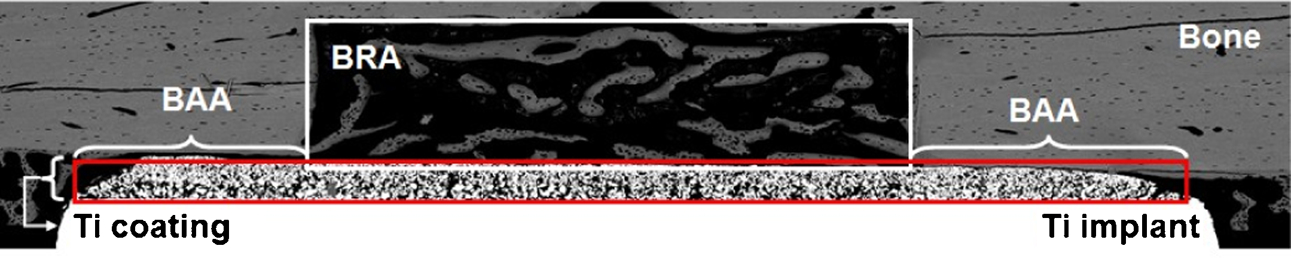
SEM micrograph of a cross-section at the implant/bone interface indicating the main regions of interest for histomorphometrical analysis: bone regeneration area (BRA), bone adaptation area (BAA) and porous Ti coating
In this bone cavity model, three main regions of interest can be determined:
the central cavity or ‘bone regeneration area (BRA)’, which is a region without any bone at the time of implantation
the periphery of the implant or the ‘bone adaptation area (BAA)’, where the implant is initially in contact with the bone at the time of implantation
Quantitative histomorphometrical analysis was performed using image analysis software (CTAn, SkyScan NV). The following parameters will be presented in this paper: the
Results and discussion
Representative cross-sections of the different coatings were obtained by SEM (Fig. 2), while the cumulative intrusion volume obtained by MIP was used to establish an IPC size distribution (Fig. 3). EPD Ti coatings are highly interconnected, exhibiting 50% porosity. While the porosity remains largely unaltered for EPD Ti+HT or EPD Ti+SGBAG, it is significantly reduced after MAO treatment to around 28·0%. SEM images indeed reveal that the MAO coating (grey phase in Fig. 2c) has filled up a large part of the available pore volume. These observations are confirmed by the MIP result (Fig. 3). For the pore size range from 300 down to 10 μm, which can mainly be associated with the surface pores, the pore size distribution for EPD Ti+MAO is comparable to the EPD Ti reference. However, for the bulk pores (<10 μm), an important fraction of the pore volume is taken up by small pores (<5 μm) in MAO, in contrast to HT and SGBAG for which the pore size distribution below 10 μm is comparable with that in the EPD Ti coating. The strong deviation seen for EPD Ti+SGBAG at low pore sizes (<0·1 μm) is most probably due to an undesirable reaction between the Hg and the BAG; it is well known that Hg forms amalgams with several metals, including sodium. 13 A possible explanation for the fraction of large pores in the EPD Ti+HT coatings is the presence of surface cracks on those samples.
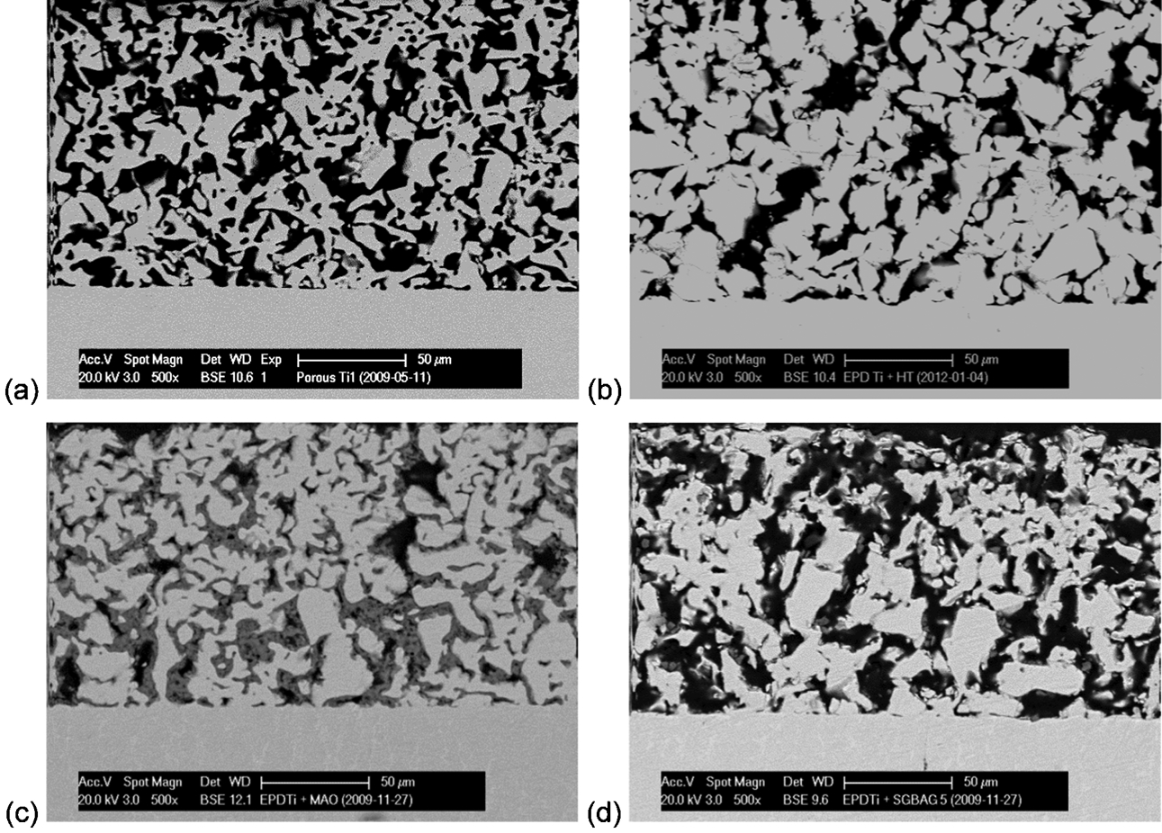
SEM micrographs of representative cross-sections of a EPD Ti, b EPD Ti+HT, c EPD Ti+MAO and d EPD Ti+SGBAG prior to implantation
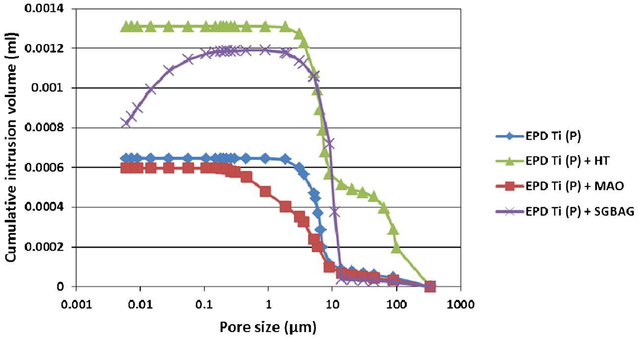
Cumulative intrusion volume as function of pore size measured by mercury intrusion porosimetry for various porous Ti coatings
The general consensus in the literature is that the ideal pore size for bone ingrowth lies in the 100–400 μm range, which represents a compromise between sufficient mechanical strength of the porous structure and adequate space within the pores to allow cell migration and vascularisation. However, it should be noted that this minimum size of 100 μm refers to the macropore size, whereas the much smaller sizes of the interconnecting pore channels (IPC), which are the actual limiting factor for bone ingrowth, are rarely mentioned in the literature. Several studies have demonstrated bone ingrowth for IPC sizes less than 50 μm, and even at 30 μm.14–17 The IPC size distribution of the porous Ti coatings presented here is in the 1–50 μm range, with an additional submicrometre distribution for EPD Ti+MAO coatings.
Representative BSE–SEM micrographs of cross-sections of coatings in the BRAs after 2 weeks (EPD Ti+SGBAG) and 4 weeks (EPD Ti+HT) of implantation are shown in Fig. 4a and b respectively. After 2 weeks, a low-mineralised bone phase (grey phase) had formed in the BRA above the coating (white phase). The bone trabeculae were irregular and highly cellular, i.e. with a high proportion of osteocytes, both distinctive features of primary bone tissue or so called woven bone (Fig. 4a). Although the bone was in contact with the Ti, only limited bone was observed inside the porous structures after 2 weeks.
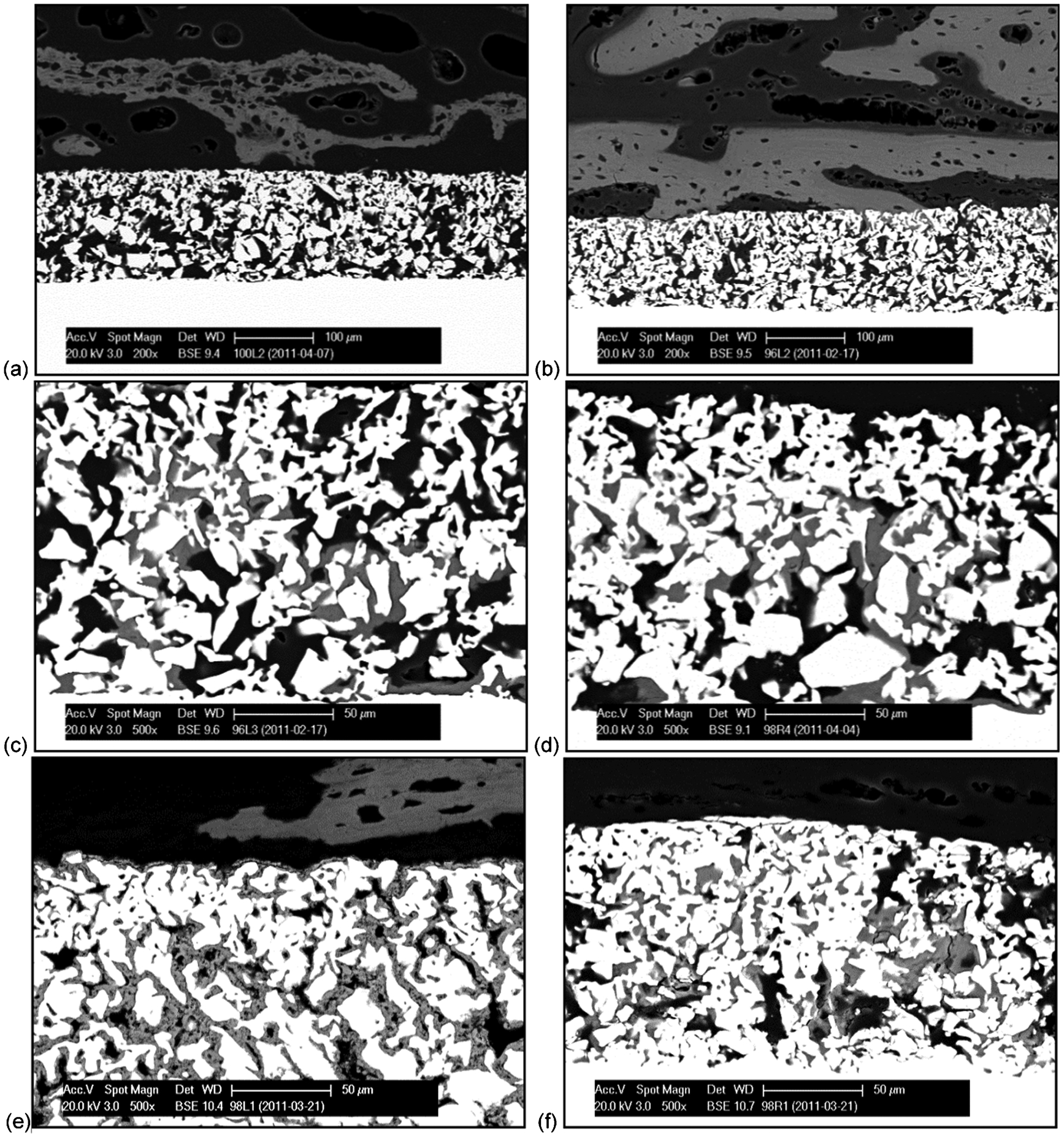
Representative BSE–SEM micrographs of cross-section through implant/bone interface: a EPD Ti+SGBAG after 2 weeks and b EPD Ti+HT after 4 weeks of implantation. Detailed BSE–SEM micrographs of porous Ti coating for c EPD Ti, d EPD Ti+HT, e EPD Ti+MAO and f EPD Ti+SGBAG after 4 weeks of implantation
After 4 weeks, the bone trabeculae were larger and the woven bone was replaced by a well mineralised bone phase consisting of a layered structure with a much lower proportion of osteocytes, i.e. lamellar bone (Fig. 4b). Bone ingrowth (including osteocytes) throughout the whole coating up to the coating/substrate interface was observed for all coatings, except in the case of the EPD Ti+MAO coatings, for which the grey phase inside the pores could be identified as the TiO2-based MAO coating already present before implantation (Fig. 4c–f). This observation confirms bone ingrowth for IPC sizes down to 5 μm, but not in the submicrometre range. Additionally, the growth pattern of the bone inside the porous coatings was visualised by binarised the BSE–SEM images. Representative images are shown in Fig. 5 for EPD Ti, EPD Ti+HT and EPD Ti+SGBAG. The bone is present throughout the whole coating thickness in the form of trabeculae which are clearly interconnected with the bone outside the porous coatings.
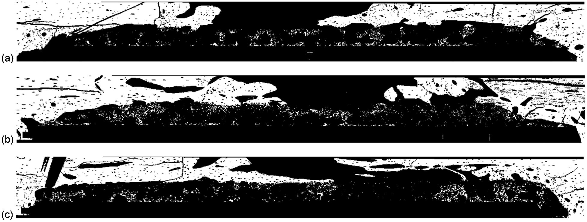
Binarised BSE–SEM micrographs of cross-sectioned implant/bone interface for a EPD Ti, b EPD Ti+HT and c EPD Ti+SGBAG after 4 weeks of implantation
The results of the histomorphometrical analysis are summarised in Fig 6. The BAF, i.e. newly formed bone in the BRA, seems to increase with healing time, confirming the histological observations above. There is a trend towards an increased BAF for all biofunctionalised coatings as compared to the unmodified EPD Ti. However, this is only statistically significant (p<0·05) for EPD Ti+SGBAG (61·8±5·6%) compared with EPD Ti (36·3±8·6%) at 4 weeks. This confirms the osteogenic potential of the sol–gel derived glass-ceramic coating.
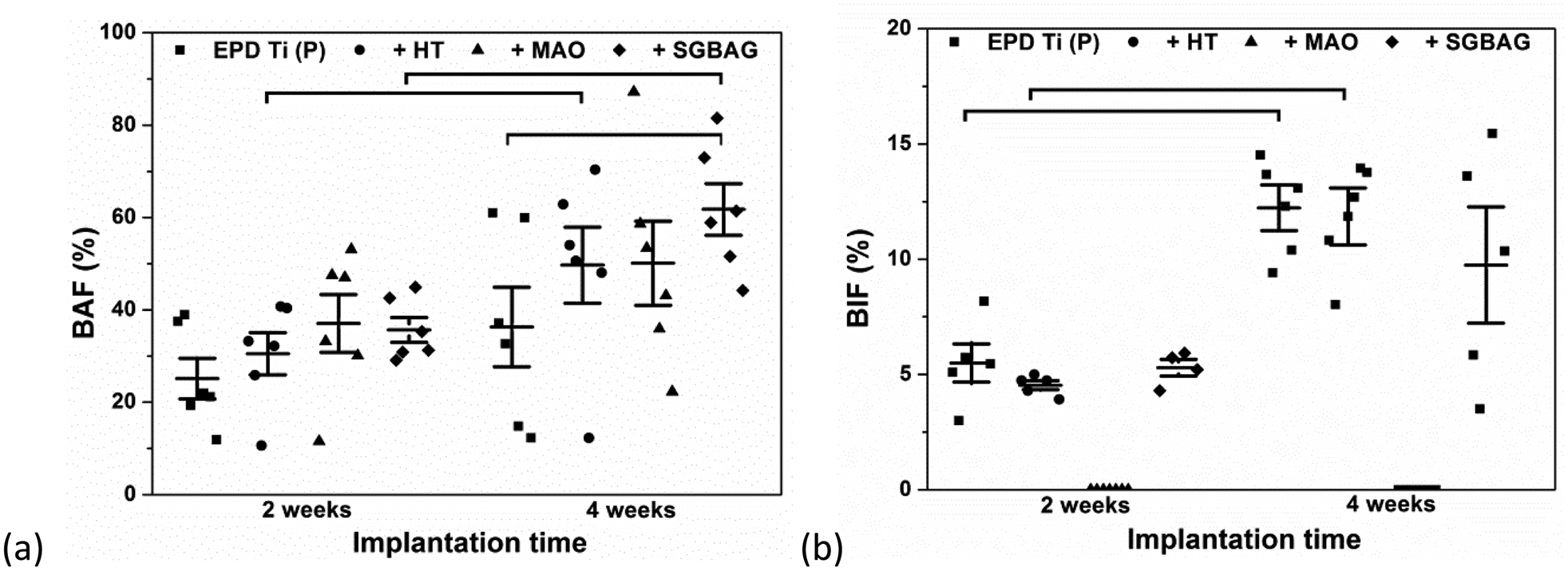
a Bone area fraction (BAF) and b bone ingrowth fraction (BIF) after 2 and 4 weeks of implantation. Results are presented as mean values ± standard errors, statistically significant (p<0·05) differences are indicated by the horizontal lines 11
The BIF, i.e. new bone established inside the porous Ti coating, increases over time, except for EPD Ti+MAO, where no bone ingrowth could be observed at all. However, no significant differences were seen on comparing the other three conditions. With respect to bone ingrowth, the current results considering the early bone response at 2 and 4 weeks do not support a hypothesis of increased bone formation inside the biofunctionalised porous Ti coatings.
Conclusion
The current data clearly demonstrate bone ingrowth into the experimental EPD Ti coatings, shifting the generally accepted minimum pore size required for bone ingrowth from 50 μm to below 10 μm. Moreover, the bone inside the coatings appears to be well interconnected with the surrounding cortical bone, enabling micro-interlocking of the porous coating to improve the long term stability of implants. However, surface functionalisation of the inner pore surface should not further reduce the pore size below this minimum value since this will obstruct colonisation of the pore structure by osteoblasts. Micro-arc oxidation processing parameters, for example, should be fine-tuned to minimise the effect on the pore characteristics.
The application of a sol–gel derived bioactive glass-ceramic in the inner pore surface significantly increased the bone regeneration outside the porous structure, although this effect was not observed inside the porous coating. 11
Footnotes
Acknowledgements
The research leading to these results has received funding from the European Commission's Sixth Framework Programme under project contract NMP3–CT–2006–026501 (MEDDELCOAT) and the Seventh Framework Programme (FP7/2007–2013) under the grant agreement COATIM (project 278425) and the Research Fund of KU Leuven under project IDO/06/013 and GOA/08/007 and the knowledge platform IOF/KP/11/007 (Flabicoat). Based on a presentation at Euro PM 2013, organised by EPMA in Gothenburg, Sweden, on 15–18 September 2013.