
Abstract
Christopher Wren's figure of the brain ‘viewed from below’ in Thomas Willis's Cerebri anatome (1664) set a precedent, not only for subsequent images of the brain, but for scientific illustration. The image is the visual proof of a Baconian experiment conducted by the Oxford dissection team, in which dye was pumped into the carotid arteries of animal and human specimens in order to imitate the natural flow of blood, thereby applying William Harvey's theory of circulation to the brain. Usually described as engravings, Wren's images are in fact etchings (produced by the acid process), significant because etching for book illustration was then in its infancy in England. In addition to considering Wren as an experimental etcher, this article frames our understanding of Wren's contributions to Willis's project in terms of his virtuosic talents as amateur draughtsman, natural historian, anatomist, and his use of optical and draughting instruments.
In William Hogarth's satire, ‘Credulity, Superstition and Fanaticism: A Medley’ (1762), a Methodist preacher is besieged by hobgoblins. In the lower right corner is a strange creature: a human brain. Viewed from below, its protuberances look like the legs of an insect. The brain sits upright on its cerebellum, cushioned by Joseph Glanvill's treatise on witches (used to justify the Salem trials) and John Wesley's Sermons (Figure 2). Listening to the preacher with an ear attached by a single auditory nerve, the brain is a warning not to believe all we hear — lest it go directly to our heads.
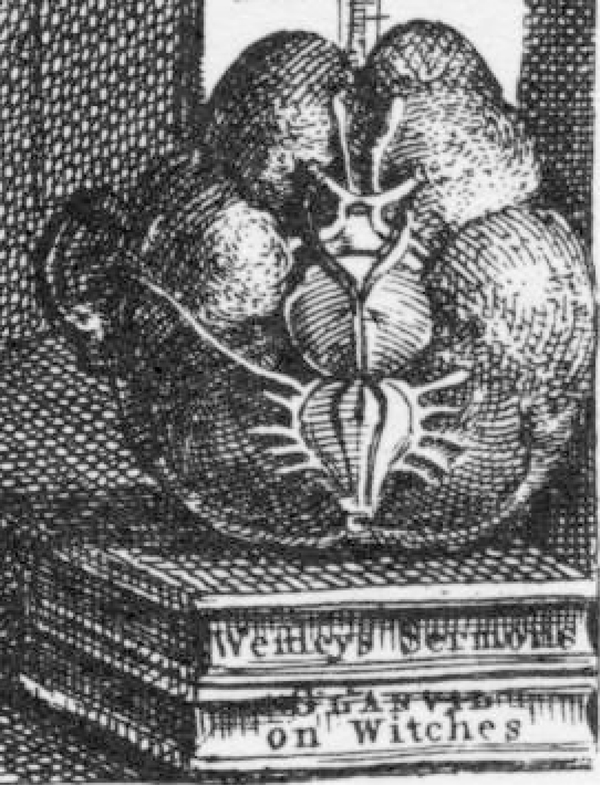
Detail from William Hogarth, ‘Credulity, Superstition, and Fanaticism: A Medley’, 1762, engraving
Hogarth's brain quotes an earlier English draughtsman: Sir Christopher Wren (1632–1723). Now remembered as the architect of St Paul's, Wren earlier contributed the figures of the brain (see Figure 1) for Thomas Willis's (1621–75) Cerebri anatome (1664).1 Willis's treatise was one of the first and most successful books published by the newly chartered Royal Society (hereafter Willis 1681; Martensen 2004, 75).2 The work is most significant for giving new prominence to the material brain (Feindel 1965; Martensen 2004; Knoeff 2004; Rijcke 2010). Invoking the image of Atlas, Willis likened the brain to man's entire world; with his treatise, he hoped to provide new shoulders to bear this great ‘Globe … built upon [or within] the Skull of a Man’ (1681, Preface). In the same century as René Descartes (1596–1650), Willis sought the seat of the ‘rational soul’. However, while Descartes believed that ‘God's regularity and perfection’ in the material world could only be deduced from philosophical principles, Willis proposed that the soul could be found by anatomizing (Martensen 2004, 56). And whereas Descartes geometrically ‘proved’ the soul's location in the pineal gland at the brain's centre, Willis believed it to be located in the grey matter of the cerebral cortex (Rijcke 2010, 34).
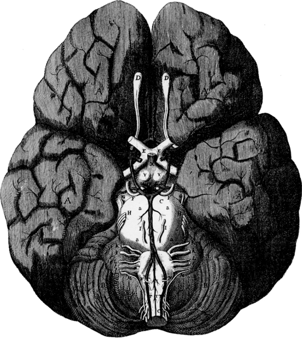
Christopher Wren's first figure (the base of the human brain), in Thomas Willis, Cerebri anatome, 1664, 16·2×15·2 cm
Along with shifting attention to the cortex, what is now recognized as the seat of higher function, another of Willis's feats was to apply William Harvey's (1578–1657) demonstration of the circulation of the blood to brain physiology (1628; 1649). Harvey's theory that the heart acts like a pump moving blood around the body (and that vessels contain unidirectional valves that control blood flow) still met with occasional opposition (Martensen 2004, 24–5). Willis believed that chemical reactions undergone by ‘animal spirits’ (carried by the blood) led to the generation of the rational soul in the cortex. He combined Harvey's anatomical method and theory with his own experimental ‘manipulation’ of the blood to determine the nature of these reactions (Rijcke 2010, 34; Frank 1980, 164–92). While Willis's physiology is no longer viable, it propelled him to describe and display, like never before, the anatomical parts that housed these transformative reactions. Even the Danish naturalist, Niels Stensen (aka Steno, 1638–86), who disagreed with Willis on many points, admitted that the ‘best diagrams of the brain that we have to date are those given to us by M. Willis’ (Stensen 1669, 1965).
Arguably, the most important figure in Cerebri anatome is the first (Figure 1), which depicts for the first time the complete arterial loop (anastomosis) now known as the Circle of Willis (Kemp and Flis 2008, 174). The anastomosis was considered to be an example of God's providential design. If there were a blockage in one part, blood would still be able to reach the brain by another route. Constanzo Varolio (1543–75) was the first to include a description of the arterial circle in his work on the human optic nerves (1573), though he did not understand its significance (Martensen 2004, 50). The engraving in Casserius's Tabulae anatomicae (1627) shows an incomplete circle, as observed by artist Odouardo Fialetti (1573–c.1638), remarkable considering that its function was still unknown. German anatomist Johann Vesling (1598–1649) was the first to provide a full textual description of the structure, but despite the profuse illustration of his anatomical atlas, he did not include an image (1653). Finally, Swiss physician Johann Jakob Wepfer (1620–95) had recently theorized that blockages in the blood vessels cause damage to the brain and other tissue, another application of Harvey's theory (1658).
With the first figure in Willis's treatise, Wren synthesized past and recent findings, and underlined the importance of the circulatory structures, a perspicacious act of ‘visual pointing’ (Kemp 1996, 83). The deeply etched parallel lines at the centre serve to darken the vessels, including the anastomosis and the basilar artery that descends from it (Figure 3). The structure stands out starkly against the white optic nerve (labelled EE) and the round medulla oblongata (C) with its protruding cranial nerves (including HH, II). The Circle of Willis is presented as a highly visible, complete, symmetrical, and robust structure, the branches of which interlace with the nerves and disappear as they permeate the surface.
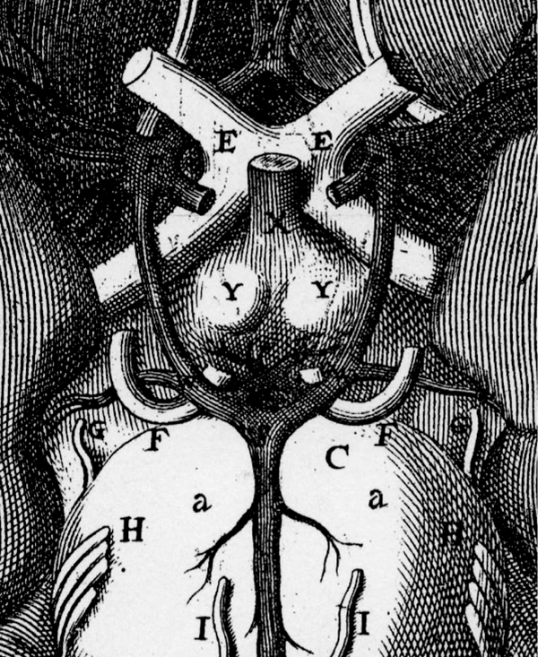
Detail of Figure 1 showing the anastomosis
Surprisingly little is known about the production of the figure. Wren's social background, perceivable spiritual beliefs, and his family connections with the Church of England, all lent credibility to his status as a witness in a culture of gentlemanly science (Rijcke 2010, 41–2; Shapin 1994, 42–125). This partly answers the question of what qualified him to contribute to Willis's investigations. But what role did Wren play exactly? To answer this question, I investigate Wren's early activities as naturalist, anatomist, draughtsman, and printmaker; and bringing to bear Kim Sloan's assessment of the role of drawing in the life of the gentleman virtuoso; and Ann Bermingham's emphasis on the changing status of drawing as a performative and ‘social’ skill (2000, 11–18; 2000, 33–73). Finally, I demonstrate how Wren's first figure functions as a work of mechanical ‘realism’, or as a ‘cultural hologram’, capturing a crucial moment in the experiment at the heart of Willis's treatise (Martensen 2004, 202; Rijcke 2010, 41). I suggest that Wren's figure is the result of an ‘artificial’ experiment in the imitation of nature that conjoins early modern ‘art’ (as craft) and ‘science’ (as the generation of matters of fact about nature) (Alpers 1983; Shapin and Schaffer 1985). By singling out Wren's contribution, and using inferential criticism (Baxandall 1985), we gain an understanding of the famous figure of the brain on its own historical terms — as a material and cultural object, and as the imprint of contemporary scientific and artistic practice.
Cerebri anatome: The time, the people, and the place
Willis's researches were carried out by members of the ‘Oxford Experimental Philosophy Club’, which existed in the 1650s not as a formal institution, but as a so-called ‘invisible college’ (Webster 1975, 57–67). This body of practitioners was devoted to what mathematician John Wallis (1616–1703) called the ‘New Philosophy’ (171–2). Under the Commonwealth, most of the members longed for the return of the monarchy, and a scientific society with royal patronage that would be modelled on what Francis Bacon (1561–1626) had described in The New Atlantis as ‘Solomon's House’, a great store of ‘Knowledge of the Causes and Secrett Motions of Things; And the Enlarging of the bounds of the Humane Empire, to the Effecting of all Things possible’, that is, the practical application of knowledge (342).
Willis's investigations are a significant episode in the early history of the Royal Society, officially founded after the Restoration (1660). Willis was granted his medical degree in the same year, and made Sedleian Professor of Natural Philosophy at Oxford. In 1661, Wren was appointed Savilian Professor of Astronomy. Willis dedicated Cerebri anatome to the cleric Gilbert Sheldon (1598–1677), whom he had recently helped recover from a stroke. Sheldon, a royalist political prisoner during the Interregnum, was, for his loyalty made Archbishop of Canterbury (1663) and Chancellor of the University of Oxford (1667). Eventually one of the most powerful men in England, Sheldon financed the building of the Sheldonian Theatre, one of Wren's earliest architectural designs. Construction had just begun when Cerebri anatome was published.
In the preface of his treatise, Willis credits: the ‘Learned Physician and highly skilful Anatomist’ Richard Lower (1631–91); the ‘Doctor of Physick’ Thomas Millington (1628–1703/4); and Wren, described as ‘Doctor of Laws, and Savill-Professor of Astronomy’ (1681, Preface). Also present were the physician Edmund King (1629–1709) and a ‘Dr Masters’ (Sprat 1667, 301). This sizable team fulfilled Bacon's recommendation for a large number of credible ‘witnesses’ in order to corroborate the evidence gathered in support of matters of fact (Dear 2001, 138–45; Shapiro 2000). Credibility and the right to hold and express opinions freely depended upon social rank; and/or proven distinction in learning; and whether or not one was a ‘Christian Gentleman’ (Shapin 1994, 65–125; Shapiro 2000; Martensen 2004, 95–127).
Wren's family connections helped. His father, Christopher Wren Sr. (1589–1658), was Dean of Windsor, and his uncle, Matthew Wren (1585–1667), had been Chaplain to Charles I, rising to become Bishop of Ely. Willis writes that Wren ‘was pleased out of his singular humanity, wherewith he abounds, to delineate with his own skilful hands many Figures of the Brain and Skull, whereby the work might be more exact’ (1681, Preface). Along with Millington and Lower, Wren also contributed to the anatomical, ‘chemical’, mechanical, and sometimes philosophical discourse (56–7).
The investigations took place at Beam Hall in Merton Street, Willis's home and an early ‘House of Experiment’ (Frank 1980, 179; Shapin 2010, p. 64). During the Interregnum, Beam Hall also served as the clandestine meetinghouse for members of the Church of England (Feindel 1996, 268–70). Willis was devoutly religious, and the nature of his Anglican beliefs shaped his search for the soul in the material of the brain. His particular brand of belief allowed him to surmise that ‘base’ flesh might house the lofty soul, an idea most contemporary Calvinists would have abhorred (Knoeff 2004, 439–40; Bynum 1973, 445–68).
Willis's attention to the solid parts of the brain and to the motion of the animal spirits resonated with Harvey's focus on the solid parts of the heart and the movement of the blood (Rijcke 2010, 34; Frank 1980, 169–88). The team's use of comparative anatomy was also strongly influenced by Harvey, who had once been criticized for attending to the ‘empty glory of vivisections’ (1980, 17). Willis had decided upon composing a ‘certain Anatomy of the Brain [based on] the frequent dissection of all sorts of living Creatures’ (1681, 56, 79). He refers to the brains of birds, fish, dogs, and ‘many other four-footed beasts, [which] were little different … from the figure of the same, and the disposition of the parts, in a man’ (56).
Cerebri anatome is divided into two parts: the anatomy of the brain; and the description of the nerves. The treatise contains thirteen plates. The first eight are labelled as ‘figures’; they depict views of the brain and skull from different angles. Several figures rely on comparative views in creatures; for example, the second shows the base of a sheep's brain with its analogous structures. The other five plates, likely based on drawings by Lower, are ‘tables’, functioning as schematic representations of the ordered pairs of nerves (Knoeff 2004). Each plate works in tandem with the text, describing the dissected parts ‘in the same order wherein they occurred in Inspection’ (Willis 1681, 68, 60), a format following Vesalius (1543), allowing the reader to reproduce the experiment, virtually or physically (Rijcke 2010, 29). However, even removed from the treatise, Wren's first figure speaks to Willis's primary anatomical findings, and displays the experiment that made them possible.
Wren: Draughtsman, naturalist, and anatomist
Wren spent the first thirteen years of his life (1632–45) at his father's rectory in East Knoyle, Wiltshire, where he was born, and at the deanery in Windsor (Davies 2008, 303–4). His earliest recorded drawing, made at age ten, depicts the ‘prospect of the parish church at Knoyle and a view in perspective of a dining-room in his father's house there’ (Davies 2008, 304). From the sixteenth to the mid seventeenth century, drawing had evolved from a ‘courtly practice into a gentleman's pastime’. It had come to be associated with ‘virtue and Protestant modes of truth-telling’ (Bermingham 2000, 33). Drawing was thus a part of the social and cultural capital of the young virtuoso. The description of Wren's early drawing suggests a geometrically principled training. Wren could have learned the ‘continental’ principles of Alberti's single-point perspective from manuals such as Henry Peacham's Art of Drawing with a Pen (1606) (Kemp 1991; Bermingham 2000, 33). Wren would have also been encouraged to draw ‘from the life’, and, alternatively, to draw from model books containing human figures in proportion, birds, beasts, and flowers (Alpers 1983; Swan 2005, 36–40; Bermingham 2000, 67–8).
Drawing was a ‘mechanical art’ that could set the problem-solving virtuoso apart from other kinds of virtuosi (Bermingham 2000, 34). Between 1646–9, Wren lived in London with the physician and surgeon Charles Scarburgh (1615–94). Scarburgh's house was a meeting place for the many intellectuals of the ‘Royalist persuasion’ associated with Gresham College (Bennett 1982, 16). Assisting Scarburgh in the dissections at Surgeons’ Hall in late 1649, Wren likely made drawings from samples of dissected material (Davies 2008, 310; Bennett 1982, 19). In Parentalia, it is recorded that Wren ‘composed a Treatise on the Motion of Muscles, explaining the whole Anatomy by Models form'd in Pasteboards … presented to Sir Charles Scarburgh, but lost at the Fire of London’, probably a description of anatomical drawings (Bennett 1982, 16; Wren Jr 1750, 221–2).
In 1650, Wren entered Wadham College, Oxford as a ‘gentleman-commoner’ (a student who paid all of his fees at once), completing his BA by 1652. Between 1653/4, when he proceeded to MA, and 1657, when he was elected a fellow of All Souls and was appointed Professor of Astronomy at Gresham, Wren was involved in a number of natural historical investigations. The clergyman and natural philosopher, John Wilkins (1614–72) introduced Wren to the meetings of the Oxford natural philosophers (Davies 2008, 311; Jardine 2004, 107–29). Wilkins also partly financed the activities of the invisible college, and his marriage with Oliver Cromwell's sister, Robina, provided political protection for an otherwise strongly royalist group (Webster 1975, 171). In 1654, John Evelyn (1620–1706) visited Wilkins's rooms at Wadham, admiring the intermingled objects, gadgets, and experiments of the New Philosophy:
[Wilkins] shew'd me the transparent apiaries, which he had built like castles and palaces, and so order'd them one upon another so as to take the hony[sic] without destroying the bees … He had also contriv'd an hollow statue which gave a voice and utter'd words, by a long conceal'd pipe that went to its mouth, whilst one speaks through it at a good distance. He had above in his lodgings and gallery a wide variety of shadows, dyals, perspectives, and many other artificial, mathematical, and magical curiosities, a way-wiser, a thermometer, a monstrous magnet, conic and other sections, a balance on a demi-circle, most of them of his owne and that prodigious young scholar Mr Christopher Wren, who presented me with a piece of white marble, which he had stain'd with a lively red very deepe, as beautiful as if it had been natural (Evelyn Diary 1654, 13 July).
Many of the wonders Evelyn observed — glass apiaries and Wren's dying of white marble to create a kind of mock porphyry — are works of ‘artifice’; they were crafted to imitate natural phenomena and in the process they aided in explaining how nature works (Alpers 1983, 27–30). Sprat describes others, such as Wren's use of the ‘Registers of Furnaces … for keeping a perpetual temperature … [for the] hatching of Eggs, insects, [the] production of Plants, [and] Chymical Preparations’ and the ‘imitating [of] Nature in producing Fossils and Minerals’ (Sprat 1667, 317). Artifice was closely related to ‘curiosity’, which described man's ability to use, restrain, fashion, or improve the raw materials of nature (Arnold 2006, 57; Whitaker 1996, 82–4).
This ‘curious’ impulse characterizes many of Wren's early projects. Wren's father was described as a ‘Learned Man, skillful in all the Branches of Mathematicks’; he encouraged his son with the gift of Bacon's natural history, Sylva Sylvarum, on his tenth birthday (Davies 2008, 304; Wren Jr 1740). Wren's father also inspired his early considerations upon the injection of antidotes into the bloodstream for poisons (Bennett 1982, 77). Extending these interests, in 1656, Wren attempted the intravenous injection of a dog with Wilkins and Robert Boyle (1627–91) (Oldenburg 1665, 7:128; 1716, 3:291). In that year, he wrote to William Petty (1623–87): ‘I injected wine and ale into the mass of blood in a living dog by a vein in good quantity till I made him drunk but soon after he pissed it out ….’ (Martensen 2004, 41). Wren is further credited with surgically ‘taking out the Spleen of a Dog without killing him’, an experiment to determine the usefulness of the organ (Frank 1980, 141). In 1665–6, Richard Lower would apply Wren and Boyle's intravenous methods to his own experiments in canine blood transfusion (Frank 1980, 169–88; Lower 1669).
Wren devised instruments to improve drawing. A 1652 list of Wren's Wadham projects includes two such devices: ‘A Perspective Box, to survey with it’; and ‘A Scenographical Instrument, to survey at one Station’ (Bennett 1982, 74; Wren Jr 1750, 198–9). The first is identifiable as a camera obscura; the second is identifiable with the machine described and illustrated seventeen years later in the Philosophical Transactions: ‘a perspectograph … [for] drawing the Out-lines of any Object in Perspective’ (Oldenburg 1669 4, 893). The perspectograph is a camera lucida equipped with a sight (for registering the position of the eye), moveable along a mechanical track or pulley system in horizontal and vertical directions. The moveable sight, attached to the drawing instrument, allows the hand to trace the outline of the object or landscape being viewed (Kemp 1990, 180–3). In 1653, the instrument maker Ralph Greatorex (c.1625–75) discussed the construction of Wren's perspectograph with Samuel Hartlib (c.1600–62) and, by 1663, Royal Society secretary Henry Oldenburg (c.1619–77) had one (Bennett 1982, 17).
Evidence of the perspectograph's use is found in Elias Ashmole's (1617–92) Institution, Laws and Ceremonies of the Most Noble Order of the Garter, for which Wren drew a view of Windsor, etched by Wenceslaus Hollar (1607–77) (Ashmole 1672, 131; Jardine 2002, 146–9). I suggest that Wren also employed his device to make drawings from brain specimens, which would explain the nuanced or irregular shape of the printed figure — as opposed to the fairly round or blob-like form in previous views, for example, those in Casserius and Vesalius. The outline of Wren's printed figure achieves a distinction between the frontal lobes at the top, and the temporal lobes at the bottom, like no image of the brain before it.
Drawings by Wren (other than architectural ones) are rare. One is interleaved in the heirloom copy of Parentalia; it depicts studies based on the vivisection of the river eel, ‘Anguilla fluviatilis’; eleven figures of various parts that make up the ‘scheme’. Apparently, there were schemes for other creatures, all of them ‘exhibited at the Meetings at Oxford’ (Wren Jr 1750, 227). The occasion was a discourse on comparative anatomy, in which it was reported that the ‘Fabrick of the Parts appear'd very often irregular, and differing much both from Brutes, and one another (227). In 1656, Wren wrote a letter to Petty again mentioning such ‘schemes’, recording other observations made with ‘Glasses’:
Some Things [we] observ'd very considerable in Fowls. Some Parts of Animals [we] more exactly trac'd by the Help of Glasses, as the Kidneys, the Plexus of the Brain, &c (227).
In 1661, Wren presented a gift of drawings made using the microscope to Charles II. They included drawings of ‘a flea, a louse, and the wing of a fly’ (Neri 2008, 84). In fact, Wren's use of the microscope may date as early as 1649 (Bennett 1982, 18, 73). As Janice Neri points out, by 1655, Wren had planned to write a book ‘with pictures of Observ[vationes] Microscop[icae]’ (2008 87, fn. 21). Accordingly, in Micrographia, Robert Hooke (1635–1703) gave credit to Wren for being the first to make such observations (1665, Preface).
When Wren was examining parts of the kidneys and brain under the ‘Glass’ in 1656, he was likely already engaged in Willis's research. Wren's figure of the brain contains, albeit in a simplified manner, a representation of the capillaries uniformly spread across the folds, a detail that reflects Wren's description to Petty: ‘The Nerves we have found to have little Veines and Arteries in them’ (Wren Jr 1750, 227). This description accords with Willis's interest in the thin tubes or ‘nerve fibres’ that were believed to be the canals through which ‘animal spirits’ passed in an ebb and flow movement from the brain to the distal points of the nerve endings via the spine. Willis conjectured that obstructions to this movement resulted in pathologies (Barcia Goyanes 1995, 479). Wren's analysis of brain tissue with the microscope also accords with Willis's description of the ‘wonderful Net’ of minute vessels (what is now called the arachnoid mater, that is, the meninges or membrane that covers the brain and spinal cord), the texture of which Willis describes as ‘shew[ing] like the picture of a fruit-bearing wood’ (Willis 1681, 59, 72).
Willis and Wren's collaboration is demonstrated in a watercolour drawing of a pathological specimen, inscribed with both of their names (Gerbino and Johnston 2009; Neyer 2009, ill. 1). The drawing shows two views of the small intestine with rounded lesions caused by ‘typhoid fever of the lower ileum’, a condition described (but not illustrated) in Willis's posthumous Pharmaceuticae rationalis (1678; Coles 1986). Wren drew the specimen very delicately, capturing the robin's egg blue discolorations of the lesions and the minute, red, spidery vessels of the intestine; he even included the pieces of string that support and display the dried specimen. The naturalistic, unmediated detail leads one to wonder about comparable lost preliminary drawings of the brain.
In contrast, instrumentation is suppressed in the figures of the brain, which are more like the ‘serenely arranged objects’ of Micrographia (Neri 2008, 83). Alternatively, Wren's drawings of the brain might have resembled those that make up the ‘scheme’ of the river eel, the quick hatching and grey washes reminiscent of the shading and ‘colouring’ in the figures of the brain. Otherwise, preliminary drawings might have taken an abbreviated form, indicating salient features only, like the sketches of insects recently attributed to Hooke by Janice Neri, dramatically different from the final figures in Micrographia (95).
Wren: Experimental etcher
There is no reason to doubt that Wren designed all eight of the figures in Willis's treatise. But a closer look at the circumstances and process by which the first one was made demonstrates that Wren also did the printmaking. The plates were etched (using acid), not engraved, as is usually assumed. Acid-etched lines can be distinguished from those engraved with a burin (incised in the metal) by their blunt ends; this is particularly visible in the annular protuberances (labelled aa) that stem from the basilar artery descending from the Circle of Willis (Figure 3). The darkest areas of Wren's first figure reveal that some of the etched lines bled together; whether or not this was intended, it added an effective contrast.
The fact that the figures are etchings means that in addition to being an amateur draughtsman, Wren was also an amateur etcher, and an early experimenter in the medium for its use in English book illustration. The first book to contain etched illustrations by an English hand was Edward Benlowes’ poem, Theophila (1652), illustrated with plates designed and etched by Francis Barlow (c.1626–1704) (Hodnett 1988, 54–6). Wren's figures set a precedent in English works of natural philosophy and natural history, which was immediately followed by Hooke's large-scale etchings for Micrographia (1665), including the foldout plates of the gnat, ant, louse, and eye of a fly, all etched in a manner similar to the Cerebri anatome plates.
The next use of etchings of comparable quality is not found until the works of Martin Lister (1639–1712) in the 1680s, and the reason for the time lag is worth further study. Lister was a strong promoter of scientific illustration, and advocated its use in the Philosophical Transactions (Unwin 1995, 209–30). Lister's earliest works with etched figures include his translation of Johannes Godartius's Of Insects (1682) with plates by another gentleman ‘amateur’, Francis Place (1647–1728). This was followed by his Historiae Conchyliorum (1685–92), a work on shells. Anna Marie Roos has ‘rediscovered’ and published some of the thousand or so copper plates for Conchyliorum in Oxford (2012). Lister's work contains a combination of engravings and etchings, for which his daughters, Susanna (c.1670–1738) and Anna (1671–bef. 1704), did the preparatory draughts and the printmaking, an instance remarkable not only for the young age of the amateur practitioners, but also for its familial collaboration (Roos 2012, 10–12). Further to this, Sachiko Kusikawa has recently elucidated the highly skilled amateur draughtsmanship and etching skills of Richard Waller (d.1714–15) and of Henry Hunt (d.1713), both active from the 1680s (2012, 273–94).
Etching is less difficult and laborious than engraving, because the acid is used to incise the lines as opposed to the force required to dig the burin into the copper. But in England, in the early 1660s, etching was still an experimental medium. Although the first etchings printed to paper date to the late fifteenth century, the first treatise to standardize the practice in Europe was French printmaker Abraham Bosse's (1602/4–76) De la manière de graver à l'eaux-forte et au burin (1645). In turn, Bosse's work was only translated into English in 1662, by printmaker and publisher, William Faithorne the Elder (1616–91). Faithorne explicitly described the best tools and their proper use; the most reliable recipes for the ground (a tar or tallow-based mixture laid onto the plate) and directions for pouring diluted ferric chloride acid (aqua fortis) onto the plate to ‘bite’ the lines. Faithorne had brought the new ideas and techniques back from royalist exile in France, during 1647/8–1652 (Griffiths 2004).
At the same time, from 1652–6, John Evelyn was compiling observations on ‘Trades. Secrets & Recipes Mechanical’, including those of etching (Sloan 2000, 14). In 1657, he wrote that his observations were intended ‘for the use of that Mathematico-Chymical-Mechanical School designed by our noble friend Dr [Wilkins], where [members] might (not without an oath of secrecy) be taught to those that either affected or desired any of them’ (Evelyn, de Beer ed. 1955, 3, 92; quoted in Sloan 2000, 14). Evelyn was concerned with not betraying the tradesmen from whom he had learned the secrets, which, if published, might be used inappropriately under the Commonwealth (2000, 14). Evelyn's main concern was to make the knowledge available to the Oxford virtuosi who could be trusted to use it as amateurs only — to improve and disseminate their research. Propriety dictated that virtuosi like Wren were only to practice a manual skill like drawing or etching as a pastime, not as a business (Sloan 2000, 15).
After the Restoration, Evelyn finally published Sculptura (1662), a history of ‘chalcography’ or methods of engraving on copper. The treatise only partly realized his larger intended ‘History of Trades’, though it is a testament to the experimental fervour of the preceding decade, and it names some of the earliest English etchers:
We are no less obliged to celebrate some of our own Country-men famous for their dexterity in this incomparable Art … such at present, is that rare and early prodigy of universal science, Dr Chr. Wren, our worthy and accomplish'd friend (1662, 133–4).
Corroborating Evelyn, Sprat ambiguously records that Wren invented a ‘curious and exceeding speedy way of Etching’ (1667, 316). An early tradition also associates Wren with the development of mezzotint (the half-tone process of that produces a softer-looking image against a dark background). The introduction of mezzotint to England from the Low Countries where it was developed is now generally credited to Prince Rupert, Count Palatine (1619–82), another virtuosic ‘amateur’ (but highly skilled) draughtsman and printmaker, though Wren (along with Francis Place) were among the earliest English practitioners (Thomas 2010, 279–96).
Given Wren's experience with printmaking, I suggest that he not only designed but also etched the figures for Cerebri anatome. This involved drawing the brain directly onto the prepared plate or else making a drawing that he would have transferred to plate (Griffiths 1996, 13–77; Stijnman 1999, 26–39). Once the preparatory drawing was transferred, or the drawing made on the plate with the etching needle, Wren added long parallel lines (some of them wavy, but confidently drawn) to emulate the brain's contours and to shade the figure. Cross-hatching was added to emulate the brain's furrows and convolutions, along with finer details like the branches of minute vessels.
A facility was required for pouring the diluted acid over the plate, or for bathing it. Like Lister, Wren would have had access to a suitable laboratory to combat the dangerous fumes produced by the chemical reaction of the copper and the acid (Roos 2012, 10). The printing of a proof followed the biting of the plate. By technical necessity, the etched figures were produced separately from the printing of the text; the etchings were later folded and interleaved. Printing of the figures required a rolling press. In 1632, in Blackfriars, Anthony Van Dyck (1599–1641) established one of the first large-scale ateliers for etching; such workshops were numerous by mid-century (Hodnett 1978, 14). Wren might have used one of the many shops around Fleet Street, not far from Gresham College, where there were then all sorts of booksellers, stationers, and printers of broadsides. Assistants could have carried out the actual inking and printing, although Wren and Willis would have been present to check the proof impressions before giving the go ahead for a large edition.
A magnified look at the first figure (Figure 4), which measures 16·2×15·2 cm overall, reveals that a preliminary outline of the brain's shape was made by gently dragging the etching needle through the ground, evidently the first step in etching the plate (Figure 4b, c, d). The long, thick parallel lines that make up the shading of the figure appear ‘open’ at some parts in the outline, so that from a distance, although the outline looks solid, it has a slightly rough, jagged, and therefore more natural appearance than it would if the outline were one continuous line (Figure 4a). At some points (e.g. Figure 4a), tiny strokes were made with the needle in between these open parallel lines, in order to give the outline more definition. There are also patches of medium-length strokes interceding the long parallel lines that create the effect of dips and contours, for example, near a small branch of vessels (Figure 4c).
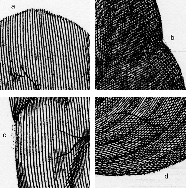
Further magnified details of figure 1
These interceding lines are only noticeable under magnification, suggesting that Wren employed a magnifying glass while etching. According to John Aubrey (1626–97), Hollar used the same ‘exacting’ method; in turn, the ‘curiosity’ of his fine lines was ‘not to be judged without a magnifying-glass’ (Pennington 1982, xlviii). The long parallel shading lines of Wren's figure occasionally overlap the outline, and in other areas the lines fail to meet it, which reveals how sketchy (and somewhat dotted) the initial outline was (Figure 4c and d). This sketchiness suggests that Wren began his drawing of the brain directly on the copper plate — that is, without a drawing with the same scale made for transfer. However, this does not exclude the possibility of other kinds of preparatory studies made from the many different brains that Willis and his team dissected. Based on the repeated procedures of dissection, Wren synthesized the figure from drawings made from drawings of many specimens, similar to the way Royal Society members Francis Willughby (1635–72) and John Ray (1627–1705) derived their contemporary descriptions of birds and fish using a large collection of specimens and pictures (Grindle 2005, 15–22; Kusukawa 2000, 179–87). Wren's final image is not one of objectivity (including all the incidental details of dissection), but a ‘truth-to-nature’ ideal (Daston and Galison 2007, 55–113).
Conclusion: The experiment
Robert Plot (1640–96), the first Professor of Chemistry at Oxford, exclaimed that ‘Dr Willis's Method of dissecting the Brain … is new, and most exact, that there is scarce any one Part in it, but what has receiv'd considerable Advancements’ (1677, 301). Willis used a method of dissecting the brain pioneered by Varolio (1573), proceeding upward through the base of the skull (Martensen 2004, 50). When the brain was extracted, cleaning and preservation had to take place quickly; when exposed, the brain loses its shape, and decays very quickly. Applying their earlier intravenous experiments, Willis and his colleagues injected the tissues with spirits (pure alcohol), and the arteries with dye, both in order to preserve the flesh, and to colour the vessels (Rijcke 2010, 34–5). The dye they used was likely made from vegetable matter (e.g. berries), or metal oxides, ingredients for dying studied by Petty (Sprat 1667, 284–306).
Once the vessels were injected, the ‘model’ could be studied, discoursed upon, and drawn. However, this was not the entire reason for the procedure. Willis's text makes it clear that he is fulfilling what he promised the reader in the preface: an ‘ocular demonstration’ or ‘curious’ experiment (in the sense that it imitates nature), repeated hundreds of times using animal and human brains:
Let the Carodick Arteries be laid bare on either side of the Cervix or the hinder part of the Head, so that their little Tubes or Pipes, about half an inch long, may be exhibited together to the sight; then let a dyed liquor, and contained in a large Squirt or Pipe, be injected upwards in the trunk of one side: after once or twice injecting, you shall see the tincture or dyed liquor to descend from the other side by the trunk of the opposite Artery: yea, if the same be more copiously injected towards the Head, from thence returning through the Artery of the opposite side, it will thorow [that is, make its way] below the Praecordia, even to the lower region of the Body … Further, in the Vessels which constitute the wonderful Net [the arachnoid], and which cover the Basis of the Brain [the anastomosis], some footsteps of the same tincture will appear (Willis 1681, 72; see also 59, 79, and 85).
Like surveying the course of a wending river, Willis and his team carefully watched as the inky substitute for blood they forced by ‘pipe’ into the vessels flowed through and traced the vascular landscape of the brain, stopping where its flow was restricted because of unidirectional valves — corroboration of Harvey's principle of circulation as applied to the brains of vertebrates.
When Wren sketched the outline on the copper plate for the first figure, synthesizing observations from drawings conceivably made after multiple preserved specimens, we can imagine him further clipping and cutting back the vessels and cranial nerves with his eyes, so to speak, removing imperfections in order to make the overall form symmetrical, ideal. These were the final touches on the ‘natural’ solution to the problem of the fast-decaying brain and the messy procedures of dissection. Wren crystallized the structures, making them appear as though they were the healthy and functioning, an artificial object imitating the ‘living and breathing Chapel of the Deity’ that Willis had sought to reveal, and produced by the ‘singular humanity’ of a virtuoso (1681, Epistle dedicatory).
Footnotes
1
The full Latin title is Cerebri anatome: cui accessit nervorum description et usus studio Thomas Willis [i.e. Thomas Willis (et al.), The anatomy of the brain: including the description and function of the nerves].
2
Translated into English by poet Samuel Pordage (1633–c.1691) in Five Treatises (1681). I rely upon Pordage's edition for this article.
Nathan Flis is a doctoral candidate in the History of Art at St Catherine's College, University of Oxford. He is currently completing his doctoral dissertation, From the Life: the art of Francis Barlow (c.1626–1704), which focuses on the life and work of England's first professional painter, draughtsman, and etcher of birds and animals, who was also one of the most prolific book illustrators and political satirists to work in seventeenth-century London.
Correspondence to: 10 Court Place Gardens, Iffley, Oxford OX4 4EW, Phone: 01865 711 709, Email: