
Abstract
Biological studies indicate that numerous materials present in living tissues owe their success to an optimal combination of properties and adaptive structures, rather than to extreme properties per se. Through studying natural tissues and by biomimesis, new polymer and composite materials may be designed to emulate the structural and functional responses of bone. These materials must ensure biochemical affinity with host tissue through judicious mixing of specific chemical cues. Also, they must mimic the response under load exhibited by natural bone through complex organisation of material phases, i.e. embedding of collagen fibres in the extracellular substance. Fibre and particulate reinforced polymers are increasingly significant in the development of new biomedical materials, since they can be engineered more accurately than monolithic structures. Meanwhile, design of nanocomposites with specific morphological and chemical signals is emerging as a powerful approach to the mimesis of extracellular matrix of natural bone. In both cases, the manipulation of the main materials features at the micro- and nano-metric scale offers an intriguing strategy for improvement of biological and mechanical response. Several biodegradable and bioresorbable materials, as well as technologies and scaffold designs, will be critically reviewed, illustrating the potential of bio-inspired composites and multicomponent platforms for bone tissue engineering.
Introduction
The design of bio-inspired materials, able to guide tissue regeneration, is a challenging goal. The ideal material must control and promote specific cellular events,1 and its properties must reflect an understanding of the structure to be substituted. How nature crafts complex tissue structures may be understood by studying her processing routes. These intricate phenomena define the final shape and structure of the tissue, from macro- to nano-scale, and the nature of its various physical and chemical interactions.2
Learning from nature is an approach which may be successfully adopted in bone and mineralised tissue regeneration. Bone is a natural, anisotropic composite structure, with higher stiffness and tensile strength than soft tissues such as skin, cartilage or blood vessels. For its replacement, the material scientist must offer smart structural components, which respond in situ to the pertinent stimuli triggering bone tissue formation mechanisms such as biomineralisation. Ideally, mimesis of the living tissue, in its mechanical, biological and functional aspects, must be achieved. As part of this strategy, open-pore biocompatible and biodegradable porous scaffolds have been used to provide a temporary substitute for the extracellular matrix (ECM) of natural bone.3, 4 It has been shown that polymeric materials may be used to build multifunctional scaffolds which satisfy two key requirements:
material components with time and space controlled biodegradability
percolative architectures, with interconnecting pores of the right scale to promote tissue integration and vascularisation.
Additionally, scaffold components should have mechanical properties appropriate to their implantation site and handling regime, and exhibit the appropriate surface chemistry to promote cell activities (attachment, differentiation and proliferation). They should also be easily formed into a variety of shapes and sizes.5
Another factor to consider is the response of the implant to specific external stimuli. It is essential that, when introduced into the body, new biologically active material stimulates a biochemical response from the living tissue in order to obtain a strong biological fixation.6 Not only must the colonisation of new bone within the material at the implant site be directed but the biological microenvironment around the material during the integration process and wound healing must be controlled, regulating changes in pH or cell-associated enzyme activity.
Correct bone growth must be coupled with efficient angiogenesis, to ensure the neovascularisation of the developing tissue absolutely essential to maintaining nutrient supply and oxygen to cells, as well as removal of metabolic products.7 Therefore, cell biology, materials and engineering aspects must be addressed together to deliver the three main components required, namely harvested cells, recombinant signalling molecules, and porous matrixes.4 These define the properties of the biohybrid which must be actively stimulated by delivery of specific inductive factors (e.g. BMP-4, VEGF).8
Human bone is essentially a three-component composite material (Fig. 1),9 comprising cellular, organic and inorganic phases, strictly assembled to form the natural tissue. The cellular phase – three different types of cells, namely osteoblasts, osteoclasts and osteocytes – synthesises and regulates ECM deposition and mineralisation, modulating the complex mechanism of bone remodelling.10 – 12 The extracellular matter embeds tissue-specific cells in a highly complex matrix consisting of the other two components – a non-mineralised phase and a mineralised (hydroxyapatite – HA) phase.13 The organic phase contains collagens, glycoproteins, proteoglycans and sialoproteins which play an essential role both in the control of growth and differentiation of osteoblasts, osteocytes and osteoclasts and in bone remodelling.14 Finally, the inorganic phase comprising HA (65–70% of the total matrix) provides adequate structural support.15 Approximately 60% of the bone graft substitutes currently available incorporate ceramics such as calcium sulphates, bioactive glasses, and calcium phosphates. Hydroxyapatite and tricalcium phosphate have been considered the major candidates among synthetic and natural inorganic ceramic materials for scaffold materials for bone tissue engineering.16 – 19 Calcium phosphates are osteoconductive, promote osteointegration and are, in some cases, osteoinductive. Several calcium phosphate-based commercial products are currently available. These include Bio-Oss (Geistlich Biomaterials, Inc., Baden Baden, Germany) and OsteoGraf (see first image, below) based on HA in particulate (Bio-Oss) or block/particulate (OsteoGraf) forms. Vitoss (Orthovita, Inc.), a formulation of β-tricalcium phosphates, has also been efficiently used for filling of bone defects.20
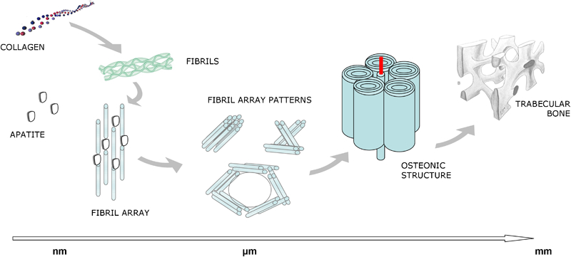
Sketch of the bone hierarchical structure (adapted from E Beniash. Biominerals: hierarchical nanocomposites: the example of bone.9)
Although these ceramic materials resemble the natural inorganic component of bone and possess osteoconductive properties,18 – 21 they are brittle and therefore do not adequately match the mechanical properties of bone.
Both natural polymers (e.g. collagen, alginate, agarose, chitosan, fibrin, hyaluronan) and synthetic polymers have been considered as materials to support cell ingrowth in diverse tissues,21 – 25 synthetic polymers generally offering advantages in versatility and processability.25 Their physical–chemical properties can be easily modified, and the mechanical behaviour and degradation rate can be suitably tailored by varying the chemical composition of the macromolecule. The incorporation of functional groups and side chains can lead to the bioactivation of synthetic polymers using specific molecules.25 Among the synthetic polymers, aliphatic polyesters such as poly(lactic acid) (PLA), poly(glycolic acid) (PGA), poly(lactic-co-glycolic acid) (PLGA) and poly(caprolactone) (PCL) are polymers commonly used for scaffold manufacturing1,3,25 – 27 and their degradation products can be removed by metabolic pathways (see Table 1). 28 28,29 However, polymeric scaffolds proved too flexible, while ceramic structures were found to be too brittle.30
Main properties of several biodegradable polymers with FDA approval
For this reason, composite materials have increasingly attracted attention in tissue engineering research.19 A composite material may be generally considered as a combination, on a macroscopic scale, of two or more materials, which are different in terms of composition and/or morphology, to obtain specific physical, chemical and mechanical characteristics. The resulting composite material may show a combination of the best properties of their components, and, in many cases, interesting properties which are not displayed by the single constituents.31 – 35 The successful application of composite materials in bone engineering requires the estimation, case by case, of chemical and/or mechanical interactions between all the composite constituents. This must include consideration of the nature and level of filler incorporated, as well as the surface topography or morphology of the reinforcing agent.36 The choice of particle fillers or fibres with micro- or nano-metric characteristic size scale is pivotal to the optimisation of mechanical/chemical interlocking, the interface and thus the ultimate properties of composite systems. The ability here to modulate final scaffolds properties down to the nano-scale enables reproduction of all the morphological signals, intrinsically present in the natural tissue microenvironment, which are determinant of defining the final fate of cells.
Scaffolds with tailored porosity and hierarchical organisation
General aspects of scaffold structure and fabrication
The morphology of human bone is characterised by a complex hierarchical structure exhibiting bone gradients of density and anisotropic properties, which confer its unique properties of lightness, stiffness, elasticity, and the ability to self-regenerate following minor trauma. The peculiar structure of bone also allows efficient distribution of the mechanical loads to the level of the smallest trabeculae. Here the strain-sensing mechanism of the osteocyte cells can participate through activation of the permanent bone remodelling process. This mechanism is associated with continuous arrangement of the bone tissue in the presence of environmentally mediated biochemical stimuli, so that local damage is minimised and the functionality of the bone as a whole is optimised. Therefore, only scaffold structures with a high degree of hierarchy could be able to provide optimal biological response, approaching that of natural tissue, after in vivo implantation. This is a particularly significant issue in the case of long bone disease, where the biomechanical loads are larger and more complex.
The development of hierarchically organised bone scaffolds would therefore represent a breakthrough, and an improvement on the current solutions for repairing bone and osteochondral tissue.37 – 41 Recent work performed on polymer based composite scaffolds has included a comparative study of three-dimensional (3D) structures with single macropore size and multiscale porosity (i.e. micro- and macro-porosity) which sought to quantify the change in their mechanical properties and biocompatibility after in vitro culture.42 A finely structured design with porosity, pore size and interconnectivity at different dimensional levels is recommended to provide a morphologically friendly environment to cells. This may be attained either macroscopically, by controlling scaffold rod spacing, or microscopically, by varying rod porosity. Macroporosity – pores greater than 50 μm diameter – is thought to contribute to osteogenesis by facilitating cell and ion transport.39 Other studies suggest that microporosity – pores less than 20 μm diameter – improves bone growth into scaffolds by increasing surface area for protein adsorption,43 increasing ionic solubility in the microenvironment,44 and providing attachment points for osteoblasts.39 Finally, pore interconnectivity is essential, in order to drive bone deposition rate and depth of infiltration in vitro 39 and in vivo.45 Regular, interconnected pores provide spacing for the vasculature required to nourish new bone and to remove waste products.43 The combination of pore size and interconnectivity required for optimal osteoconductivity has yet to be determined, making the ability to adjust these parameters an important capability for scaffold fabrication.
Several techniques have been used to fabricate 3D porous scaffolds, each characterised by its own advantages and limitations (Fig. 2). Conventional methods allow construction of scaffolds which are characterised by a continuous and uninterrupted pore structure. However, their use does not provide any long range channelling microarchitecture. These techniques include gas foaming, solvent casting/particulate leaching, phase separation, melt moulding, freeze drying, solution casting, and emulsion freeze drying.17,19,46 – 48 The internal architecture of scaffolds, pore shape, pore size and interconnectivity result are critical for in vivo performance, influencing tissue regeneration and determining the mechanical properties of the structures.49 – 57 Recently, we proposed designing porous scaffolds made of PCL57, 58 by phase inversion/salt leaching. The phase inversion/particulate leaching method is one of the most commonly used in fabrication of scaffold for tissue engineering. The method involves mixing water-soluble, or easily removable, particles into a solution comprising biodegradable polymer in a suitable solvent. Several materials have been used as porogen agents, to generate the requisite porous architecture within a polymer scaffold. The selection of templating agent is constrained by the compatibility of the particle removal method – e.g. solvent, heating, or chemical treatment – with polymer characteristics. Sodium chloride, sugar, gelatin or hydrocarbon particles are widely used in particulate leaching process,59 with salt crystals the most widely used. The polymer solution/particles mixture is then cast into the desired shape mould and the solvent is removed by solvent/non-solvent exchange. This technique has been recently used to develop a composite scaffold of PCL/HA. The optimisation of single process steps allowed the conferment of a bimodal distribution of pore sizes, with larger pores – i.e. from 100 to 300 μm in size – through the dissolution of sodium chloride crystals in water, and smaller pores – from 1 to 10 μm – induced by a thermodynamically driven polymer/solvent separation mechanism. The literature suggests that smaller pores can play an influential role in cell adhesion and proliferation, whilst the larger pores are able to promote the nutrient supply and waste removal through the improvement of scaffold vascularisation.59, 60 Several studies have demonstrated that scaffolds with bimodal micrometre scale (l-bimodal) porosities may often be necessary for the regeneration of highly structured biological tissues, such as bone and cartilage.42 On the other hand, transport issues, 3D cell colonisation and tissue ingrowth would be inhibited if the pores are not well interconnected, even if the porosity of the scaffolds is high.61
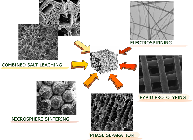
Summary of main scaffold preparation techniques
However, fine control of the internal architecture is strongly limited using scaffold conventional manufacturing techniques;18, 62 the interconnectivity and the pore shape and size obtained by using these methods are related to many processing variables.63, 64 All of this suggests that conventional scaffold manufacturing techniques do not allow the precise control over the internal architecture and interconnectivity. In addition, the highly porous scaffold (exceeding 70% porosity) obtained by the sole phase inversion/particle leaching method shows drastically lower ultimate mechanical performance in comparison with scaffolds obtained by other techniques with the same porosity characteristics58,65 – 68 (Table 2).
Summary of basic morphological and mechanical properties of composite scaffolds for bone regeneration
The introduction of solid freeform fabrication has led to the division of scaffold fabrication techniques into two groups, defined as ‘conventional’ and ‘novel’ or more advanced methods.18, 47 Solid freeform fabrication encompasses a group of technologies that can be employed to manufacture devices in a layer-by-layer fashion from the 3D computer design of the object. Solid freeform fabrication was initially developed for manufacturing prototype engineering parts and, consequently, the name ‘rapid prototyping’ (RP) is also widely used. The RP techniques, based on computer-aided design (CAD)/computer-aided manufacturing (CAM) methods, allow fabrication of well organised 3D structures with 100% interconnectivity and specific pore shape and size, enhancing cell activities through the improvement of the mass transport of nutrients and oxygen. The RP comprises three steps – data input, data file preparation and model building46 – with magnetic resonance imaging or computed tomography as possible data sources.69, 70 These data can be used first to fabricate a customised CAD model, then a scaffold which accurately reproduces the shape of the defect. Over the past years, several rapid prototyping techniques have been modified and developed to fabricate scaffolds for tissue engineering. Among these are fused deposition modelling, 3D printing, stereolithography, ink-jet printing, selective laser sintering and many other extrusion-based technologies (e.g. multiphase jet solidification, fused deposition modelling, 3D Bioplotting) have been considered.71 – 74
Microstrands and microdroplets can be deposited and combined with in situ bonding, to obtain layer-by-layer deposition patterns and structures similar to non-woven materials.75, 77 This enables realisation of 3D scaffolds with interconnecting pores, so enhancing cell activity. The biggest hurdles to the adoption of RP techniques are the restrictions relating to material selection and inner scaffold structure.52
3D fibre deposition
3D fibre deposition78, 79 is the most recently examined RP technique for use in scaffold manufacture. In this fused deposition technique, a molten polymer or a polymer/solvent solution is injected through a needle, then deposited through a servo-mechanically controlled syringe.80 – 83 Here, CAD/CAM techniques are integrated with image analysis, and 3D scaffolds with specific size and shape and 100% interconnectivity can be formed. The dispensing machine used in this technique – a 3D plotter, also known as a Bioplotter – was initially developed by Landers et al. 75 – 77 to fabricate scaffolds from hydrogels for soft tissue engineering. However, a wide range of materials may be processed.76, 77
In designing advanced multifunctional scaffolds, the aim should be to create hierarchical porous structures with the desired mechanical performances and mass transport properties (permeability and diffusion), while reproducing the complex 3D anatomical shapes. This means that mechanical and transport mass properties should be properly combined. The performance of 3D rapid prototyped scaffolds strongly depends not only on the intrinsic properties of the material, but also on the different 3D architectural and geometric features (Fig. 3), highlighting the possibility of tailoring mechanical and mass transport properties.
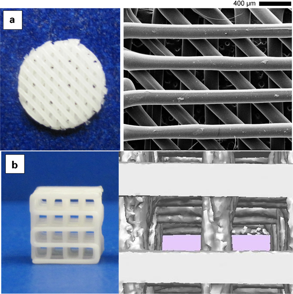
Examples of scaffold architecture: images of 3D fibre-deposited PCL scaffolds characterised by two different geometries and lay-down patterns: a 0°/45°/90°/135° and b 0°/90°
A key aspect is the evaluation of mechanical and mass transport properties at a given scale, and the consideration of these in the context of microscopic properties and scaffold structure; also, the elastic properties and the permeability of the scaffold can be assessed through a computational implementation of the homogenisation theory.84, 85 For a fixed scaffold design, it appears clear that by increasing the amount of material incorporated, scaffold porosity and permeability decrease, whilst the elastic properties increase. However, at a given level of porosity, it is possible to tailor the microstructure to achieve different values of effective stiffness and permeability. In designing a specific scaffold architecture, it would be possible to optimise these properties (i.e. stiffness, permeability) by adjusting process parameters, even taking into account the requirement of the target scaffold.
Following this approach, microstructures were designed with maximised permeability, promoting mass transport and cell migration, while displaying effective linear elastic properties which matched those of natural bone. Another promising strategy involves the initial use of a non-porous block, then optimising the distribution of the material within the scaffold volume according to the desired porosity level and mechanical properties.86, 87 Another way of computing the optimal topology of the scaffold microstructure is by the identification of discrete domains, using finite elements characterised by density values, ranging from 0 to 1, and including specific penalisation factors (solid isotropic microstructure with penalisation).72 In this case, the local material laws for both stiffness and diffusivity have been defined, the optimisation process then involves the minimisation of a specific objective function.
In summary, hierarchical scaffolds with desired 3D anatomical shape and specific mechanical and mass transport properties can be effectively designed by integrating image capture techniques, analysis, topological optimisation, materials selection/preparation and the 3D fibre deposition technique.
Particulate and/or fibre reinforced composite scaffolds
Composite materials are generally composed of two phases – a continuous and a dispersed phase separated by an interface with intermediate properties. The continuous phase is referred to the matrix and polymers represent the material mostly used as its component. The dispersed phase can be discontinuous (i.e. platelet or filler) or continuous (i.e. fibres) and it is usually stiffer than the matrix. It is considered as the reinforcing component of the composite, since it enhances mechanical properties of the matrix (i.e., stiffness, strength). The interface between matrix and reinforcement plays the major role in determining the mechanical performances and environmental stability of composite materials.
It is well known that if the matrix/reinforcement interface is not suitably tailored, it can provide a decrease in the composite properties. The physical and mechanical properties (i.e. coefficient of thermal expansion, stiffness, strength, fatigue behaviour) of the composite materials are strongly related to the interfacial features. For example, from a mechanical point of view, the interface has to transfer a part of the stress to the reinforcement, making a contribution to the composite stiffness and strength. Consequently, an important aspect in designing composite materials is related to the stress transfer from matrix to reinforcement, trying to avoid discontinuities in the stress transfer and generation of stress concentration at the matrix/reinforcement interface. All the problems related to the matrix/reinforcement interface may be clearly considered as system-specific and this leads to great difficulties in designing optimised interfaces that would be adequate and common for all the different system. Accordingly, to obtain suitable interfaces, several techniques have been employed involving the reinforcement coating, the modification of the matrix composition, the chemical functionalisation of the constituents.88
During the last decade, polymer based composites have gained an increasingly important role in the development of new generation biomedical materials. They can be engineered more accurately than monolithic phase materials so that the effective tissue functionality (i.e. mechanical response) can be faithfully reproduced. As nature teaches, connective hard tissues like bone are constituted by the ECM (i.e. collagen, apatite and water) and cells, namely osteocytes in bone and odontoblast in dentine, which control and adapt the structure performance based on a mechano-sensitivity processes.89 Collagen fibrils of ∼100 nm diameter, reinforced by platelet apatite crystals at the nano-structural level,90 are distributed in an organised manner in mineralised tissues. Since the elastic moduli of collagen and HA show an appropriate gap from 1·5 to 110 GPa, the optimum combination of soft and ductile collagen with the stiff and brittle apatite crystals into a synthetic composite material may confer upon it a range of properties emulating that found in natural tissue.91 In this context, fibre and particulate reinforced polymeric composites represent the ‘engineering response’ to develop mineralised tissue analogues. Similar to the natural tissue, the interaction between the polymeric matrix and a reinforcing phase may be modulated to tailor the mechanical performance in terms of material strength and anisotropy. In particular, an adequate support of the mechanical stimuli by proper mechanical features (i.e. suitable stiffness, elasticity) is necessary to ensure a progressive transfer of stress to the newly forming bone tissue as the scaffold degrades. However, a designed pore architecture remains a basic prerequisite for assuring sufficient porosity and permeability to promote efficient cell colonisation, complete vascular invasion and satisfying all the main transport demands, i.e. nutrients and oxygen trafficking, of the remodelling tissue58, 92 Hence, there is often a conflict between maximising surface-to-volume ratio, to enhance cell colonisation and fluid transport, and optimising the mechanical response of the scaffold. This conflict often leads to a compromise in the optimal design solution.93 Today, several technologies based on templating strategies allow optimisation of the biomechanical response of the scaffolds through correctly balancing porosity and mechanical properties. In order to guarantee the desired mechanical response, the highly porous network may be further sustained mechanically through the integration of ceramic particles94 or continuous fibers65 to prevent the collapse of the pore architecture during load application.
Filler reinforced composites
Common alloplastic materials including PLGA, poly(L-lactic acid) (PLLA) and PCL have been combined twith HA and tricalcium phosphates (α-TCP, β-TCP) paricles to develop composite scaffolds for hard tissue replacement.4 Such calcium phosphate-polyester combinations show good bone-bonding properties, due to the osteoconductivity of the calcium phosphate. They also exhibit controlled degradation kinetics of the polymer phase, forming low molecular weight products which are easily eliminated in the physiological environment. A further advantage is the high bioactive potential induced by the employment of ceramic materials, which promotes formation of mineralisation sites. Also, the addition of materials based on degradable natural polymers95 and synthetic polymers,65 in conjunction with HA particles, improves mechanical response. The brittle behaviour, directly ascribable to the ceramic phase can be softened by the coupling with polymeric phase, with an overall increase in the composite toughness. This provides a more valid basis for mimesis of the complex behaviour of natural tissues to external mechanical stimuli.96
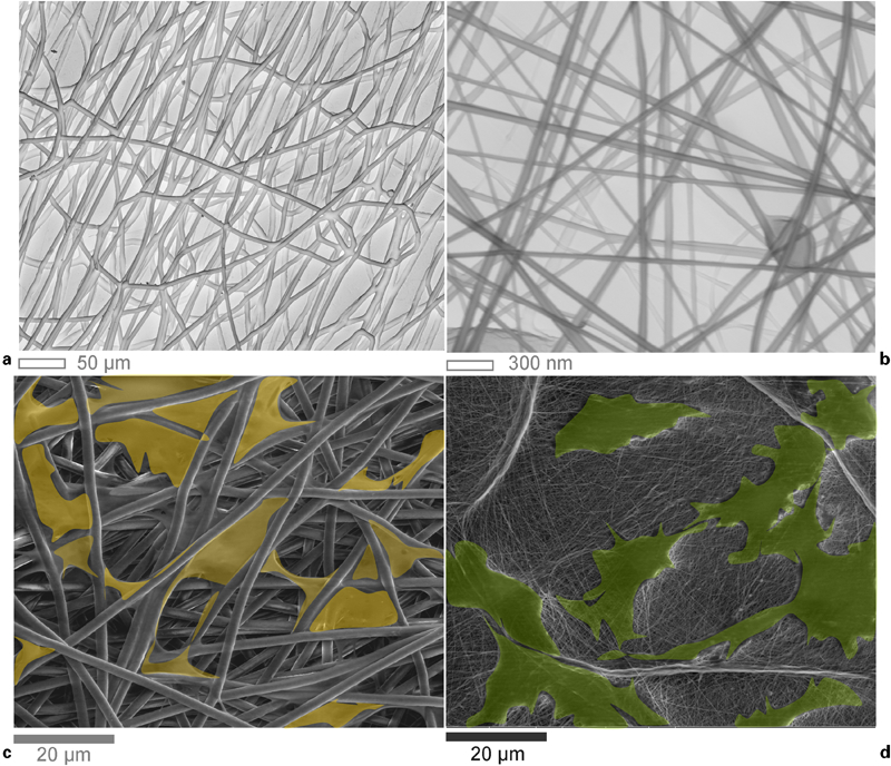
Although polymer matrixes reinforced by ceramic fillers (e.g. HA, tricalcium phosphates) are able to mimic the collagen/HA micro-/macro-morphology of native bone material,97 – 99 polymer based composite scaffolds with particulate reinforcement represent a less successful strategy in terms of mechanical response and chemical stability. Causa et al. 97 proposed a particulate reinforced composite formed from a PCL matrix loaded with micrometric HA rigid particles. The addition of ceramic particles to the polymer strongly enhances the mechanical performance of the PCL scaffold. This is due to the hardening function offered by the fine dispersion of particles inside the polymer matrix of PCL, which shows a significant increase in mechanical resistance, and up to a threefold increase in the elastic modulus.1 In this case, the strengthening mechanism of HA within PCL samples might firstly be ascribed to the stiffer mechanical properties of HA in the composite material. However, studies on macroporous composites demonstrated that HA particle clustering, due to non-homogeneous distribution of rigid particles within the polymer matrix generally, has a negative effect on the mechanical performance of the composite materials (Fig. 4). Also, the use of physical mixing methodologies to prepare the scaffold may favour masking of ceramic particles in the polymer matrix. This may limit the effective exposure of ceramic particles on the scaffold pore surface and hinder their direct contact with cells, thus compromising their osteoconductive/osteoinductive recognition.100, 101
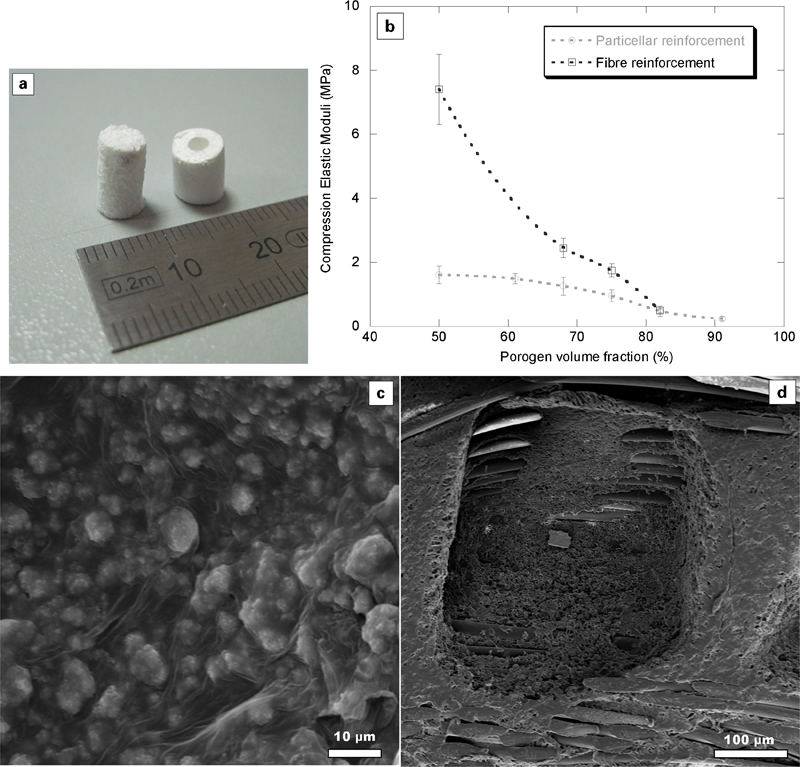
a PCL porous scaffolds with HA particle or PLLA fibres as reinforcement; b comparison of compressive response as a function of porosity degree and used reinforcement systems; c, d details of reinforcement/matrix interface
Finally, it is well known that the efficiency of rigid ceramic particles as reinforcement system may be strongly conditioned by the interaction of the composite scaffolds in the biological environment. A critical aspect is the chemical modification of ceramic particles in contact of culture media. Among calcium phosphates, HA is the most stable phase, being less resorbable than other bioceramics,97 although its resorbability can be slightly improved by addition of ceramic oxides and ionic doping agents.100 Other calcium phosphates, e.g. α- or β-tri-calcium phosphates, are highly reactive with biological fluids via chemical reactions such as hydrolysis, and are able to influence the final properties of the polymer matrix and the local microenvironment. Once TCP-polyester composites have been conditioned in biological media, the phosphate ions readily dissolve and buffer the acidity of the carboxylic endgroups produced by polyester chain cleavage.102 Consequently, the composites degrade more slowly and maintain their shape longer than the pure polymer. On the basis of these considerations, recent studies have examined composites in which degradable polymers are coupled with reactive calcium phosphates;101 for example, the in vitro performance of α- and β-TCP in PLGA103, 104 scaffolds has been recently investigated. In both cases, the average molecular weight of the polymer decreased more slowly in the composite than in the pure polymer, indicating the influence of the ceramic phase on the degradation kinetics of the polymer. Recent modelling studies confirmed these results by identifying both the saturation behaviour and the buffering effect of incorporating β-TCP in the various polyesters, and also underlining an upper limit of the β-TCP weight percentage (about 30%) above which improvements in buffering effects may be neglected.104 The inclusion of low ceramic fraction – lower that 24% by weight – is generally sufficient to assure efficient osteosynthesis,105 – 107 and so combine the advantages of biocompatibility and osseointegration in the resulting composite.
Continuous fibre reinforced composites
An alternative strategy to improve the mechanical response of composite materials traditionally involves the inclusion of fibres – in a continuous or chopped form – with preordered spatial organisation into the polymer matrix. In the past, a multitude of partially resorbable composites has been obtained by combining a degradable polymeric matrix with high modulus slowly resorbable fibres (glass, carbon, amides). However, the long-term effect of bioinert, biostable or slowly degradable fibres is not well documented in living tissue, and recent studies have demonstrated the presence of an acute or chronic inflammatory reaction in some cases under specific conditions, with the detection of a thin encapsulating membrane of mature connective tissues around the implant region. Alternatively, totally degradable reinforced composites may represent the main goal in the design of new fixation materials, because of the drastic decay of long-term problems induced after their digestion by living tissue.
Recently, fibre reinforced composite have been obtained by filament winding technologies,108 with improved mechanical properties but constrained process costs. One of the seminal studies was the development of composites by merging a matrix of Hydrothane – a hydrophilic polyurethane – with continuous fibres of PLA and PGA, helically wound by the filament winding technique. This method was used to make porous and non-porous tubular constructs for ligament-tendon application. The approach consists of applying composite theory to design biodegradable composite systems which are able to mimic the structural organisation and performance of the living tissue.109 Similarly, fibre reinforced composite scaffolds may be fabricated by the integration of hydrophilic PLA continuous fibres into a hydrophobic PCL matrix (Fig. 4), to obtain highly porous scaffolds for bone regeneration.110 In these fibrous composites, degradation preferentially occurs at the fibre/polymer interface, resulting in a higher rate of degradation than for either material alone. Usually, the characteristic degradation rate of composites is too high, and therefore not ideal for clinical applications such as bone fracture fixation which require strength retention in the long term (i.e. a few weeks up to several months). However, a continuous fibre-reinforced composite made of two interconnecting phases, which better mimics the bone structural organisation, assures a strong mechanical interlocking between two phases which assures the support for retaining some of its properties if breakdown occurs at the interface.
The main difficulty in the design of composite materials is the optimisation of the adhesion between matrix and reinforcement. Ideally, interfacial bonding will not only promote a more rapid diffusion of fluids at the matrix/fibre interface, but also not limit the mechanical strength and fatigue, which is the optimal compromise condition for improving final composite properties. The biological behaviour is mainly determined by the presence of a multi-scale porosity with tailored characteristics in terms of pore interconnections and pore size as the degradation mechanisms proceed. A well-organised pore network within the scaffold may potentially control cell colonisation and fluid transport through its peculiar geometry,110 with the combined effects of the reinforcing action of PLLA fibres and the slow degradation rate of the PCL matrix undoubtedly contributing to the improved mechanical performance under compression of the composite system. Then, changes in fibre and matrix features, such as diameter and fibre chemistry, during degradation could provide a guide for the bone remodelling process by achieving a composite structure with morphological and structural properties which then evolve, during degradation, towards a softer and more porous material needed in the long term. However, an excessive presence of open spaces may have led to premature fibrous tissue formation before new bone had a chance to infiltrate the scaffold and could drastically compromise the interface efficiency with a decay of the mechanical performance of the scaffold as porosity increases (Fig. 4b ).
The inclusion together of particulate and fibre reinforcement may further improve the mechanical properties in the presence of high pore fraction, reaching an intermediate level of mechanical response, which is the result of a synergistic effect of continuous fibres and ceramic needle-like crystals amplifying the reinforcing role of the single elements in highly porous structures.65 The precipitation of calcium-deficient HA from α-TCP assures the formation of needle crystals with a high surface/volume ratio which offer a significant contribution to the mechanical response of the scaffold, due to the large surface area available physically to interact with the polymer matrix.
A significant increase in elastic modulus – up to an order of magnitude higher (2·21±0·11 MPa – Table 2) – was obtained by integrating both PLA fibres and α-TCP particles with the PCL matrix. Despite the preferential distribution of these crystals along the inner pore surfaces, a dramatic synergistic effect between the two reinforcement systems may be recognised. This may be attributable to the highly ordered interfacial interactions which occur between the organised pre-tensioned continuous fibres and the growing needle-like crystals during the salt leaching. These interactions result in a progressive increase in the stiffness of the final composite scaffolds, up to the values necessary to mimic the mechanical behaviour of trabecular bone.
Bioactive nanocomposites
Over the last two decades, the concept of cell guidance has also been progressively revised, as new knowledge has been gained about the cell–material interaction in tissue regeneration.111 A major challenge remains the need to identify materials which will promote differentiation of immature progenitor cells to the osteoblastic lineage (osteoinduction), encouraging the ingrowth of surrounding bone (osteoconduction), and integrating it into the surrounding tissue (osseointegration) to prevent micromotion and stabilise the forming bone/surrounding bone interface.112 In the bone, HA particles at nano-level play a significant role in bone restoration, owing to unique functional properties – related to high surface area to volume ratio and ultrafine structure, similar to biological apatite – having a great impact on cell–biomaterial interaction.113 Mineralised bone is preferentially formed in close contact with the nano-metric HA, which is coated on the sub-micrometric scale.114, 115 For these reasons, HA is largely used in the particulate form for the treatment of periodontal osseous defects and alveolar ridge augmentation because of its easy fabrication, handling and close surface contact with the surrounding tissue.116 HA particles, being similar to bone mineral in physicochemical properties, are well known for their bioactivity, and their osteoconductivity in vitro and in vivo.117
In the light of the above, it may be easily understood how the concept of nanocomposites has been rapidly extended to the design of 3D multifunctional scaffolds for bone tissue engineering. All the foregoing considerations related to the materials properties have led to a design approach aimed at materials with nanostructured surface features, or reinforced with nanomaterials, the latter here referring to fibres, grains or particles which present at least one dimension in the range 1 to 100 nm. Generally, nanocomposite materials show performance that is higher than that of conventional composites, due to the novel physical properties related to nano-scale features.118, 119 For this reason, nanocomposite materials appear to be natural choices for creating scaffolds for advanced bone tissue engineering.119
Nanocomposites made of a natural or synthetic polymer reinforced with an inorganic phase are considered an appropriate strategy for bone tissue regeneration since they can better reproduce the composite structure of natural bone. Nanocomposite materials induce better cell response than conventional composites, as they are similar to the natural structure. In addition, the mechanical performances of nanocomposites can also be superior to that of conventional composites.19 It has frequently been reported that better osteoconductivity can be achieved through the use of synthetic composite materials showing size and morphology similar to both the inorganic particles and to the organic phase in bone.119 – 121 The interactions of bone cell functions with ceramic nanophases and nanostructured polymers are improved.119,122 – 125
The use of carbon nanotubes and carbon nanofibres may be an interesting strategy to reinforce polymers, especially for manufacturing scaffolds for bone tissue engineering,126 – 128 taking into account their excellent mechanical properties. In particular, using single-walled carbon nanotubes, lighter scaffolds with high strength may be obtained.128, 129 Carbon nanotubes and carbon nanofibres can be also functionalised using several side groups, trying to improve the mechanical strength and/or biocompatibility of the composite scaffolds. However, the biocompatibility and biodegradation of these inorganic nanomaterials need more investigation. Besides, in a composite material, the constituent materials combine to produce scaffolds with improved properties, which reproduce more faithfully the key features of bone structure. Clearly, the presence of bioactive phases may cause changes in the degradation behaviour, and composite scaffolds consisting of polymers reinforced with HA nanoparticles appear to be promising substrates for bone tissue. In this case, HA nanoparticles (HANPs) should be well recognised due to the chemical similarity of the bone mineral phase. However, recent biological works130, 131 demonstrated that the HANPs toxicity frequently differs from that of the bulk materials even if this behaviour is still unknown.132 Hence, it is strongly necessary the implementation of new composite process technologies which assure an optimal entrapment of HANPs in the polymer matrixes by proper chemical or physical routes.
Composites with nano-metric bulk signals
Novel scaffold materials have been developed, informed by both the cell guidance concept that and other contemporary advances in materials science and biology.133 Recently, natural hybrid materials are attracting great interest as innovative nanocomposites for tissue engineering applications.133 – 135 These materials incorporate a fine, homogeneous dispersion of bioactive nano-sized particle in a polymer matrix. Because bone is a hierarchical material, with its lowest structural level in the nano-scale range, materials with nanometre structures appear to be appropriate choices for creating better scaffolds for bone tissue engineering.128 Improved osteoconductivity can be obtained by using synthetic composite materials which are similar in the size and morphology to both of the inorganic particles and organic phase of bone.120, 121 Bone cell functions can be enhanced by interacting with nanophase ceramics and nanostructured polymers collectively, rather than individually.122, 125, 136
Numerous routes exist for nano-HA synthesis, using wet chemical precipitation,137 in synthetic body fluid,138 reverse microemulsion,139 sol–gel reaction140 or electrophoretic deposition.141 HA may be synthesised at room temperature, providing the ability to control directly particle and grain sizes.142, 143 A wet chemical method yields a composite system consisting of HA and poly(ϵ-caprolactone), wherein the HA particles are uniformly dispersed within the polymeric matrix. The use of a low temperature approach to HA synthesis, in the presence of a suitable polymer solvent, would provide a novel pathway to generate homogeneous composite materials preserving the microstructural features of HA crystals. However, differences in the chemistry of hydrophilic HA and hydrophobic PCL may lead to inadequate homogenisation of the bioactive phase into the polymer matrix, favouring cluster formation on the micrometric scale.142 In order to overcome these limitations, the wet chemical precipitation method includes precipitation of HA in tetrahydrofuran, the same solvent utilised to dissolve the polycaprolactone.144 The use of amphiphilic surfactant (Span85, 75% oleic acid) improved the dispersion of HA in the PCL matrix, since, oleic acid is a fatty acid with a long hydrocarbon tail and a hydrophilic head.145 Oleic acid is believed to mediate the homogenisation of hydrophilic HA powders when dissolved in a hydrophobic organic solvent, such as tetrahydrofuran.146 The morphology of composite material and the dispersion of ceramic phase in polymeric matrix were investigated by SEM analysis and Mapping test, respectively. In the first case, the SEM image of the composite without the mediation of a surfactant shows that the HA particles were severely agglomerated within the PCL matrix as confirmed by the mapping test. This means that it is able to mediate the interaction between the hydrophilic ceramic (HA) and the hydrophobic polymer PCL-dissolved solution, and thereby create a homogeneous population of ceramic nanoparticles within the polymer matrix and, consequently, the resulting HA/PCL composites.147 X-ray diffraction analysis showed that HA with low crystallinity could successfully be synthesised at room temperature. Many tests have shown that there is no new bone formation in highly crystalline porous HA ceramics, but bone generation has been observed in poorly crystalline HA ceramics.148 Therefore, the use of poorly crystalline HA should increase the bioactivity of the composite, which in turn, could lead to the faster establishment of a strong bond between the composite and tissue.149 HA/PCL porous scaffolds for bone tissue engineering have also been developed by combining chemical synthesis and phase inversion/salt leaching. This strategy allows to reach sub-micrometric HA particles uniformly dispersed within the polymer matrix.144
The use of low temperature approaches to synthesise HA in the presence of a solvent in which the polymer is soluble provides a novel pathway to generate homogeneous composite materials preserving the microstructural features of the HA crystals. In this case, the pore architecture was assured by the removal of solvents and used porogens (i.e. NaCl crystals), of predefined particle size. Both chemical and morphological cues are necessary to ensure an adequate biological response following implantation into bone tissue. Indeed, osteoconductivity is essential for the recruitment of cells capable of forming bone matrixes and ultimately for successful bone in growth into scaffold. These composites frequently show interactions among constituent phases at the micrometric scale, which can provoke differential resorption rates during dissolution, and, ultimately, material instability in vivo. However, in the case of hybrid materials, the inorganic and organic components are not distinguishable at the micrometric scale and the constituent phases interact chemically on a nano-scale. Hence, a successful approach for the chemical synthesis of composite material in bone regeneration is the sol–gel approach. A sol–gel method enables the powderless processing of glasses, ceramics and thin films or fibres, directly from solution. Precursors are mixed at the molecular level and variously shaped materials may be formed at much lower temperatures than it is possible by traditional methods of preparation. One of the major advantages of sol–gel processing is the possibility to synthesise hybrid organic–inorganic materials with chemical interaction between the phases. The ability to incorporate coupling agents,(e.g. 3-(glycidoxypropyl)trimethoxysilane150) into the system allows obtaining a covalent bonding between constituent phases, more stronger that weak forces (i.e. electrostatic interaction) which typically occur in the not functionalised systems. The amount of the bridging bonds or cross-link density gives the chance of controlling the degradation rates of organic components (i.e. Gelatin151) into the hybrid system.
The organic–inorganic composite materials may be prepared in various ways. The simplest route relies on dissolution of organic molecules in a liquid sol–gel.150 – 157 An alternative uses the impregnation of a porous gel in the organic solution. In the third type, the inorganic precursor either already has an organic group or reactions occur in a liquid solution to form chemical bonds in the hybrid gel. The sol–gel process itself leads to formation of gels from mixtures of liquid reagents (sols) at room temperatures. It involves several steps: the evolution of inorganic networks, formation of colloidal suspension (sol) and gelation of the sol to form a network in a continuous liquid phase (gel). During the ‘aging’ step (after gelation and before drying) the sol–gel derived material expulses the liquid phase (solvent which can be water or alcohol) in the process called syneresis. Drying of the obtained gels, even at room temperature, produces glass-like materials called xerogels (i.e. xeros). The process generates a porous material, where the pore size depends on such factors as time and temperature of the hydrolysis and the kind of catalyst used. The diameter of the pores is directly related to the shrinkage of the ‘wet’ gels. During the drying process the gel volume decreases even several times (which is the main reason of cracking).
The sol–gel process for HA preparation typically produces a fine-grain microstructure containing a mixture of nano-to-submicrometre crystals (Fig. 5), the size range most acceptable to the host tissue.158 The sol–gel product is characterised by nano-size dimension of the primary particles. This small domain is a very important parameter for improvement of the contact reaction and the stability at the artificial/natural bone interface. Also, the high reactivity of the sol–gel powder allows a reduction of processing temperature, and any degradation phenomena occurring during sintering.159 The major limitation of the sol–gel technique application concerns the possible hydrolysis of phosphates and the high cost of the raw materials.158 Also, most processes require strict pH control, vigorous agitation and a long time for hydrolysis. These problems were solved by using a non-alkoxide based sol–gel approach were the calcium and phosphate precursors are calcium nitrate tetrahydrate and phosphorous pentoxide, respectively.160 More importantly, gel formation is achieved without the need for any refluxing steps. In this case, the P2O5 reacts with alcohol to form P(O)(OR)3 oxyalkoxide with the liberation of water, which is in turn partially hydrolyses the oxyalkoxide precursors. The presence of phosphorus hydroxyl alkoxide is not sufficient in itself to form a gel, indicating the important role of Ca(NO3)24H2O. It is likely that Ca(NO3)24H2O leads to the formation of alkoxy-nitrate salts, which participate in a polymerisation reaction with the partially hydrolysed phosphate precursors,161 the polymerisation reaction so resulting in the gel. For the synthesis of the organic–inorganic composite material PCL/HA, the polymer may be added during the production of inorganic phase in order to allow the chemical interactions between the components. Molecular-level mixing of calcium and phosphorous precursors with the polymer chains derived from the sol–gel process resulted in composites having enhanced dispersion and exhibiting good interaction between the inorganic phase and the polymer matrix. Several studies have demonstrated by Fourier transform infrared spectroscopy and atomic force microscopy analyses the presence of hydrogen bond in the composite materials synthesised by sol–gel method.162, 163 The presence of HA particles in the composite material beneficially offsets the acidic release from the polymer through the alkaline calcium phosphate164 and mitigates erosion problems associated with the release of acidic degradation products. In vivo and in vitro measurements of pH in bone chambers have shown that the pH drop is 0·2 units near the eroding polyesters.165 Furthermore, by a sol–gel process it is possible to obtain a homogeneous distribution of n-HA crystals with 10–30 nm as diameter and 40–50 nm as length was detected into the composite. A homogeneous distribution of nano-scale HA particles in the polymeric matrix facilitates an increase in bioactive potential of the materials. Many investigations of nanophase materials to date have illustrated their potential for bone repair. For example, increased osteoblast adhesion on nano-grained materials in comparison to conventional (micrometre grained) materials has been reported.166, 167 Osteoblast proliferation in vitro and long term functions were also enhanced on ceramics with grain or fibre sizes <100 nm.168 In addition to osteoblast responses, modified osteoclast behaviour has also been documented on nanophase ceramics, and in vivo studies have demonstrated increased new bone formation on metals coated with nHA compared to conventional apatite.169
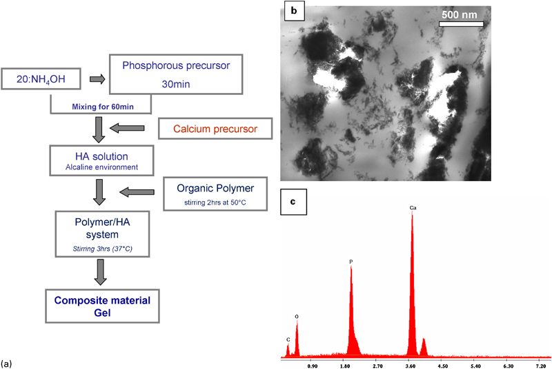
HA/PCL nanocomposites: a sketch of chemical synthesis; b TEM analysis of HA particles dispersed in PCL matrix; c EDS analysis of HA nanoparticles
Magnetic nanocomposites
An innovative approach to the design of magnetic scaffolds for bone tissue engineering has recently been proposed,170 involving the use of appropriately functionalised magnetic nanoparticles. With traditional scaffolds, pre-loading techniques are usually considered since specific biomolecules (i.e. growth factors) are seeded in the scaffolds. However, these approaches strongly reduce the controlled delivery of localised long-term biochemical stimuli,171 – 173 how difficult it is to obtain a temporal control which should favour the tissue regeneration process.174 – 176 A great challenge would, therefore, be the achievement of controlled delivery which reproduces the production of endogenous growth factors through the design of advanced multifunctional scaffolds. Accordingly, a novel concept involving the use of magnetic nanoparticles, which can be suitably functionalised and bioactivated, to design advanced polymer-based composite scaffolds has to be considered. These magnetic scaffolds should be able to attract and take up in vivo growth factors and other bio-agents through the magnetic particles. Acting as carriers, when a magnetic field is applied these functionalised magnetic nanoparticles may transport the bioagents towards and inside the magnetic scaffold. These bioagents may then be released, so improving the tissue regeneration process. To reproduce natural production of endogenous growth factors, a tuned delivery will be considered for improving the control of tissue regeneration and angiogenesis processes. Magnetic scaffolds could represent an interesting alternative to conventional scaffolds, and could provide considerable benefit to patients. Even though the magnetic guiding process is already known in nanomedicine, its results have not yet been applied to scaffolds. Hyperthermia treatment of tumours, and drug delivery are typical examples of processes that are guided by an external magnetic field enhancing the efficacy of therapies.177 – 180 An interesting method to prepare a magnetic scaffold was proposed, involving the dip-coating of collagen/HA scaffolds in aqueous ferrofluids containing iron oxide nanoparticles coated with different biocompatible polymers, hence directly integrating the nanoparticles into the structure.170 As a result, magnetisation values capable of creating magnetic gradients were obtained.138
Taking into consideration materials properties and the availability of rapid prototyping technologies, we proposed a further innovative approach in developing multifunctional nanocomposite scaffolds for advanced bone tissue engineering may be to design magnetic PCL/iron oxide scaffolds manufactured through 3D fibre deposition technique.19 These 3D nanocomposite magnetic scaffolds clearly showed a morphologically controlled structure (Fig. 6), which means a well organised and reproducible internal architecture with desired porosity, pore shape and size. Nanocomposite scaffolds were built layer by layer by injecting the heated nanocomposite material through a suitable needle and, then, depositing the PCL/iron oxide fibres according to specific lay down patterns.19 Preliminary analyses evidenced that the inclusion of iron oxide nanoparticles enhances biological and mechanical performances. Confocal analyses highlighted an increase in adhered number and a more evident spreading of human mesenchymal stem cells (MSCs) in comparison with the results obtained from the neat PCL fibres. This could probably be related to the synergic contribution of the presence of iron oxide nanoparticles and of the surface topography and roughness of the nanocomposite fibres. The mean value of tensile modulus (640·0 MPa) obtained from the preliminary measurements on the nanocomposite fibres is close to that of cancellous bone,75 thus supporting the possibility of obtaining 3D fibre-deposited scaffolds for advanced bone tissue engineering by suitably selecting specific patterns and architectures.181 The results obtained would seem encouraging, as these nanocomposite magnetic scaffolds can be attracted and fixed by an external magnetic field (Fig. 6). Furthermore, they may be also capable to attract inside their fully interconnected structure bioagents.
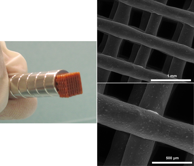
3D nanocomposite magnetic scaffold via 3D fibre deposition technique
Composites with micro/nanostructured bioactive surfaces
The induction of new bone formation seems also to be dependent on the presence of apatite crystals on the scaffold surface, which contributes to making the scaffold more bioactive and osteoconductive. Frequently, induction of the apatite crystals deposition on the scaffold surface was stimulated by using the basic Kokubo treatment, which consists of immersion in simulated body fluid (SBF) with a specific ion concentration, not less than that of the human blood plasma.160, 182 In vitro studies have demonstrated that SBF can be used to reproduce in vivo apatite layer formation on the surface of various materials after implantation,183 so confirming its efficacy in improving surface osteoconductivity. Recently, Barrere et al. 184 have proposed accelerating the classical biomimetic process, reducing its duration from 7–14 days to a few days by immersing substrates into supersaturated SBF. This illustrates the interdependence of ionic strength and pH, as well as the influence of the substrate chemistry on the formation of the resultant apatite. The controlled deposition of HA crystals by accelerating simulated body fluid (5xSBF) allows formation of a biologically active surface which can drastically improve the bonding to living bone.185 The proposed integrated approach enables efficiently exploitation of the inherent features of composite material bulk and surfaces by modulating the spatial distribution of bioactive signals, moving toward a more efficient replacement of bone tissue. Because the apatite particles are generated from aqueous solution, this technique can be used for various complex porous scaffolds, unlike many other surface modification methods that are limited to flat surfaces or to very thin porous layers. The HA generated is more similar to natural bone mineral in its low crystallinity and nanocrystal size, which is beneficial to its degradation and remodelling properties. In vitro, mineral crystals were observed to grow on composite materials after 7 days in supersaturated biological solution (5xSBF).160 It was not surprising that higher concentration facilitated apatite formation, since it was reported186 that increasing the ionic concentrations of an SBF upper the saturation limit is a practical way to facilitate apatite nucleation and growth (Fig. 7).
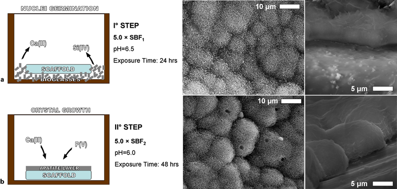
Biomimetic coating of PCL substrates by two-step procedure: evaluation of crystal evolution in a nuclei inducing solution at day 7 and b crystal growth inducing solution at day 14
Recent studies have reported that formation of apatite on artificial materials is induced by functional groups which could reveal negative charge and further induce apatite via the formation of amorphous calcium phosphate. The SEM/image of composite material after treatment in supersaturated solution 5xSBF is shown in Fig. 7. The image shows that after 7 days of soaking in 5xSBF, the composite material was uniformly covered with a calcium phosphate crystals layer with a thickness of few micrometres. Analyses show that the rose petal-like apatite crystallites are composed mainly of HA, with a Ca/P molar ratio of 1·67; the energy dispersive X-ray spectra clearly show Ca and P peaks which are typical of HA. It was also possible to elucidate the presence of HA deposition on the internal pore walls, confirming that the high concentration of SBF solution induced a fast and uniform deposition of HA on the scaffold. In this case, the copious formation of apatite like globular crystals on the exposed surfaces of the composite scaffolds may be directly correlated to the powerful contribution of nanoscale HA particles.186 However, long treatment times (7–14 days) are often not completely compatible for tissue engineering scaffolds, which are composed of highly degradable polymers, such as hyaluronic acid derivates (e.g. HYAFF11). It has been observed that the benzyl ester HYAFF11, underwent a spontaneous hydrolytic degradation of the ester bonds, even in the absence of any enzymatic activity, with complete dissolution occurring after just 1 week.187 That study also demonstrates the real effect of esterification degree on degradation mechanism, as well as on the hydrophilic behaviour of the final material. As the esterification degree increases, polymer chains not only become progressively less flexible and hydrated, but also more rigid and stable. This limits the hydrolytic attacks, reducing the density of nucleation initiation sites which are more reactive than microscale particles. In this case, Kokubo-like strategies rapidly fail, due to a lack of crystal deposition within the timescale of the available experimental window. It means that a drastic reduction of exposure time is mandatory to prevent a catastrophic uncontrolled material loss during the conditioning via SBF solution. Figure 9
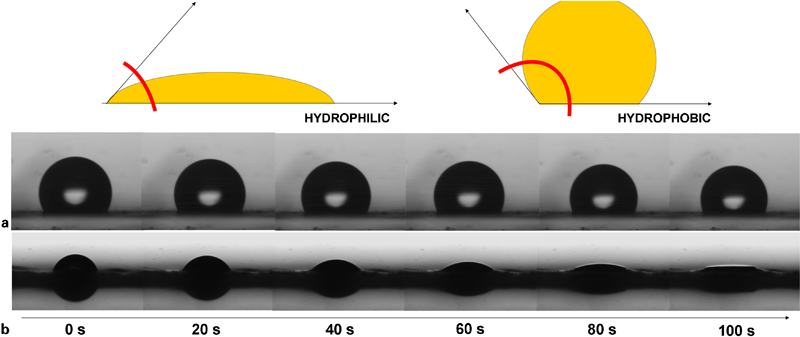
Difference in wettability in a PCL and in b PCL/gelatin electrospun membranes
Several studies have demonstrated that an effective apatite growth can occur in the presence of specific environmental conditions which influence the chemical solubility as well as the material surface activation. A novel treatment, combining the preliminary use of bioglasses with supersaturated SBF solutions at different salt concentrations, has been optimised182 to overcome the applicability limitations of traditional treatments on highly degradable polymers like HYAFF11. The analysis performed on this biomimetic HA-based scaffold showed that mineralisation had already occurred at day 21 and significantly increased at day 35.182 Apatite crystals on HA-based scaffold seem to act as nucleation sites for the deposition of inorganic bone components, as we showed by light and electron microscopy. On non-biomimetic HA-based scaffold an immature mineralisation started to occur only at day 35, where we have also found a lower collagen type I expression and increased cell proliferation, indicating that the biomimetic treatment reduced the time taken to induce the process. Biomimetic HYAFF11 scaffold promotes the precipitation of apatite crystals, inducing the formation of new bone. There was copious mineralisation apparent as early as day 21, which increased up to day 35. In these culture conditions, mineralisation was not observed among the cells, as previously reported with rat MSC grown on non-woven HA-scaffold.188 This result was not dependent by the biomimetic treatment, and even on non-biomimetic HA-based scaffold, mineralisation occurred in the same way. Probably, the different structure of the scaffold (sponge instead of non-woven), composition, porosity, pore size and total surface available for the cells favoured the deposition of mineralised zone along the backbone, rather than among the cells. The cells were well retained inside the scaffold, as also previously reported,189 but h-MSCs establish only a few focal contact with the scaffold indicating that the mineralisation processes were probably mainly induced by the released soluble factors rather than by cell-attachment properties. These data suggest a possible in vitro trophic effects of h-MSCs190, 191 in inducing mineralisation processes, which can be also influenced by the composition192 or elasticity193 of the scaffold. It is known that HA not only enhances HA crystal proliferation and growth, but that it can also promote mineralisation194 since it is a prominent ECM component during the early stage of osteogenesis.195
Nanotechnology: the new frontier for composites and multifunctional materials
The capability of controlling physical and chemical interactions at the level of natural building blocks, from proteins to cells, may favour a more efficient exploration, manipulation, and application of living systems and biological phenomena. Nanotechnology has made great strides towards the creation of new materials, surfaces, and bulk architectures which find numerous applications in the biomedical area.196 In particular, nano-structured biomaterials, in the form of nanoparticles, nanofibres, nanosurfaces and nanocomposites have gained increasing interest in regenerative medicine.197 These materials are able to mimic the physical features of natural ECM at the nano-scale level. Although different experts define ‘nanomaterial’ in different ways, the most commonly accepted definition is that a nanomaterial is a material with a basic structural unit in the range of 1–500 nm. When the dimensions of a material are decreased into the nano-scale, some unusual changes in physiochemical properties occur. This can be attributed to the size (i.e. size distribution), chemical composition (i.e. purity, crystallinity, electronic properties), surface structure (i.e. reactivity, surface groups), solubility, shape, and aggregation of nano-metric materials compared to their micrometric equivalents.198 Additionally, nano-scale materials have more surface area, surface defects, density of atoms at the surface, and altered electron distributions. These differences also cause changes in the surface properties of the material, and in its reactivity further affecting the interactions between the surface and proteins.
In recent years, nanotechnology has been recognised as an important tool in scaffold design to reproduce the features of microenvironment-mediated signalling which determine the tissue specificity and architecture of native tissues.199 It is well known that HA crystals, the main constituent of the inorganic phase of bone ECM, are 2–5 nm thick while collagen type I, the main constituent of the organic phase of bone, is 0·5 nm in diameter. This implies that cells are naturally accustomed to interacting with nano-scale features and surfaces. In native tissues, nano-scale protein interactions are crucial for controlling cell functions such as proliferation, migration, and ECM production.200 Protein adsorption characteristics are, in turn, dependent upon the surface features of the implanted biomaterials (roughness, charge, chemistry, wettability).201 Recent reports have also demonstrated that the unique properties of nanobiomaterials offer advantageous interactions with the proteins that control cellular function.202, 203 Particle or fibre dimension may strongly influence these surface properties and the corresponding protein interactions. It means that changes in material properties may manipulate protein characteristics, so altering protein adsorption behaviour which further influences cell adhesion onto biomaterial surfaces.204 Hence, the development of structurally-motivated approaches based on organised nano-fibrous assemblies rises in the attempt to reach a more aware engineering of natural tissues.
Recent approaches to design responsive scaffolds for bone have concentrated on finding chemical composition and morphological features which are able to stimulate cells and, by appropriate tissue remodelling, yield tissues with different functionalities.205 This ability to engineer complex morphologies and the precise spatial distribution of bioactive signals, with molecular to macromolecular precision, has lead to the creation of nanoscale materials and devices which can not only change the ability of a cell to attach to them but also, to some extent, regulate cellular functions such as growth, differentiation, and apoptosis.206, 207 In particular, the ultimate performance of such composite ‘bone’ will be the result of these complex interactions taking place across all levels of organisation, either mediated by chemical signals of materials (collagen, apatite) and biomolecules (i.e. growth factors) then a rigid spatial organisation of materials phases. Among the novel technologies in materials science available for scaffold design, electrospinning is the most promising for production of fibres on the micro- or nano-length scale with tailoring physical, chemical and biological properties, and which are generally adaptable to various cellular environments.208, 209 Currently, micro- or nano-scale textured fibrous platforms are attracting more attention because their ability to reproduce the multi-scale organisation of collagen fibre – from the micrometric – and nano-metric level – hierarchically assembled into dense bundles as in the natural ECM. Meanwhile, multicomponent fibres including various bioactive phases are largely establishing due to their extreme similarity of chemical composition to the natural bone.
Micro- and nano-metric fibres
The micro- and nano-patterning and the texture size scale of electrospun membranes may be critical to guiding the cell–materials interaction mechanisms, and therefore critical also to improving cell adhesion and spreading. The basal lamina membrane, which is ubiquitous within tissues, has unique nano-fibrous characteristics,210 suggesting the importance of substrate topography on biological behaviour. To date, a growing body of evidence has demonstrated that micro-to-nanoscale topography of electrospun substrates plays an important role in controlling adhesion, proliferation and survival of adult and embryonic stem cells in culture. The characteristic size scale of fibre meshes significantly influences the cytocompatibility,211 also producing several effects on the cell shape, morphology, adhesion and proliferation. Osteoblast adhesion was improved when the cells were cultured on nanotextured surfaces, rather than on conventional microsurfaces.212 NIH-3T3 cells cultured onto PGA/collagen fibre with submicrometric size scale appear to be fully attached, elongated and more fibroblastic than those seeded on micrometric fibres.213 bMSC showed that fibre size scale influenced cytoskeletal organisation and cell viability,214 whereas evidence of osteoblast differentiation has been observed in the case of MC3T3-E1 preosteoblast cell culture on nanofibrous PLLA meshes.215 – 217 Finally, recent studies also indicated a strong effect of the architecture of scaffolds on cell metabolism, offering new insights into the role of materials in specific cell activities, also implying the existence of very interesting criteria for the control of tissue growth through the tuning of scaffold architecture.216 Despite these studies, it remains unclear how topographical features (specifically, nanofibre diameter and alignment) influence stem cell proliferation and differentiation, partly due to the lack of a reliable method for producing fibres with well-defined diameters.
The current advances in materials science and biotechnologies give fresh impetus to prove the effect of micro/nanotopography on the evolution of cellular mechanisms (i.e. adhesion and cell differentiation). Recently, the hMSC biological response on cast films and PCL electrospun fibres with nano-texture was compared.217 In particular, it has been shown that cell adhesion and viability of human MSCs significantly enhance by the nanopatterning provided by the fibre network, which offers a more effective anchorage to cells. Nanotopography also improves proliferation and differentiation efficiency of MSCs, offering an active topographical signal able to influence cell differentiation. Fibre nanotexture is also capable of supporting differentiation of MSCs into osteogenic phenotype, consistent with other literature which shows the relevance of nanotopography on guiding the fate of chondrogenic and neurogenic lineages of MSCs.218, 219 The consensus of this evidence is that there exists a strong correlation between fibre architecture and biological response. A greater understanding of material/property relationships is a powerful tool in the design of fibre electrospun meshes, enabling the conferment of optimum morphological and physical properties on these constructs. These fibre electrospun meshes will then exhibit enhanced performance in directing the basic mechanisms of cell–material interaction at the preliminary stage and, ultimately, determining the final fate of cells. An accurate selection of material properties (i.e. polymer concentration, solvent chemistry) as well as the fine control of process parameters (i.e. voltage, flowrate) may be a prerequisite for obtaining the characteristic mesh size able to improve the cell recognition.
Cell adhesion represents the first aspect of cell–material interaction which may drastically determine the response of cell physiological and biological functions, so determining the future fate of the biohybrid system (material and cells).220 In particular, the choice of polymer solution with tailored dielectric properties allows modifying the influence of the adhesion mechanisms of MSC, due to the effect of solvent permittivity on fibre morphology (i.e. size scale)221 and macromolecular assembly (i.e. crystallinity).222 It is notable both that cells boundaries are stretched when in close proximity to the fibres, and that hMSCs preferentially spread and grow on nanotextured fibre meshes (Fig. 8), showing some filopodias and higher numbers of intercellular connections maintained through the filopodia.216
PCL fibres with different mesh size from polymer solution solvent with different permittivity: a, c PCL/CHCl3 and b, d PCL/HFP. a, b inverse SEM images and c, d adhered hMSC cells after culture at 24 h
Multicomponent and bioactive fibres as bone ECM analogue
The incorporation of bioactive agents – either by mixing, encapsulation or covalent attachment – to electrospun fibres could lead to the realisation of advanced biofunctional TE scaffolds. Biofunctionalisation of electrospun fibres will determine the efficiency of these fibres for regenerating biological functional tissues.223 A wide array of literature contributions has clearly demonstrated that cell–substrate interaction is drastically affected by the presence of chemical cues, able to support all the main cell functions including adhesion, proliferation and differentiation.224, 225 An interesting and potentially highly fruitful strategy for bone tissue engineering is certainly one which is based upon the capability to reproduce the native microenvironment of tissues, providing all the biochemical stimuli which characterise the ECM of natural tissues. In order to achieve the effective biomimesis of the natural ECM both from a morphological and biochemical point of view, the design of micro- and nano-fibre polymers scaffolds must also involve the integration of natural biopolymers which are able to enhance the recognition of the fibre surface through their innate hydrophilic nature. A range of natural polymers, of great interest because of their high biological recognition, have recently been fabricated into 3D nanofibre scaffolds for tissue engineering applications; these include collagen, gelatin, elastin, silk fibroin, fibrinogen, hyaluronan, and chitosan. Based on this approach, various polymer compositions such as synthetic/natural polymer blends were electrospun to produce new scaffolds with the required bioactivity and mechanical properties suitable for vascular, dermal, neural, and cartilage tissue engineering.226 For example, PCL may be successfully processed with gelatin polymer solution (50/50 wt/wt) in flourinated solvents (e.g. TFE, HFP) for obtaining a fibre network with submicrometric sizes and random fibre distribution.217,227 – 229 In this case, nanofibres exhibited good biocompatibility with MSCs in terms of adhesion, spreading and proliferation. Although synthetic polymers like PCL are intrinsically able to promote cell adhesion and to direct the growth of cells, their cell affinity is partially compromised by their low hydrophilicity and lack of surface cell recognition. Variation in surface charge has been shown to affect cellular spreading and affinity for the surface of the material.230 Charge makes a surface more conductive to tissue integration, with both a net positive/negative charge being shown to promote bone formation.231 The surface energy and surface charge are directly related to the affinity of a surface for liquids.
The integration of a gelatin phase into a composite significantly improves its hydrophilicity and also supports the more common strategies of encoding cell-recognition domains.232 In the case of PCL/gelatin membrane, the droplet spreads out due to the hydrophilic behaviour of protein (Fig. 9). In contrast, hydrophobic PCL membranes show only a stable beading up of the droplet. It is also well-known that bone formation is increased on wettable, hydrophilic surfaces.233 Therefore, the presence of gelatin molecules improves the cell affinity with the hydrophobic PCL, due to the exposure of preferential integrin sites able to favour cell adhesion and differentiation, thus creating a favourable environment in which to reproduce the chemical cues of natural ECM. These results are in agreement with several studies on the nanofibre blends obtained by mixing gelatin with synthetic polymers. For example, PCL/gelatin nanofibres promote BMSC growth and migration.234 Bone cells cultured onto both PLA/gelatin and PLA fibres clearly indicate a higher viability of osteoblasts (MC3T3-E1) in the presence of protein cues.235 PCL nanofibres with heparin sulphate, plus MSCs pre-committed to an osteogenic lineage, promoted the secretion of bone matrix and the formation of new bone tissue under a subcutaneous model in nude mice.236 Also, different approaches based on the mixing of natural and synthetic polymers have been investigated with successful results. For example, synthetic nanofibres coated with natural polymers, such as collagen and gelatin, offer an active surface able to promote a good initial adhesion and growth of osteoblasts.237, 238
The further integration of calcium carbonate, calcium phosphate and hydroxyl-apatite clays into nanofibrous polymer matrix allow mimesis of the natural composition of the bone ECM. Nano-HA particles also slow down the degradation process, by neutralising or buffering the pH changes caused by the typically acidic degradation products of polyesters; this potentially enhances the mechanical and biological response of the scaffolds during in vitro and in vivo implantation. Synthetic polymers, decorated with nano-sized HA particles, offer a chemical cue capable of affecting the cell behaviour, thus catalysing bioactivity and mineralisation processes.239 Although the underlying mechanism for this phenomenon is not clear, bioactive HA particles obtained by the dissolution and precipitation of calcium and phosphate ions out of the fibre surface layer enable creation of a microenvironment that resembles the natural bone and stimulates osteogenic differentiation of MSCs. The limitation of nanofibrous systems consists in their relatively poor mechanical properties. However, their combination with an appropriate polymer matrix may lead to the realisation of a unique nanocomposite system for 3D matrices. Alternatively, they may be used as coatings for composite scaffolds, as previously described (Fig. 10). Both strategies will provide technologies to design multifunctional and cell instructive composite platforms, which are able to mimic the ECM structure, function and microenvironment and so to foster tissue organisation.
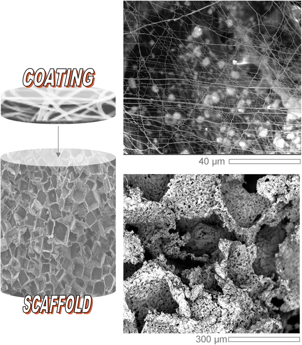
HA/PCL porous scaffold with nanofibrous coating
Future trends
In this century, materials scientists and engineers engaged in biomaterials research have started to address a formidable challenge the mimicking of nature itself. Today, the principal inspiration for designing new synthetic high performance and multifunctional materials is drawn from the observation and study of their biological counterparts. A key learning from this is that numerous materials present in living tissues owe their success to an optimum combination of properties and the possession of adaptive structures, rather than to extreme properties per se. This must now be developed and illuminated by gaining an understanding of the intricacy of the interface between biomaterials and living tissues, and applying this to the characterisation of the interactions occurring on the nano-, micro- and macro-scales. These endeavours will lead to the rational, biologically informed development of current, bottom-up approaches based on molecular self-assembly of small building blocks (amphiphilic peptide molecules, nanofibres) to deliver optimal 3D scaffolds with nano-porosity, high pore interconnectivity plus outstanding biological recognition.
A future challenge lies in the reappraisal of the traditional concepts of composite materials in the light of recent advances in nanotechnology. Successful implementation of nano-inspired strategies will be absolutely essential to the creation and manipulation of local microenvironments, as will productive investigation of the relevant biological mechanisms, including tissue morphogenesis, differentiation and maintenance. Current progress in composite materials and nanotechnology indicate a bright future for their use in bone tissue engineering. However, further significant advances are necessary in order to realise the full potential of nanobiomaterials for clinical use.
Footnotes
Acknowledgements
This study was financially supported from the MIUR funds – Rete Nazionale di Ricerca TISSUENET (n. RBPR05RSM2), MERIT (n. RBNE08HM7T) – and EC projects – IP STEPS (FP6-500465), MAGISTER (FP7-NMP3-LA-2008-214685).