
Abstract
This work proposes a method for preparing metallic Cu nanoparticles using CuO nanoparticles as a precursor, and performs metal–metal bonding by using the metallic Cu nanoparticles. Colloid solution of CuO nanoparticles with a longitudinal particle size of 13·0±3·0, a lateral particle size of 8·4±2·2 and a crystal size of 7·8 nm was prepared with salt base reaction using Cu(NO3)2 aqueous solution and NaOH aqueous solution. Preparation of the metallic Cu particle colloid solution was performed in water using the CuO nanoparticles, hydrazine and cetyltrimethylammonium bromide, which resulted in production of the metallic Cu nanoparticles with a particle size of 50·6 nm and a crystal size of 30·5 nm. Metallic copper discs could be bonded using the metallic Cu nanoparticles under annealing at 400°C and pressurising at 1·2 MPa for 5 min in H2 gas. A shear strength required for separating the bonded discs was recorded as high as 39·6 MPa.
Introduction
Metal–metal bonding can be performed efficiently with the use of solder.1– 4 In a conventional solder based bonding process, metallic alloys composed mainly of lead and tin have been used as solders or filers put between the materials to be bonded, because such metallic alloys have low melting points so that metallic materials can be bonded using the alloy at low temperature. Since it has been pointed out in recent years that the lead is harmful to living bodies,5– 7 limitation tends to be imposed on its use. For releasing the limitation, various tin based, lead free alloys have been developed as new solders.8– 12 Low temperature metal–metal bonding can be achieved using the solders previously developed. However, the bonded materials may be released with use at temperatures higher than their melting points due to remelting of the solders, which will fall short of reliance of the solders.
Since metallic materials such as Au, Ag and Cu are conductive electrically and thermally, metal–metal bonding will be satisfactorily achieved using those metallic materials as filers. Owing to their melting points higher than those of the conventional lead and tin based solders, successful metal–metal bonding requires high temperature annealing. High temperature annealing during the bonding process causes to damage thermally on the materials near the bonded place.
However, the metal–metal bonding process using such metallic materials has an advantage: the bonded materials will be durable with the use at a high temperature compared with that for the materials bonded using the lead and tin based solders.
A decrease in metallic particle size down to several nanometres provides a decrease in their melting point.13, 14 According to the decrease in melting point, temperature in metal–metal bonding process can be decreased by using metallic nanoparticles as a filer inserted between metallic materials. Studies on metallic Ag nanoparticles as the filer have been performed by various researchers.15– 20 The use of metallic Ag nanoparticles not only provides high electric and thermal conductivities for bonded materials, but also stabilises chemically the materials. Besides the metallic Ag nanoparticles, Ag oxide nanoparticles have been also examined for filers in metal–metal bonding, which was performed in reducing gas such as H2.21– 27 Those Ag based nanoparticles well helped the metal–metal bonding. However, the Ag based nanoparticles cost a great deal, and migration of the Ag filer that deteriorates the bonding takes place with use.28, 29
Metallic Cu is also a candidate as the filer for bonding, since it also has high electric conductivity and high thermal conductivity. In addition, it is available at a low cost, and superior in migration property to the Ag. Several methods have been proposed for preparing nanoparticles of metallic Cu.30– 35 They are based on direct reduction of copper ions with reducing reagents. Our group has studied on metal–metal bonding process using metallic Cu nanoparticles prepared with the direct reduction,36, 37 in which a shear strength was achieved 37·7 MPa.37 In the direct reduction methods, the metallic nanoparticles form simultaneously with action of the reducing reagents on the ions. As a consequence, product derived from reducing reagents will be contained as impurity inside the obtained metallic nanoparticles after the reduction. Because impurity in the filer may spoil the bonding properties, pure nanoparticles are desired as the filer. Apart from the direct reduction, methods using copper oxide nanoparticles as a precursor are also promising for preparing metallic Cu nanoparticles.38– 41 In the methods, few impurity derived from reducing reagents will be contained inside the obtained metallic nanoparticles, because the reducing reagents act only on the oxide particle surface.
In our previous work,42 CuO nanoparticles were prepared with a salt base reaction in aqueous solution, which concluded that the reaction temperature was an important factor for the morphology of CuO particles and CuO particle aggregates. In the present work, the CuO particles, which are prepared according to our previous work,42 are used as a precursor for metallic Cu nanoparticles; the aim of the present work is to propose an alternative method for fabricating metallic Cu nanoparticles with the use of the CuO nanoparticles as a precursor. The knowledge obtained in the fabrication of CuO particles will be useful for the synthesis of metallic Cu particles. Their metal–metal bonding properties were also studied.
Experimental
Chemicals
Copper(II) nitrate trihydrate (Cu(NO3)2.3H2O) [Kanto Chemical Co. Inc., 77·0–80·0%, as Cu(NO3)2] and 1M sodium hydroxide solution (NaOH) [Kanto Chemical Co. Inc.] were used as CuO precursors. Cetyltrimethylammonium bromide (CTAB) [Kanto Chemical Co. Inc., 99%] was used as a stabiliser. Hydrazine monohydrate [Kanto Chemical Co. Inc., >98·0%] was used as a reducing reagent. All chemicals were used as received. Water that was ion exchanged and distilled with Yamato WG-250 was used in all the preparations, and was deaerated by bubbling with N2 gas for 30 min before preparation of aqueous solutions of Cu(NO3)2 and hydrazine.
Preparation
The precursor of metallic Cu nanoparticles was copper oxide nanoparticles. Colloid solutions of the copper oxide nanoparticles were synthesised using a salt base reaction in a way similar to our previous work.42 An aqueous solution of NaOH was added to Cu(NO3)2 aqueous solution under vigorous stirring at 80°C. Initial concentrations of Cu(NO3)2 and NaOH were 0·01 and 0·019M respectively, which resulted in Na/Cu molar ratio of 1·9 in the final solution. The reaction time was 3 h.
Colloid solutions of metallic Cu nanoparticles were synthesised by reducing the obtained copper oxide particles. Freshly prepared CTAB aqueous solution and hydrazine aqueous solution were in turn added to the copper oxide particle colloid solution under vigorous stirring at 25°C in air. Initial concentrations of Cu, hydrazine and CTAB were 8·0×10−3, 0·2 and 5·0×10−3M respectively. The reaction time was 3 h.
After the reactions, a process composed of centrifugation at 10 000 rev min−1, removal of supernatant, addition of water, and shake of the mixture with a vortex mixer for dispersing the particles was repeated several times to wash the particles. Powder of the particles was obtained drying the particles at room temperature in vacuum after the final removal of supernatant.
Characterisation
The particles were characterised by transmission electron microscopy (TEM), X-ray diffractiometry (XRD) and thermogravimetry differential thermal analysis (TGDTA). Transmission electron micrographs were taken with a JEOL JEM-2000FX II microscope operating at 200 kV. Samples for TEM were prepared by dropping and evaporating the particle colloid on a collodion coated copper grid. Dozens of particle diameters in TEM images were measured to determine number averaged particle size and standard deviation of particle size distribution. X-ray diffractiometry measurements were carried out with a Rigaku RAD-B X-ray diffractometer at 50 kV and 150 mA with Cu Kα1 radiation. For preparing a powder sample for the XRD measurement, supernatant of the particle colloid was removed with decantation, and then residue of the colloid was dried at room temperature for 24 h in vacuum. Thermal analysis was performed in air or 3% (v/v) H2/N2 gas at a heating rate of 10°C min−1 with a Mettler-Toledo TGA/SDTA851 thermal analyser. Samples for thermal analysis were obtained in the same manner as that for the XRD samples.
Metallic bonding property was investigated by the same set-up as used in our previous works.18, 36, 42 Powder samples were sandwiched between copper discs (a stage: diameter: 10 mm, thickness: 5 mm; and a plate: diameter: 5 mm, thickness: 2·5 mm), and pressed at 1·2 MPa under annealing in H2 at 400°C for 5 min with a Shinko Seiki vacuum reflow system. For examination of bonding strength, shear strengths, which were required to separate the bonded plate and stage, were measured with a Seishin SS-100KP bond tester. The surface of copper plate was observed with a JEOL JSM-5600LV microscope after the measurements of shear strengths.
Results and discussion
Morphology of copper oxide nanoparticles
An inset of Fig. 1 shows a photograph of the colloid solution of copper oxide nanoparticles. The colloid solution was black. Since CuO has a colour of black, this observation indicated production of a large amount of CuO. Figure 1 shows a UV-vis extinction spectrum of the colloid solution. Extinction of the spectrum gradually increased as the wavelength became shorter, and no dominant peaks were detected.
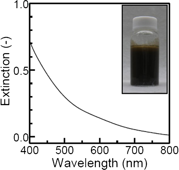
UV-vis extinction spectrum of particle colloid solution prepared with salt base reaction: initial concentrations of Cu(NO3)2 and NaOH were 0·01 and 0·019M respectively; inset shows its photograph
An inset of Fig. 2 shows a TEM image of the CuO nanoparticles. The CuO particles appeared to be distorted: a longitudinal size and a lateral size of the particles were 13·0±3·0 and 8·4±2·2 nm respectively. These sizes were somewhat small compared to those for our previous work.42 To simplify the preparation steps, the reaction time was shortened to 3 h, whereas the reaction time was 12 h in the previous work.42 Accordingly, the particle growth was controlled in the present work because of the short reaction time. The particles partially aggregated. According to our previous work,42 the CuO nanoparticles prepared with the salt base reaction at 80°C have an isoelectric point of ∼10·9. In the preparation of CuO nanoparticles, pH of the solution increased from an initial value of 4·5–5·6 with the addition of NaOH aqueous solution. The increase in pH to 5·6 meant an approach of pH to the isoelectric point. This approach of pH diminished electrostatic repulsion between the particles. Consequently, the particle aggregation took place. The aggregates had a longitudinal size of 51·0±11·2 and a lateral size of 22·4±3·8 nm, which were a little larger than those for our previous work.42 In general, small particles tend to aggregate because of large surface energy of the particles. Accordingly, the small Cu particles obtained in the present work probably formed the large aggregates. Figure 2 shows an XRD pattern of the copper oxide nanoparticles. Dominant peaks were detected at 35·6, 38·8 and 48·9°. They were assigned to those of monoclinic CuO (JCPDS card no. 5-0661). An average crystal size of the CuO particles, which was estimated from the XRD line broadening of the 35·6° peak according to the Scherrer equation, was 7·8 nm. Because the particle size observed with TEM was quite close to the crystal size, the particles consisted mainly of single crystals.
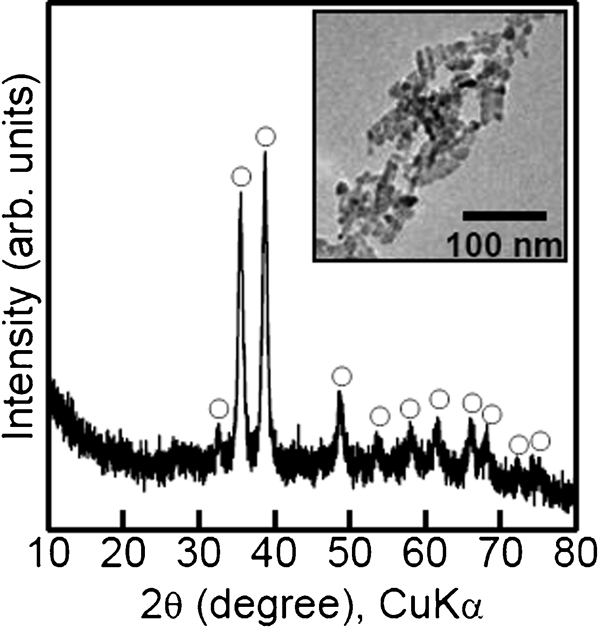
X-ray diffractiometry pattern of particles prepared with salt base reaction: sample was same as in Fig. 1; 0:CuO; inset shows their TEM image
Morphology of metallic copper nanoparticles
The CuO particle colloid solution with black became gradually dark red and muddy with the hydrazine addition. An inset of Fig. 3 shows a photograph of the colloid solution of the CuO colloid solution after the hydrazine addition. Figure 3 shows a UV-vis extinction spectrum of the colloid solution. The background intensity of the solution itself was as high as ∼0·2, which was derived from the muddiness of the colloid solution. This result provided speculation on production of aggregates or flocculates in the solution, which scattered UV-vis light. A peak was observed at 597·5 nm on the background. According to Refs. 43 and 44, the peak was attributed to surface plasmon resonance of metallic Cu nanoparticles, which indicated production of metallic Cu nanoparticles.
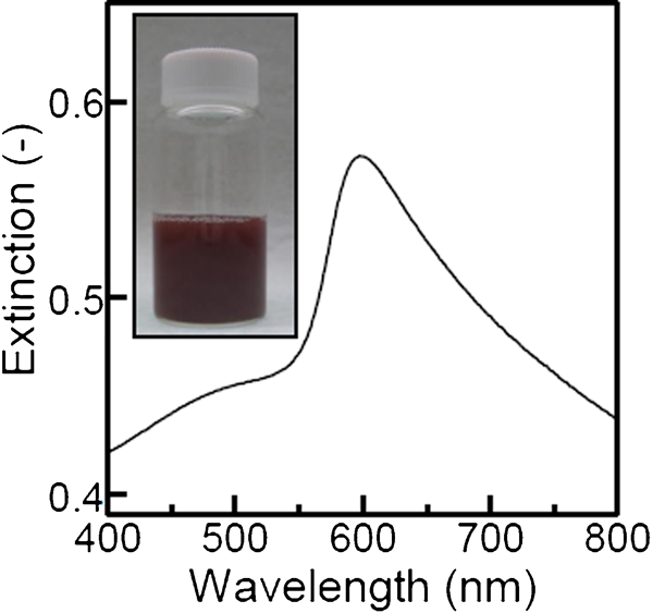
UV-vis extinction spectrum of particle colloid solution: initial concentrations of Cu, CTAB and hydrazine were 8·0×10−3, 5·0×10−3 and 0·2M respectively; inset shows its photograph
An inset of Fig. 4 shows a TEM image of the metallic Cu nanoparticles. The particles with rough surface were produced. Their average particle size was 50·6±5·9 nm. Figure 4 shows an XRD pattern of the metallic Cu nanoparticles. The pattern showed peaks at 43·3, 50·3 and 74·2°, which were attributed to those of cubic metallic Cu (JCPDS card no. 4-0836). Besides those metallic Cu, a faint peak was also detected at 36·6°. This peak was assigned to cubic metallic Cu2O (JCPDS card no. 5-0667). The detection of Cu2O indicated that the particles were partially oxidised. An application of the Scherrer equation to the XRD line broadening of the 43·3° peak provided an average crystal size of the metallic Cu particles of 30·5 nm. Because the particle size observed with TEM was larger than the crystal size, the particles were polycrystalline.
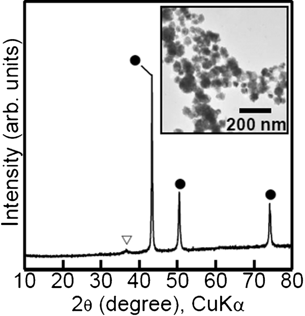
X-ray diffractiometry pattern of particles synthesised reducing obtained copper oxide particles: sample was same as in Fig. 3; •: metallic Cu, ▽: Cu2O; inset shows their TEM image
Bonding property of copper oxide nanoparticles
Thermogravimetry differential thermal analysis curves for the CuO particles are shown in Fig. 5. With the use of air as atmosphere (Fig. 5a), the particle weight did not vary significantly, and no dominant DTA peaks were detected in a range of temperature examined. This result indicated that the pure CuO particles were produced in the present process. For the atmosphere of H2/N2 gas (Fig. 5b), exothermic peaks and a weight loss, which would be assigned to reduction of CuO to metallic Cu, were detected in a range of 200–300°C. The weight did not change above 300°C. This result indicated that the reduction of CuO was completed around 300°C. Therefore, a bonding temperature was adjusted to 400°C for completing the reduction.
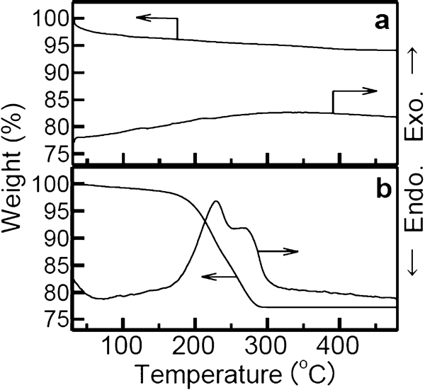
Thermogravimetry differential thermal analysis curves in a air and b 3% (v/v) H2/N2 gas for CuO nanoparticles
The shear strength for the CuO powder was as low as 4·5 MPa: the copper discs could not be strongly bonded using the CuO powder. Figure 6a shows a photograph of the copper stage after the measurement of shear strength. The pressed powder was still blackish, though its circumference gave faintly metallic luster. This observation indicated that the CuO was not completely reduced with the present bonding conditions.
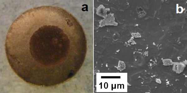
a photograph and b SEM image of copper stages after measurement of shear strength: powders used were CuO particles
Figure 6b shows an SEM image of the copper disc surface separated with the shear stress. The powder was not densely stuck on the copper disc, since the CuO does not have a strong affinity for the metallic copper. The low shear strength was supported with this observation.
Bonding property of metallic copper nanoparticles
Figure 7 shows TGDTA curves for the metallic Cu particles. In the case of atmosphere of air (Fig. 7a), a weight gain accompanying an exothermic peak was dominantly detected at ∼200 and ∼350°C. The weight gains were probably resulted from oxidation of the metallic Cu. The weight did not change above 350°C. This result indicated that the oxidation of metallic Cu was completed around 350°C. In the thermal analysis in H2/N2 gas (Fig. 7b), no weight gain was measured in the temperature range examined, which indicated that oxidation of the metallic Cu particles did not take place in the reducing gas. An exothermic peak and a weight loss were detected remarkably around 250–300°C. The as prepared metallic Cu particles contained copper oxide a little, as shown in Fig. 4. The copper oxide was probably reduced to form metallic Cu dominantly around 250–300°C in the reducing gas. The reduction resulted in removal of oxygen from the copper oxide, which corresponded to the weight loss. At the same time, oxygen molecules were generated from the reduction of copper oxide, and the oxygen oxidised a trace of organic compounds left on the particles such as CTAB, which provided the exothermic peak. No large weight loss was detected above 400°C, which indicated that the reduction was almost completed around 400°C. Therefore, the metal–metal bonding was also performed at 400°C, so that copper oxide would be enough reduced.
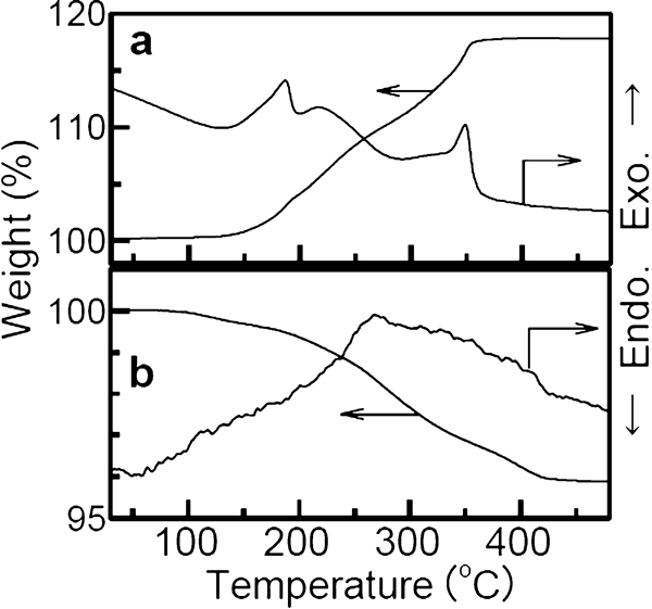
Thermogravimetry differential thermal analysis curves in a air and b 3% (v/v) H2/N2 gas for metallic Cu nanoparticles
The metallic Cu powder revealed a shear strength as high as 39·6 MPa: the copper discs could be strongly bonded using the metallic Cu powder. Figure 8a shows a photograph of the copper stage after the measurement of shear strength. The whole area of the pressed powder was lustrous, which indicated that the powder was still metallic after the bonding process. Consequently, the metallic Cu powder bonded the copper discs strongly.
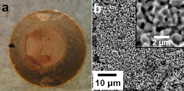
a photograph and b SEM image of copper stages after measurement of shear strength: powders used were metallic Cu particles
Figure 8b shows an SEM image of surface of the copper disc separated with the shear stress. Some dimples were observed accompanying with sharp tips on the surface. Dimples are formed in bonded region when metals that are strongly bonded are separated with shear stress. Such dimples were produced in Morisada et al.'s work, which performed strong bonding with the use of Ag nanoparticles.28 Accordingly, this observation supported that the coppers could be strongly bonded using the metallic Cu particles.
Conclusions
The metallic Cu nanoparticles were fabricated by using CuO nanoparticles as the precursor that were prepared by our previously developed method. The colloid solution of CuO nanoparticles that had the longitudinal particle size of 13·0±3·0, the lateral particle size of 8·4±2·2 and the crystal size of 7·8 nm was prepared by reacting 0·01M Cu(NO3)2 with NaOH in aqueous solution at the Na/Cu ratio of 1·9 and 80°C. The preparation of the metallic Cu nanoparticle colloid solution was performed by reducing the obtained CuO nanoparticles with the concentration of 0·01M CuO with 0·2M hydrazine in the presence of 5·0×10−3M CTAB in water at 25°C, which produced the metallic Cu nanoparticles with the particle size of 50·6 nm and the crystal size of 30·5 nm. The metallic Cu nanoparticles were used as filers for metal–metal bonding. The bonding examination was performed by annealing the metallic copper discs with the metallic Cu nanoparticles at 400°C under a pressure of 1·2 MPa for 5 min in H2 gas. The metallic Cu nanoparticles revealed the bonding property worthy of mention: the shear strength as high as 39·6 MPa was required for separating the bonded discs. Further study on practical use of the metallic Cu particles is in progress.
Footnotes
Acknowledgements
This work was partially supported by Hitachi Ltd. We express our thanks to Professor T. Noguchi in College of Science of Ibaraki University, Japan for his help for TEM observation.