
Abstract
Biomimetic calcium phosphate coatings have been studied to improve the corrosion resistance of biodegradable magnesium alloys. The corrosion resistance of these coatings is often limited by defects during creation. A method for improving the corrosion response of these coatings is therefore needed if biomimetic coatings are to be used for corrosion protection. In this study, a calcium hydroxide underlayer was applied to improve the properties of these biomimetic coatings. The in vitro corrosion response was studied using hydrogen evolution and electrochemical techniques. It was found that the calcium hydroxide layer increased the corrosion resistance of the coatings. The coatings created had fewer defects than the unmodified biomimetic coatings. Over time, the calcium hydroxide layer also prevented the defects in the coating from growing, leading to longer lasting protection. The results of this study suggest that calcium hydroxide coatings can significantly improve the corrosion protection of a biomimetic coating.
Keywords
Introduction
Magnesium is promising as a degradable biomaterial.1,2 Magnesium has the strength and toughness properties that make it desirable for orthopaedic applications. Magnesium can degrade in vivo without any acute or long term toxic effects. Unfortunately, unprotected magnesium degrades too quickly in the body for most applications. This results in a loss of mechanical strength before the bone has healed enough to take the load, andthe release of H2 gas at rates above which thesurrounding tissue can safely dispose of it. The degradation rate needs to remain very low during theinitial phase of tissue recovery and then degrade completely at a rate low enough that hydrogen gas does not accumulate. The exact rates will depend on the type and site of the implant; however, the degradation rate of pure magnesium is too high. To be useful, it is necessary to slow the corrosion rate of magnesium.
So why does magnesium corrode too quickly in the physiological environment? First, magnesium is a very active metal,3 with a standard reduction potential of −2·37 V. When magnesium corrodes in water, the net reaction is Mg(s)+2H2O→Mg(OH)2+H2(g).4,5 Mg(OH)2 is fairly insoluble in water. If part of this layer dissolves or is removed by mechanical means, the metallic magnesium beneath corrodes and repassivates the surface, leading to good protection. However, Cl− acts as a catalyst to speed up the reaction.4 Cl− replaces OH− in the layer but is much more water soluble than Mg(OH)2.6 The high concentration of Cl− in vivo means the passive layer is not protective in the body, leading to fast degradation rates. Thus, some mechanism of protection is needed for this material to be used in vivo. A protective coating that reduces the corrosion rate of the implant is one method of solving this problem.
Calcium phosphate compounds (CaP) are attractive elements for corrosion protection due to their biological properties. They are insoluble in physiological fluids.7,8 They are non-toxic and biocompatible.9 – 13 CaP compounds are a major component of bone. Therefore, for orthopaedic applications, CaP is promising.8 CaP compounds are used to improve the biocompatibility of metallic implants.8,9,14 CaP compounds have been studied to improve the corrosion properties for biomedical applications, including magnesium and its alloys.12,15 – 19 One coating method is a biomimetic treatment using solutions similar to physiological fluid to create coatings that mimic and persist in vivo.7,9,11,20–22 Biomimetic coatings are low temperature, contain no toxic elements and can coat complex shapes and porous structures.20 Their properties are useful for enhancing the biocompatibility of the coated surface.9
Unfortunately, biomimetic coatings are fraught with problems. During the coating process, adhesion of the coating to the surface and complete coverage are necessary to prevent internal corrosion of the implant. Any defects can lead to corrosion underneath the coating, leading to lower adhesion of the coating and increasing corrosion rates.23 CaP compounds are much more brittle than the underlying substrate. As such, the likelihood of defects is high for a load bearing implant. Therefore, a coating that is not as sensitive to defects is desirable. Furthermore, the coating process in chemical solutions often allows,15 and in some cases, partially relies on, the corrosion of Mg to deposit the CaP.15 Magnesium corrosion releases Mg2+ ions in solution, and these ions can have a negative effect on the solution coating by inhibiting the formation of crystalline apatite,24 favouring more soluble phases.7 To ensure good corrosion protection, a method to improve these biomimetic coatings must be developed.
Calcium hydroxide [Ca(OH)2] is often used in biomaterials for dental applications.25 The application of a calcium hydroxide coating can be used to provide a pretreatment or sublayer for the ultimate CaP layer. This can be used to increase the protective properties of biomimetic coatings. By depositing a Ca(OH)2 layer, the deposition of CaP has plenty of Ca2+ to form CaP as this layer dissolves. The other product of the dissolution is OH−, which raises the pH and promotes deposition. The created coating therefore can be more coherent and have better properties than the biomimetic coating would otherwise.
Once the final CaP coating is applied and immersed into the intended corrosive environment, a slightly soluble calcium base layer exists just below the surface. Any cracks that form, or defects from the original coating, will lead to corrosion of this layer. In an in vivo solution, the phosphate ions present can react with the calcium ions to precipitate a new CaP layer at the defects. This study shows the improvement of a biomimetic CaP coating using an electrochemical assisted deposition (ECAD) coating of calcium hydroxide for corrosion protection of biomedical magnesium.
Materials and methods
Preparation
Pure Mg (99·98% Timminco) samples were cut to 15×15 mm sections and mounted in epoxy with a copper wire attached at the back for electrical connection, and one face exposed for deposition. Samples were polished to 1200 grit with SiC. Samples were then ultrasonically cleaned in ethanol for 5 min and allowed to dry in air.
Calcium hydroxide coating
The ECAD samples were coated using a standard three-electrode set-up with a saturated calomel electrode (SCE) as the reference and a flat Pt plate as the counter electrode. The sample face was mounted parallel to the counter electrode at a distance of 1 cm in a 2M Ca(NO3)2 solution. Electrostatic coating was performed with −3·2 V(SCE) for 10 min. Once deposition was complete, samples were rinsed with ethanol and allowed to dry in air.
Biomimetic CaP coating
After ECAD coating, the samples were biomimetically coated using a modified ×5 concentrated simulated body fluid (SBF) in the process described in Ref. 23. The pH of the solution was controlled during the process by dissolving carbon dioxide gas (CO2) in the solution. One litre of biomimetic coating solution was heated to 37°C, and CO2 (g) was bubbled through for 15 min to reach a pH of 6. The CO2 was then removed, samples were immersed in the solution and air was bubbled through the solution for 24 h. The solution was stirred with a magnetic stirrer to ensure uniform ionic concentrations. After 24 h, the solution pH rose to ∼8. The samples were removed and rinsed with distilled water and allowed to dry in air at room temperature.
Characterisation
The coatings created were characterised by scanning electron microscopy (SEM) (JEOL 7000F FE-SEM), energy dispersive X-ray spectroscopy and Glancing angle X-ray diffraction spectroscopy (GA-XRD) (PANalytical X'Pert-Pro MPD PW3040/60) to determine the composition.
Corrosion testing
Corrosion tests were carried out to measure the corrosion rate. Experiments were carried out in Hank's balanced salt solution (HBSS) at 37±0·5°C and pH of 7·4±0·05, buffered with 4-(2-hydroxyethyl)-1-piperazineethanesulfonic acid (HEPES) (25 mM). Five hundred millilitres of solution was used per sample for each hydrogen evolution test. Glass burettes were used to collect the hydrogen gas and measure the total hydrogen evolved with a precision of ±0·1 mL. Corrosion solution was refreshed every 72 h to keep the pH and ionic composition relatively constant. Five samples of each biomimetic, ECAD+biomimetic and uncoated Mg were measured. The H2 evolution was monitored for a total of 14 days.
Electrochemical tests were performed by potentiodynamic polarisation (PDP) and electrochemical impedance spectroscopy. A three-electrode set-up was used with a Pt counter electrode and an SCE. Three hundred millilitres of HBSS was used for each test. The area of the working electrode was 1 cm2. The PDP tests were carried out after 20 min in solution to allow the open circuit potential to stabilise. The PDP tests utilised a scan rate of 1 mV s−1 and scanned over the range open circuit potential (OCP) −0·100 V to Ref +0·500 V. Electrochemical impedance spectroscopy scans were performed once every hour over a period of 72 h to evaluate the change in corrosion resistance with time. An AC 10 mV peak to peak signal from OCP across a frequency range from 50 kHz to 20 mHz was used.
Results and discussion
Calcium hydroxide layer formation by ECAD
Electrodeposition proved to be useful for depositing a calcium hydroxide coating on the samples. The coating produced by ECAD in concentrated 2M Ca(NO3)2 solution was dense and covered the entire surface more or less uniformly (Fig. 1a). The coating exhibited no areas that were not covered. The coating consisted of Ca(OH)2. This was determined through XRD (Fig. 2) and energy dispersive X-ray spectroscopy. The ECAD process required an overvoltage to create a calcium hydroxide coating. The surface of the magnesium was held at −3·2 V(SCE). The current through the cathode reduced water in the solution (Equation (1))

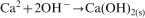
a scanning electron microscopy of ECAD Ca(OH)2 coating and b cross-section of bubble defect
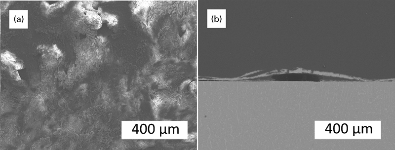
GA-XRD of a calcium hydroxide coating by ECAD, b biomimetic coating and c ECAD+biomimetic coating
Biomimetic coating process
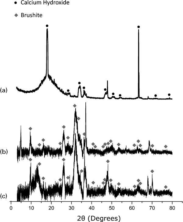
After the Ca(OH)2 layer was deposited on magnesium, a CaP layer was applied to the surface. The biomimetic method was similar to the process described by Habibovic et al.20 The process was chosen because it creates CaP coatings at low temperature in SBF that contain no ions not found in the body. This means that it is safe to use as no toxic chemicals are used during the coating process.20 The biomimetic coating created a flake-like structure of CaP compound on the surface (Fig. 3). According to the glancing angle XRD (Fig. 2), the coating formed mainly dicalcium phosphate dihydrate or brushite. The biomimetic layer produced on the surface of the ECAD coated material is slightly different to the biomimetic layers produced on bare magnesium substrates in morphology (Fig. 3a and b). The difference can be attributed to the differences in crystal growth and nucleation as well as small changes in composition due to the Ca rich ECAD layer.
a Biomimetic coating; b ECAD + Biomimetic coating; c Biomimetic coating after corrosion in HBSS for 72 hours and d ECAD + Biomimetic coating after corrosion in HBSS for 72 hours</author-query>
Biomimetic coatings can protect against magnesium corrosion by themselves; however, the coatings tend to have a few problems. The coatings are deposited in solutions similar to body fluids and therefore often include additional phases and ionic substitutions in the crystal lattice. While these increase the biocompatibility compared to crystalline synthetic CaP,10,21 these phases tend to increase the solubility as well8 and thus decrease the corrosion resistance. Substitutions in the lattice with Mg are typically high, especially on an Mg substrate, which tends to corrode during the process, creating a high Mg2+ concentration near the surface of the substrate. Mg2+ has the effect of decreasing the crystallinity of CaP.24 For ECAD samples, the corrosion of magnesium during the biomimetic coating was mitigated by the calcium hydroxide coating. Rather than the Mg substrate corroding in the coating solution, the Ca(OH)2 protected it. The pH rise that helps form coatings in the solution due to magnesium corrosion15 can instead be created by the dissolution of calcium hydroxide. This could help to prevent so many Mg ions from getting into the coating, breaking up the lattice and ultimately creating a more soluble and less protective coating.27
Additionally, the Ca(OH)2 layer provided a surface of calcium that is slightly soluble for the CaP layers to convert to and nucleate on. The phosphate groups in the biomimetic solution can replace hydroxide groups on the surface of the ECAD layer to form the more insoluble CaP. These sites are then locations for nucleation of the rest of the biomimetic coating. This could account for the differences observed in the two coatings’ morphology (Fig. 3) and composition (Fig. 2).
Corrosion results
Corrosion protection of coatings was assessed in HBSS at 37°C to simulate the chemical conditions found in the human body fluid. The corrosion mechanism to be studied was the protection against chloride ion attack as well as the deposition of CaP compounds on the surface from the SBF.15 Hence, HBSS was chosen for the tests because it contains these salts without the complication of other compounds such as proteins. The use of HEPES buffer prevents the corrosion reaction from changing the pH of the solution without the need for an active pH control system such as that found in the body. The corrosion solutions were used for up to 72 h, after which the pH of the solution was kept within ±0·1 for all samples by the HEPES buffer.
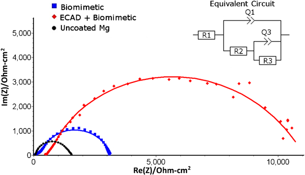
The corrosion rate was monitored by hydrogen gas evolution in order to examine the longer term corrosion behaviour of these coatings. The uncoated samples exhibited rapid corrosion (Fig. 4). For uncoated samples, the curvature of the early part of the graph showed an accelerating corrosion rate. This is due to the local pitting of the Mg surface.28 This effectively increased the surface area available to the solution, leading to an accelerating corrosion rate. Later, this rate slowed as the CaP was deposited on the surface of the Mg.29 This layer begins to protect the surface. As such, past ∼250 h of corrosion in SBF, the corrosion rate decelerates. However, already significant amounts of H2 gas had been generated; by 350 h, the average amount of gas per sample is 35·6 mL over the 2·25 cm2 surface (Table 1). Therefore, to form this protective layer in SBF, a large amount of corrosion is required to occur. This is unacceptable for degradable implants as the mechanical strength of the implant needs to survive during the initial stages of healing.
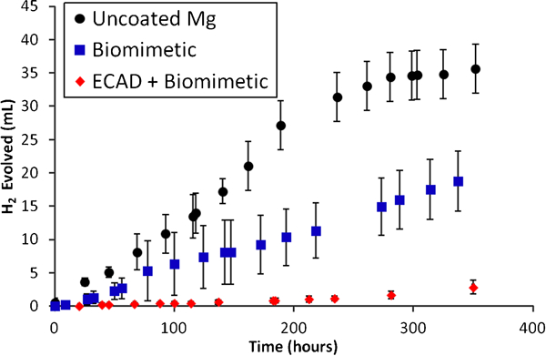
Total hydrogen evolved over 14 days
Comparison of corrosion values
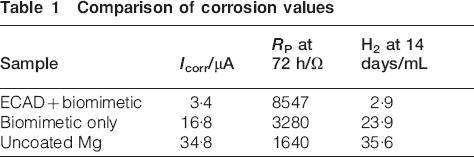
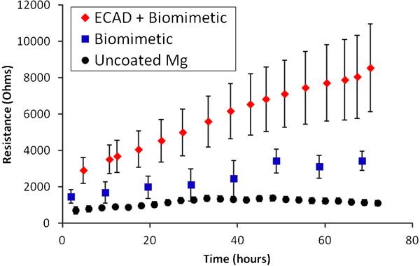
The biomimetic coatings are applied to simulate this process and create the protective coating before it is exposed to the solution. The biomimetic coatings protect the substrate from corrosion by blocking the corrosive environment from the underlying Mg. Indeed, early in the life of the coating, the simple CaP provided much greater protection from the corrosive environment than the uncoated counterparts (Fig. 4). The PDP tests showed that the biomimetic coating had a lower corrosion current density (Fig. 5) than the uncoated Mg and a corresponding higher polarisation resistance (Fig. 6). The lower current density was due to the smaller portion of exposed area to the solution. Thus, there was a decrease in anodic reaction rate. The corrosive solution cannot attack the magnesium where it is protected by the CaP coating. However, there were cracks and defects in the coating where the corrosion could continue, as seen in Fig. 3a. These cracks create an imperfect coating that allowed the corrosion reaction to progress. This effect can be seen by the two time constant frequency responses to polarisation that correspond to a porous coating (Fig. 7). The biomimetic coating exhibits a resistance and capacitance of the coating itself and one for the electrolytic double layer (EDL) that forms the interface with the metal to solution. This is in contrast to the uncoated Mg that exhibits a single semicircle corresponding to the EDL without significant contribution of a coating layer. Thus, the corrosion rate and corresponding H2 generation are lower for the biomimetic coated samples, but the defects in the biomimetic coating still allowed corrosion to occur.

Potentiodynamic polarisation curves for selected samples after 72 h immersion
Total coating polarisation resistance over 72 h immersion in HBSS
Nyquist plots of impedance data for representative samples at 72 h immersion and equivalent circuit fit
Over time, these biomimetic coating properties changed. While the polarisation resistance was relatively steady over the first 72 h, the corrosion rate remained relatively low. Later on, the coatings begin to fail. The size of the defects increased, as seen in Fig. 3c. The brushite coating created was not as stable as the hydroxyapatite coatings as they can partially dissolve in SBF.7 Furthermore, as the substrate was attacked underneath, the coatings would delaminate from the surface, exposing more area for corrosion.23 As this happened, the average amount of corrosion increased. The variability of the coating increased as well as coatings delaminated non-uniformly across the sample population. Figure 4 shows this trend of increasing corrosion, although the rate is still less than the uncoated samples. Clearly, the biomimetic coatings protect from corrosion, but not enough or for long. If coatings such as these are to be used for slowing the corrosion rate, this problem of corrosion through defects in a metastable biomimetic coating is a significant challenge.
The ECAD coatings attempt to solve this problem using the Ca(OH)2 underlayer to enhance the corrosion resistance of biomimetic coatings. Ca(OH)2 is slightly soluble in the biomimetic coating solutions, leading to better coverage and conversion of the coating. The initial polarisation resistance of ECAD coatings is higher than the biomimetic coatings (Fig. 6). There is a corresponding decrease in corrosion current density measured by PDP in Fig. 5 as well. Compared with the uncoated Mg, the ECAD coating decreases both anodic and cathodic reaction rates. While both coatings slow the anodic reaction, the ECAD coating also affects the cathodic reaction, leading to the much lower H2 formation rate. This indicates that the layer formed after the biomimetic coating process, while similar in composition, leaves a smaller area of defects though which to continue the corrosion reaction. The polarisation resistance over the first 72 h displayed an interesting trend. The ECAD coated samples actually increased in polarisation resistance as time passed in the corrosion solution. This indicates that as the samples were immersed, the protective qualities of the coating actually increased. The mechanism of this can be explained by the high concentration of calcium in the layer under the biomimetic coating. As calcium hydroxide dissolves, the ions combine with phosphate ions in solution to form insoluble CaP phases at the corrosion sites. This effectively blocks corrosion and preserves the integrity of the coating, which exhibits much less damage after exposure to the solution (Fig. 3d). This is in contrast with the corrosion of the Mg substrate on the biomimetic only coated samples. The subsequent release of free Mg2+ ions inhibited the crystalline formation of CaP24 and does not repassivate the defects (Fig. 3c). This benefit was very pronounced over the 14 days of corrosion. While H2 generation is large for the other samples, the ECAD coatings preserve their integrity and only allow a small amount of corrosion, only evolving an average of 2·9 mL of H2 over the test period (Table 1). The improvement in coating properties over time allowed this coating to remain intact and protective for a much longer period of time than the biomimetic coating by itself.
The ECAD coating polarisation resistance was dominated by a single obvious time constant (Fig. 7). The decreased amount of defects made the coating resistance dominant and the corresponding effect of the EDL less prevalent. At low frequencies, the noise on the ECAD coatings was relatively large, which may have obscured the secondary time constant that the coating was expected to exhibit. From the polarisation behaviour over time (Fig. 6), we can see that the ECAD coatings are quite variable in performance compared to the biomimetic coatings. This suggests that the coatings are very sensitive to coating creation parameters. The volcano shaped defects due to the ECAD process may have left areas of lower calcium hydroxide coverage that adversely affected the performance of the coatings. Further investigations into controlling the properties of this layer are therefore needed. However, the effect on the stability and corrosion protection of the biomimetic coatings suggests that this coating method is promising as a step to overcoming the barriers to using biomimetic coatings for corrosion protection of magnesium implants.
Conclusions
The ECAD of calcium hydroxide can increase the corrosion protection of the biomimetic coating. The ECAD coating provides a more resistant biomimetic coating with fewer defects. The corrosion protection did not degrade over time for the ECAD coatings as it did for the biomimetic coatings. As corrosion progressed, the protectiveness of the ECAD coatings increased due to the calcium rich layer depositing additional protective CaP at corrosion sites. For a biodegradable implant application where corrosion behaviour is critical, a self-healing coating such as the one describe here may provide much better corrosion properties than typical biomimetic coatings by themselves. Therefore, the calcium hydroxide layers are promising as a route to improving the corrosion resistance of biomimetic coatings.