
Abstract
The corrosion behaviour of cast and hot isostatically pressed Ti–6Al–4V–B alloys was studied for several compositions in the <0·001-1·09 wt-%B range. Passivity and corrosion resistance were observed in all of the alloys subjected to cyclic potentiodynamic polarisation (CPP) in phosphate buffered saline. The CPP tests were also conducted in 0·9 wt-% sodium chloride (saline) over a wide potential range. The pitting potentials generally decreased with increasing boron content with the notable exception of the 0·01B alloys, which did not pit. The repassivation potentials generally increased with increasing boron content. When osteoclast precursor cells (macrophages) and osteoclast activating cells (fibroblasts) were cultured on Ti–6Al–4V–B alloy discs, higher levels of macrophage receptor activator of nuclear factor κB (RANK) and fibroblast RANK ligand were expressed in comparison with CP Ti, 316L and Ti–6Al–4V. This suggests that boron containing alloys may facilitate differentiation of osteoclasts derived from macrophages that interact with the prosthetic surface. However, it is not yet known whether the upregulation of RANK or RANK ligand is sustainable over longer periods of time.
Introduction
The US population in the 65 and older age group is projected to double by 2050.1 Musculoskeletal issues, especially knee and hip fractures, are expected to grow correspondingly.2 Thus, there is a need to develop longer lasting and durable alloys that can be used as structural implant materials. The greatest impediment to durable prosthetics is the loosening of the implant over time.3 – 5 This process, known as aseptic loosening, is caused by the inflammatory response against the prosthetic metal and metal debris produced by its corrosion.3 Aseptic loosening can be attributed to the erosion of the bone anchoring the metallic prosthetic. Bone erosion occurs when osteoclast cells in the body are differentiated and become activated.6 Monocyte and macrophage cells are the precursors of osteoclasts in the body and can be induced to become osteoclasts when the receptor activator of nuclear factor κB (RANK) on their surface is bound and stimulated by its ligand (RANKL). Stimulation of RANK occurs when monocytes or macrophages interact with RANKL expressing cells, such as fibroblasts and osteoblasts.7 Overexpression of RANK on monocytes/macrophages or overexpression of RANKL on fibroblasts may induce a higher rate of osteoclast formation, thus causing a higher rate of bone erosion.6
One of the most commonly used alloys in biomedical applications, Ti–6Al–4V, forms a passive oxide layer that protects against corrosion. A relatively new class of titanium alloys containing boron has been developed for its enhanced mechanical properties. Extensive work has been carried out on the characterisation of the mechanical properties of Ti–6Al–4V boron containing alloys.8 There is a general increase in yield strength and stiffness for the alloys with increasing amounts of boron, e.g. significant increases in yield strength are achieved even with additions as low as 0·5 wt-%B to Ti–6Al–4V.9 The effects of heat treating Ti–6Al–4V–B alloys on their mechanical properties have also been investigated.10 The performance of Ti–6Al–4V–B alloys in physiological environments has previously been reported.11 – 13 In the present study, this work has been extended further, and, in particular, attention was paid to very low B additions, i.e. on the order of 0·02 wt-%B or lower.
The corrosion susceptibility of Ti–6Al–4V alloys with varying boron contents was determined in phosphate buffered saline (PBS) solutions under deaerated conditions at a temperature of 37°C. Alloy corrosion behaviour was differentiated using cyclic potentiodynamic polarisation (CPP) tests from −1 to 8 V in 0·9 wt-%NaCl. For the biocompatibility tests, osteoclast precursor cells (macrophages) and osteoclast activating cells (fibroblasts) were cultured on various Ti–6Al–4V–B alloys, as well as traditional prosthetic alloys Ti–6Al–4V, commercially pure titanium (CP Ti) and stainless steel (316L). The cells were then analysed for induction of macrophage RANK or fibroblast RANK ligand in order to better understand how the new boron containing alloys compared to traditional alloys in their potential to activate osteoclasts.
Experimental
Ti–6Al–4V alloys with B contents ranging from <0·001 to 1·09B (all alloy compositions are in weight per cent) were received in the cast and hot isostatically pressed conditions.
Corrosion test samples were prepared using standard metallographic methods to a 0·05 μm final surface finish. They were first ground using 240, 320, 400, 600 and 800 grit silicon carbide papers. They were then polished using 6, 3 and 1 μm diamond pastes and finish polished with 0·05 μm alumina suspension. Before the tests, the samples were ultrasonically cleaned using deionised water, methanol and then deionised water again for 180 s each.
Two different potentiostats (Biologic SP-200 and Gamry Reference 600) were used to run the direct current CPP tests. Corrosion tests were conducted using three-electrode flat cells with a sample testing area of 1 cm2. One of these used an Ag/AgCl reference electrode in 3M NaCl and a platinum coated niobium mesh counter electrode (used in conjunction with the Gamry Reference 600), and the other used a saturated calomel electrode in saturated KCl and a platinum gauze counter electrode (in conjunction with the Biologic SP-200). The flat cells were cleaned before each test using soap and water, rinsed with deionised water and dried using absorbent, low lint wipes. Once the sample was attached to the flat cell, the set-up was placed in a grounded copper Faraday cage and connected to the potentiostat.
Two types of cyclic polarisation tests were conducted. The first closely paralleled the ASTM F2129 standard14 and served to establish a baseline for the viability of samples for potential service in physiological environments. The solution, PBS, was prepared using 0·8 wt-%NaCl, 0·02wt-%KCl, 0·115 wt-%Na2HPO4 and 0·02 wt-%KH2PO4 and 18 MΩ cm pure water. Nominally, 250 mL of PBS was deaerated with nitrogen gas for 30 min before heating to 37°C. The solution was maintained at this temperature throughout the test while being continuously deaerated. Open circuit potentials (OCVs) were monitored for 1 h. Measurements of OCV were immediately followed by the CPP test, polarising the sample up to 800 mV versus reference potential (REF) and back to rest potential at a scan rate of 0·1667 mV s−1. The second type of CPP test was conducted in 0·9 wt-%NaCl solution at room temperature. Measurements of OCV were taken for 28 h, after which the CPP test was started by polarising the sample at a scan rate of 0·1667 mV s−1 from −1 V(REF) to an apex potential of 8 V(REF) or apex current density of 10 mA cm−2 and back down to −1 V(REF) at a reverse scan rate of 10 mV s−1. The conditions of this test were far more aggressive than those typically encountered in physiological environments15 and thought to be more likely to elicit differences in performance among the different alloys being studied.
After patient implantation, macrophages are able to interact with the prosthetic metal in two different ways. They can interact through direct contact with the alloy surface or by phagocytising (engulfing) debris particles corroding off of the prosthetic.16 In order to mimic these conditions in culture, approximately one million macrophages were cultured on alloy discs of various compositions or cultured in the presence of alloy particles and incubated for 72 h.17 After incubation, macrophages were examined for RANK protein expression by staining their cell surface with anti-RANK antibodies and analysing for surface fluorescence using a FACScaliber flow cytometer (BD Biosciences).
Fibroblasts do not have the ability to phagocytose particles or debris. They can only interact with the prosthetic alloy by physical interaction with the alloy surface. To examine the effect of the alloys on fibroblast RANK ligand activation, 250 000 cells were cultured on each alloy for 72 h. After incubation, fibroblasts were stained for RANK ligand surface expression with anti-RANK ligand antibodies and analysed by flow cytometry.
Results and discussion
Corrosion tests
Figure 1 shows the CPP tests, following ASTM F2129, for Ti–6Al–4V–B alloys ranging in boron content from <0·001 to 1·09 wt-%. All of these alloys survive this test with no evidence of pitting. The CPP tests performed in the −1 to 8 V range for alloys in the same composition range as above are shown in Fig. 2. This figure shows differences in the behaviour of the different boron containing alloys. The pitting potentials relative to the standard hydrogen electrode (SHE) are plotted as a function of the boron content of the alloys in Fig. 3. From this figure, it is seen that, in general, the pitting potential decreases as the boron concentration increases. However, a very interesting observation was made, i.e. in the very low (trace) boron addition range, the pitting potential actually increased. In fact, no pitting was observed for alloys containing nominally 0·01B, and this data point is therefore absent from the figure. This phenomenon was observed not only in cast and hot isostatically pressed alloys but also in heat treated alloys.18 This is an important observation considering that pitting was observed in all other compositions with boron contents above and below this composition. When the boron content was increased to 0·04B, pitting was observed again; however, the pitting potentials were quite high. The significance of this observation is that alloys with B compositions between 0·01 and 0·04 wt-% may also not pit under the same test conditions. Figure 4 shows that the repassivation potentials (versus SHE) generally increase with increasing boron content. Although the addition of boron reduces the pitting potential in general, these potentials are much larger than the anticipated rest potential in physiological conditions.15
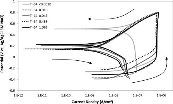
Cyclic potentiodynamic polarisation data from OCV to 800 mV to OCV (versus Ag/AgCl 3M NaCl reference) in nitrogen purged PBS at 37°C, performed in accordance with ASTM F2129: arrows depict progression of each scan from start to finish
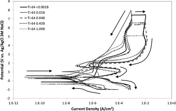
Cyclic potentiodynamic polarisation data from −1 to 8 to −1 V (versus Ag/AgCl 3M NaCl reference) in 0·9wt-%NaCl at room temperature, after 28 h OCV scan: arrows depict progression of each scan from start to finish
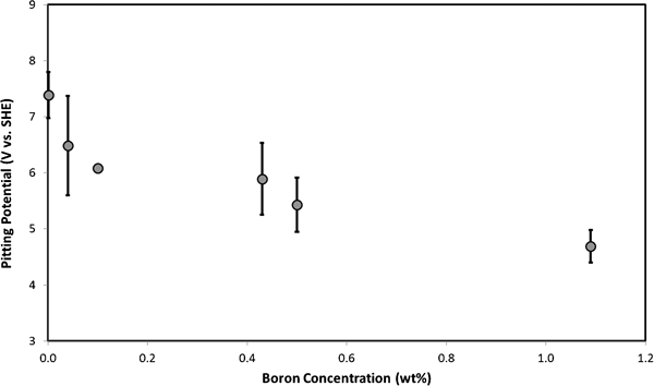
Pitting potential [V(SHE)] as a function of weight percentage of boron in cast and hot isostatically pressed Ti–6Al–4V–B alloys: no pitting was observed for all Ti–6Al–4V–0·01B alloy samples (absent from figure)
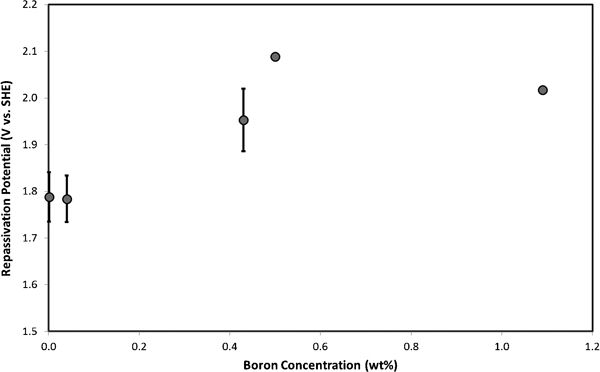
Repassivation potential [V(SHE)] as a function of weight percentage of boron in cast and hot isostatically pressed Ti–6Al–4V–B alloys: no pitting was observed for all Ti–6Al–4V–0·01B alloy samples (absent from figure)
Scanning electron microscopy reveals that the beta phase appears to be narrower and longer for the 0·01B alloy in comparison to that in <0·001B and 0·04B alloys (Fig. 5). It is to be noted that, in general, as the boron content increases, the matrix grain size decreases. This observation is consistent with findings in the literature19 that demonstrate grain size refinement for trace boron additions. One possible explanation for the increased corrosion resistance of the 0·01B alloy is that boron is present as a solute in Ti–6Al–4V at these low alloying levels. This composition is below the 0·02 wt-% boron content proposed as the limit of solid solubility in titanium alloys like Ti–6Al–4V alloys.19 While the literature is sparse in the area of boron additions to titanium alloys, boron is known to increase corrosion resistance in iron based alloys.20 Increased corrosion resistance has also been observed in aluminium metal matrix composites containing boron.21 While the exact details of the mechanism for the improvement in corrosion resistance of 0·01B alloys are not yet known, this paper is the first to report this effect.
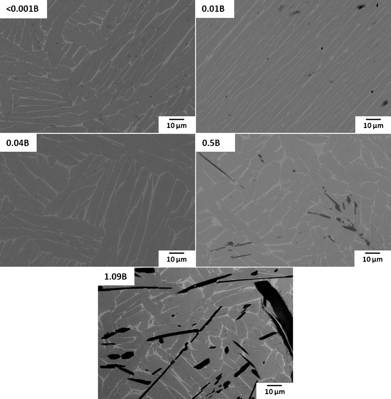
Backscattered electron images (×1000) of cast and hot isostatic pressed Ti–6Al–4V–B alloys: α phase is grey, β phase is bright and TiB needles appear black on micrographs
Biocompatibility tests
When exposed to alloy particles, it was observed that macrophages expressed little to no RANK on their surface (Fig. 6). This demonstrated that the uptake of metal debris, regardless of the alloy composition, does not increase the expression of RANK in macrophages and, thus, would not contribute to osteoclast formation in the microenvironment surrounding the prosthetic.
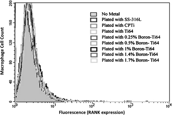
Macrophage cell count RANK expression after metal particle exposure. Human HL-60 cells were differentiated into macrophages using 10 nM phorbol-12-myristate-13-acetate and 25 nM vitamin D3. Cells were cultured with 2000 μg mL−1 of stainless steel 316L (SS) or various titanium alloy particles. After 72 h, cells were analysed by flow cytometry for RANK expression on cell surface. All cells showed little to no expression of RANK after exposure
However, when exposed to the surface of the alloys by culturing the macrophages on the metal discs, the macrophages were observed to induce the expression of cell surface RANK (Fig. 7). Both CP Ti and Ti–6Al–4V induced no expression of RANK. Stainless steel (316L) induced RANK on the macrophage surface. Notably, the titanium–boron alloys induced more RANK on the surface of macrophages than the traditional metals: stainless steel, CP Ti and Ti–6Al–4V. These results suggest that the incorporation of boron may increase the possibility of osteoclast differentiation and activation by inducing RANK expression on the surface of osteoclast precursor cells (macrophages).
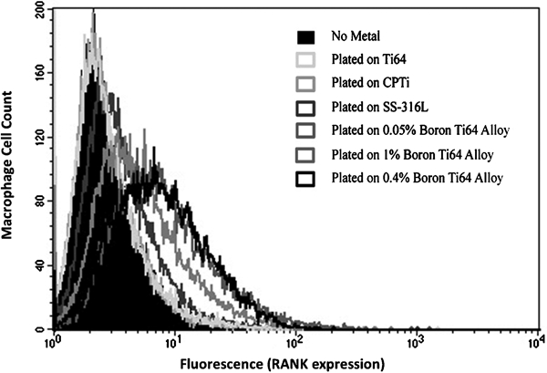
RANK induction after surface alloy exposure. Human HL-60 cells were differentiated into macrophages. Macrophages were cultured on stainless steel or various titanium alloys. After 72 h, cells were analysed by flow cytometry for RANK expression on their cell surface. Cells grown on no metal, Ti-64 and commercially pure titanium (CP Ti) expressed no RANK. Stainless steel 316L (SS) induced low levels of RANK expression. Exposure of macrophages to titanium–boron alloys induced greater amount of RANK
Although macrophages upregulated the expression of surface RANK, it was not known whether cells expressing the ligand necessary to activate RANK were induced by the alloys to express RANKL. Fibroblasts have been known to express the ligand that is complementary to RANK. When the two cell types come into contact, fibroblasts may activate macrophages to progress through differentiation into osteoclasts.22 Therefore, fibroblasts cultured on various metal alloys were tested for induction of RANKL on their surface.
It was observed that when fibroblasts were exposed to traditional metals (stainless steel, CP Ti and Ti–6Al–4V), there was some RANKL induction on the fibroblast surface (Fig. 8). However, the incorporation of boron induced RANKL expression at much higher levels. Taken together with the RANK expression observations, this suggests that macrophages and fibroblasts exposed to the novel titanium–boron alloys may induce higher levels of osteoclast activity.
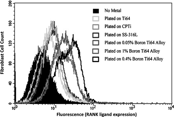
RANKL induction on fibroblasts after alloy exposure. KHOS fibroblast cells were cultured on various titanium alloys, commercially pure titanium (CP Ti) and stainless steel. After 72 h, cells were analysed by flow cytometry for RANKL expression on cell surface. Cells not cultured on metal showed little to no RANKL expression. Exposure to Ti–6Al–4V–B alloys induced most RANKL
Conclusions
The corrosion behaviour of a series of Ti–6Al–4V alloys containing boron in the composition range of <0·001-1·09B was determined in PBS and 0·9 wt-%NaCl solutions. The PBS solution was maintained at test conditions that closely paralleled the ASTM F2129 standard for simulated physiological environments. The tests conducted in 0·9 wt-%NaCl solution were subjected to an aggressive CPP scan from −1 to 8 V to differentiate alloy behaviour.
No pitting was observed in any of the alloys tested under ASTM F2129 specified conditions. For the −1 to 8 V CPP tests, the behaviour of the boron containing alloys can be divided into two distinct categories: alloys with compositions from <0·001 to 0·04B and those in the 0·43-1·09B range. The pitting potentials generally decreased with increasing boron content. The exception to this trend was the 0·01B alloys, which did not pit. The repassivation potentials generally increased with increasing boron content. The absence of pitting in the 0·01B alloy, a composition lower than the limit of solid solubility of B in Ti–6Al–4V, suggests that the presence of boron as a solute in the Ti–6Al–4V lattice may be responsible for making the alloy more noble. Further study is warranted in this area and is continuing.
Owing to the potential of prosthetic loosening caused by biometal induced bone erosion, macrophage and fibroblast cells were tested for RANK/RANKL activation when cultured on boron containing Ti–6Al–4V alloys. The results demonstrated that when cells were allowed to culture on alloy discs for 72 h, macrophage RANK and fibroblast RANK ligand were induced more by the boron containing alloys than by the traditional alloys, commercially pure titanium, 316L stainless steel and Ti–6Al–4V. This lends support to the hypothesis that boron containing alloys may facilitate the differentiation of osteoclasts derived from macrophages that interact with the prosthetic surface. However, it is yet to be determined whether the upregulation of RANK or RANK ligand is sustainable over longer periods of time beyond 72 h, which may be required to gain enough osteoclasts necessary to achieve aseptic loosening. Additionally, cytokine secretion from local inflammation is also necessary to induce osteoclast formation23 along with RANK/RANK ligand interaction. Therefore, more comprehensive studies are required to understand the impact of the new alloys on the microenvironment surrounding the implant.
Footnotes
Acknowledgements
We gratefully acknowledge the financial support from the LA Section of NACE International, Western States Corrosion Seminar, Western Area of NACE International, the NACE Foundation and Ms. S. Hall. The scanning electron micrographs used in this article were generated at the Center for Electron Microscopy and Microanalysis, University of Southern California (J. Curulli). We would also like to thank U. Ekerman (Cal Poly Pomona), B. Ulgut (Gamry Instruments) and B. Eggers (Bio-Logic). This work was supported, in part, by a Research, Scholarship, and Creative Activity (RSCA) award from the California State Polytechnic University, Pomona, to S. Alas.