
Abstract
Microbiologically influenced corrosion (MIC) is a corrosion phenomenon that is destructive to many industries. This research aims to investigate on the MIC behaviour of a 304 stainless steel (SS) substrate in a nutrient rich simulated seawater inoculated with Pseudomonas aeruginosa bacteria. Atomic force microscopy, scanning electron microscopy and energy dispersive spectroscopy (EDS) techniques were used to analyse MIC behaviour of 304 SS. Atomic force microscopy was used to observe the degree of pitting corrosion on 304 SS due to the presence of P. aeruginosa bacteria. Scanning electron microscopy and EDS were used to analyse the biofilm layer formed on 304 SS. The considerable feature was the severe pitting corrosion of 304 SS due to presence of P. aeruginosa in biofilm state.
Introduction
Microbiologically influenced corrosion (MIC) is a form of electrochemical corrosion that can be observed in many environments. It is initiated, facilitated or accelerated by the presence of micro-organisms such as bacteria and mostly appears in the form of localised pits and crevices on metal surfaces.1 – 4 It has been found that MIC is extremely harmful to engineering materials. Many industries, including power generation, water cooling system, pulp and petrochemical industries, could be damaged by this type of corrosion. Microbiologically influenced corrosion is believed to account for 20% of the damage caused by corrosion.5
The formation of a differential aeration cell is one of the most common mechanisms of MIC in the presence of Pseudomonas aeruginosa bacteria. In this mechanism, anodic and cathodic reactions occur on the metal surface because of the presence of a biofilm layer. The anodes are located underneath the biofilm where the oxygen concentration is low, and the cathode is placed at the metal surface, which has an enriched oxygen concentration.6,7
One group of metals that is less resistant to MIC is active–passive metals, such as aluminium and stainless steel (SS). These metals are mostly used in industries because of their good mechanical and anticorrosive properties. However, their common limitation is that they are not immune to MIC.8,9
Among the active–passive metals, SS has excellent anticorrosive performance because of the chromium oxide layer that forms naturally on its surface. Nevertheless, SS is more susceptible to MIC, and pitting is a major damage mechanism that affects the integrity of this alloy in industrial applications.10,11 Generally, SS is susceptible to MIC as shown by their interaction in media containing sulphate reducing bacteria, iron reducing bacteria, etc.3,12 These bacteria cause pitting corrosion on the steel surface through biofilm formation and further colonisation. P. aeruginosa, which is dominant in marine environments, is among the aerobic bacteria and forms a biofilm layer on metal surface. In addition, under aerobic conditions, biofilm layer usually leads to the production of differential aeration and concentration cells. The generation of these concentration cells is detrimental to the integrity of the passive film and enhances the susceptibility of SS to corrosion. Biofilm layer could lead to thinning the passive chromium oxide layer on the SS surface. This could help corrosive ions such as chloride ions in seawater environments to further damage the passive chromium oxide layer and induce pitting.13 – 15 Microbiologically influenced corrosion behaviour of SS in the presence of an anaerobic bacteria such as sulphate reducing bacteria has always been of an interesting research area,16 – 25 and relatively fewer studies were performed to investigate MIC behaviour of SS in the presence of aerobic bacteria.3,15,26 – 28 This study aims to gain better understanding on MIC behaviour of 304 SSs in the presence of P. aeruginosa.
Materials and methods
Materials
Standard 304 SS substrate, with chemical composition of 71·61Fe–8·53Ni–17·67Cr–0·05Cu–1·57Mn–0·033C–0·047N–0·037P–0·007S–0·06M–0·39Si (wt-%), was used as a test material. Samples of 304 SSs with a diameter of 20 mm and a thickness of 5 mm were prepared according to ASTM G1-72. The samples were ground with a series of SiC grit papers (180, 600, 1000 and 2400) to remove the scratches from the surface. The samples were polished using 0·5 μm alumina powders to obtain a mirror finished surface. The polished samples were rinsed with deionised water and degreased with acetone. Finally, the prepared samples were exposed to the control and P. aeruginosa inoculated nutrient rich simulated seawater (NRSS) medium to compare their respective corrosion behaviour.
Preparation of NRSS medium
All of the tests were carried out in NRSS medium (Table 1).
Chemical composition of NRSS powder per litre of NRSS medium
Culturing of P. aeruginosa bacteria
P. aeruginosa was prepared and cultured on agar plates with the following steps:
P. aeruginosa bacteria+100 mL NRSS medium (batch 1)
↓
10 mL NRSS from batch 1+90 mL NRSS medium (batch 2)
↓
10 mL NRSS from batch 2+90 mL NRSS medium (batch 3)
↓
0·6 mL NRSS from batch 3+299·4 mL NRSS medium (prepared batch for immersion test)
To prepare each batch, the mixture was shaken well in a shaker for 24 h with a speed of 150 rev min−1 at 30°C. The bacterial cell concentration was estimated from the optical density (OD) values. Based on the standard calibration, an OD of 1·0 is equivalent to ∼109 cells mL−1. When the OD value was close to 1·0, the prepared specimens hung on Nylon strings were aseptically introduced into the inoculated medium.
Immersion test
For the immersion tests, the 304 SS samples were immersed into two types of NRSS medium: (i) biotic medium and (ii) abiotic (control) medium. All the samples were kept at room temperature under stagnant conditions. For the NRSS medium inoculated with P. aeruginosa, 75% (225 mL) of the NRSS medium were drained out and replaced with an equal amount of fresh NRSS medium every week. This was carried out to keep the bacteria density near the steady state growth phase throughout the experiment.
Atomic force microscopy (AFM) study
An AFM was used to capture images of pits on the sample surfaces. Different areas were randomly chosen on the sample surfaces that were representative of the pitting growth in the presence and absence of P. aeruginosa.
Scanning electron microscopy and energy dispersive spectrometry (EDS) analysis
Scanning electron microscopy was used to observe the biofilm layer formed on the SS substrate, and EDS was used for elemental analysis of the biofilm and corrosion products formed on the sample surfaces.
Results and discussion
Scanning electron microscopy EDS analysis of coupons in presence and absence of bacteria
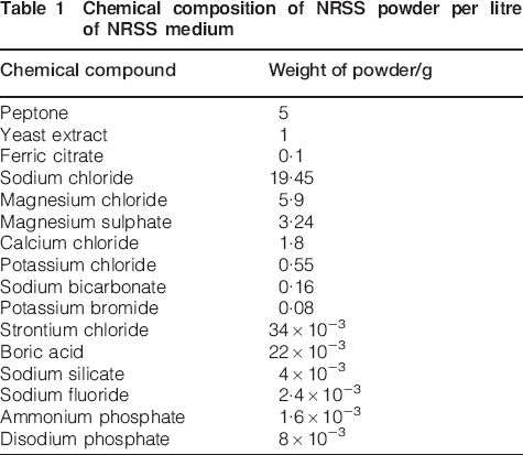
Figure 1 shows the scanning electron microscopy images of 304 SS surface exposed to the control NRSS medium. There is no biofilm formation on the surface because of the lack of bacteria in the control NRSS medium. However, there are some pits on the steel surface formed by chloride ions. The chloride ions come from the NaCl in the NRSS medium and attack at weak sites (grain boundaries, inclusions) on the steel surface. They eventually lead to the breakdown of the oxide film, thereby causing pitting corrosion. Breakdown of the oxide film results from the chemical interaction of chloride ions (Cl–) with the oxide film.29 Reactions (1)–(4) describe the reaction of steel with Cl− ions13


a image (SEM) and b EDAX spectra of 304 SS samples in NRSS medium without P. aeruginosa after 21 days immersion; micropitting on surface is observed
The interaction of Cl− ions with the hydroxide layer leads to the formation of a soluble FeCl3 product, and FeCl3 is further hydrolysed to produce a very porous Fe(OH)3.19 The Fe(OH)3 product is not stable and could not protect the 304 SS against corrosion.
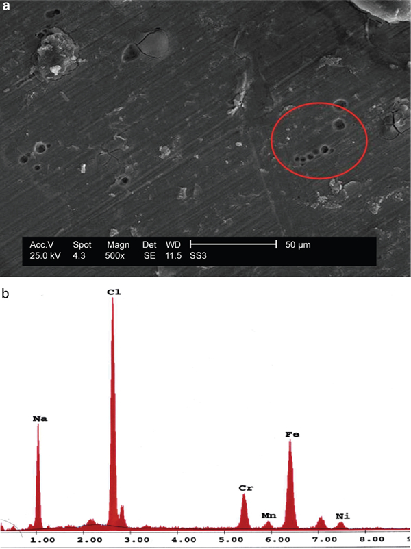
A morphological study of coupons immersed in the NRSS medium in the presence of bacteria is shown in Fig. 2. Scanning electron microscopy images confirmed the presence of P. aeruginosa on the steel surface and their extracellular polymeric substance.29
a image (SEM) and b EDX spectra of representative pits after removal of biofilms on 304 SS sample surface after 21 days of exposure in P. aeruginosa inoculated NRSS media
Previous studies,6,27,30 have described the mechanism of biofilm formation by P. aeruginosa. The bacterial cells preferentially attached themselves to the surface to form patchy or blotchy biofilms. The biofilm formed on the steels becomes larger, thicker and more heterogeneous with exposure time. Patchiness and the heterogeneous nature of the biofilm generate a condition on the steel surface promoting local differences in pH, corrosion products and dissolved oxygen (i.e. differential aeration cells). Active electrochemical corrosion cells could be created, thus resulting in the deterioration of the steel substratum underneath the biofilm in the form of pitting corrosion.
There are some prerequisites for the initiation of pitting corrosion by Cl− ions, including (i) the presence of Cl− ion in the system, (ii) the potential differences on the metal surface and (iii) the reaction temperature must exceed a critical temperature. The role of P. aeruginosa is to create a potential difference between the areas under the biofilm (anode) and the area on the metal surface (cathode). Thus, the synergistic effect of active Cl− ions and the colonised P. aeruginosa can cause the partial loss of passivity of the 304 SS samples and the initiation of pitting corrosion.27,30 The EDS results show that the oxygen content is more on biotic 304 SS than abiotic one. This indicates the aggressive role of P. aeruginosa bacteria to further oxidise the substrate, thus leading to severe pitting corrosion.
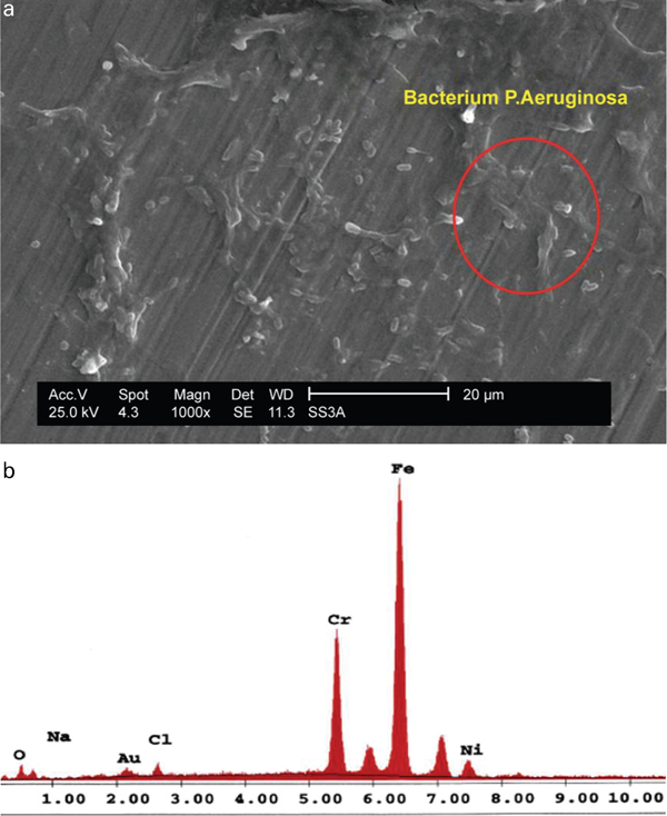
Topography study of pitting corrosion of 304 SSs
The two-dimensional and the corresponding three-dimensional renditions are shown together with the sectional analysis in every group of AFM images. Figure 3 shows the AFM images of 304 SS samples in NRSS with and without P. aeruginosa. As shown in Fig. 3a, there are some pits on the surface of the control sample (without the bacteria). The pits are initiated by aggressive chloride ions coming from NaCl salts, which they attack at weak sites on the steel surface, causing pitting corrosion. However, as shown in Fig. 3b, the more severe pits appeared when the samples were immersed in NRSS medium containing bacteria. The synergistic effect of Cl− ions and the colonised P. aeruginosa cause the partial loss of passivity of the 304 SS coupons and the initiation of deeper pitting corrosion.
Images (AFM) of 304 SS samples after 14 days of exposure to a sterile NRSS medium and b P. aeruginosa inoculated NRSS media
Figure 4 shows the AFM images of 304 SS samples in the NRSS medium containing bacteria at different exposure times.
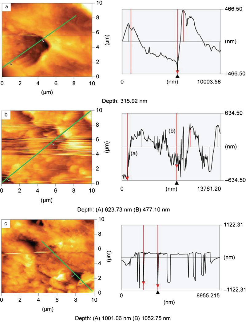
Images (AFM) of 304 SS samples after a 14, b 35 and c 49 days of exposure in P. aeruginosa inoculated NRSS medium
The bacteria tend to form a thicker and more heterogeneous biofilm on the sample surface with time. Thus, the anodic and cathodic areas increase in number, consequently leading to the creation of additional active electrochemical cells. Eventually, they were able to create severe pits on the sample surface. Thus, the AFM results confirmed the aggressive role of bacteria in promoting the corrosion process on the steel surface and causing more severe pits.
Conclusions
The following conclusions can be drawn from the research work.
The scanning electron microscopy and AFM results show that pitting corrosion is more severe on 304 SS surfaces in the presence of P. aeruginosa. The combined effect of active chloride ions and the colonised P. aeruginosa causes severe pitting corrosion.
Biofilms generate a condition on the metal surface that creates differential aeration cells. The area under the biofilm, which lacks oxygen, becomes an anodic area, and the steel surface that is enriched in oxygen becomes a cathodic area. Thus, electrochemical cells are created, resulting in pitting corrosion.
Biofilm can help concentrate chloride ions, and this can lead to the breakdown of the passive oxide film and thus increase the likelihood of corrosion.