
Abstract
Coronary heart diseases result from the blockage of one of the coronary arteries, which nourishes the heart muscle. This process leads to ischaemia of a segment of the heart and death of the contractile tissue. As cardiac tissue is unable to regenerate itself, heart function is impaired. Tissue engineering (TE) is a field of science that integrates knowledge from biology, materials sciences, engineering and medicine to develop artificial, functional tissue constructs to replace defected tissues. In cardiac TE, contracting cells are seeded within supporting biomaterial scaffolds that provide them with the essential microenvironment for functional tissue assembly. Various strategies and methods for fabricating these scaffolds have been proposed and tested in the last decade, some of which combine multiple elements that altogether contribute to the formation of an improved functional tissue. This review summarises the unique properties of various composite biomaterial scaffolds and highlights their advantages over other pristine scaffolds for engineering functional three-dimensional cardiac patches.
Introduction
The heart is the strongest muscle in the human body. Starting from the very onset of foetal development, the heart beats 24/7, pumping blood that nourishes the body's cells and removes waste products. The human heart beats approximately 70 times per minute, which adds up to more than 100 000 times a day. Considering the absolute reliance of the cells in the body on a continuous supply of oxygen, any severe interference with the physiological function of the heart might be lethal.
Cardiovascular diseases (CVD) are the main cause of death globally, with more than 17 million casualties per year. 1 Coronary heart diseases, which are responsible for a large propo rtion of deaths caused from CVD, 2 result from a partial or full blockage of one of the coronary arteries, which supplies blood to the heart muscle. This process leads to ischaemia of a segment of the heart and eventually causes the death of contractile tissue (myocardial infarction, MI) and scar formation. As adult cardiomyocytes cannot proliferate, and since cardiac stem cells are scarce, the cardiac tissue is unable to regenerate itself, resulting in a chronic cardiac dysfunction. Complications following an initial MI include heart failure, recurrent ischaemia and arrhythmias; together, they manifest a 5-year mortality rate of near 50%. 3 Unfortunately, at present, cardiac transplantation is the only cure for end-stage patients. As cardiac donors are scarce, there is an urgent need to develop alternative strategies for heart regeneration.
Tissue engineering (TE) is a field of science that integrates knowledge from biology, materials sciences, engineering and medicine to develop artificial, functional tissue constructs to replace or support defected tissues (Fig. 1). In the context of coronary heart diseases, heart patches, either populated with contracting cells, or a- cellular, may be implanted to patients that undergo MI. By replacing the scarred, non-functional infarcted area of the heart with functional, biocompatible contracting tissue or a supporting material, heart function could be restored (for recent reviews on cardiac TE, see Ref. 4–7). The concept of tissue engineering for regenerative purposes. First cells are isolated from the patient (A) whereupon they can be cultivated and expanded by standard tissue culture methods (B). The cells can then be seeded into the appropriate composite porous scaffold and supplemented with growth factors, small molecules, and/or nanoparticles (C). In order to provide the optimal conditions for the cells to organise into a functioning tissue, these constructs are then further cultivated in bioreactors providing essential conditions (D). After successfully engineering the tissue, it can be transplanted or instead of the defected tissue to restore function (E) (Reproduced with permission from Ref. 8)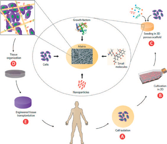
One of the major pillars of TE is realising the importance of the extracellular matrix (ECM) for functional tissue assembly. In the living body, the cells are usually embedded in a three-dimensional (3D) matrix that is primarily made of secreted fibrous proteins. Aside from being an anchoring substrate, the ECM provides physical, chemical and mechanical cues that affect and regulate cell behavior. 9,10 Acknowledging the pivotal contribution of the ECM to tissue organisation and function, researchers began to engineer biomimetic substances that recapitulate the composition and architecture of the natural ECM. Porous, ECM-like synthetic scaffolds were fabricated by various methods, such as self-assembly, phase-separation, freeze drying, particulate leaching, gas foaming, electrospinning and additive manufacturing. The matrices were successfully tested for their biocompatibility, biodegradability, capacity to support cell culture and ability to induce differentiation of stem cells towards a desired fate. 7,8,11 – 14 Nonetheless, it soon became clear that further improvements in cell viability, tissue morphogenesis and more importantly tissue function could be achieved by the generation of ‘composite scaffolds’. That is to say, scaffolds that integrate multiple elements altogether contribute to the formation of a functional tissue. In many cases, the composite is designed to provide the cells with an atmosphere that mimics their natural microenvironment.
Properties of the reviewed composite scaffolds
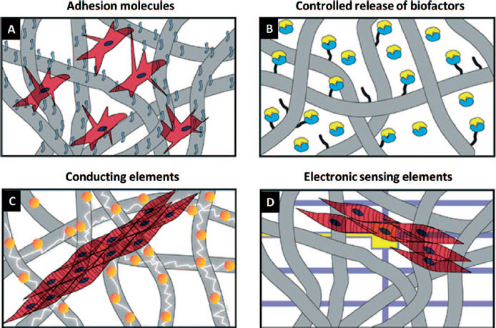
Schematic representations of different approaches for fabricating composite scaffolds in cardiac tissue engineering. a Cardiac cells seeded onto a scaffold covered with adhesion molecules (for example: fibronectin, RGD peptides, etc.). Adhesion molecules are represented with blue wavy lines. b A scaffold with attached biofactors to control tissue maturation and function (for example: vascular endothelial growth factor (VEGF), angiopoietin-1 (Ang1), basic fibroblast growth factor (bFGF), etc.). Biofactors are represented by blue/yellow complexes. c Gold nanoparticle-decorated scaffold improves the assembly and function of seeded cardiac cells. d Cardiac cells seeded onto a scaffold with integrated sensing elements. Blue perpendicular lines represent the supporting material for the sensing electrodes (in yellow) that are able to report the activity of the engineered tissue
The native heart ECM
The ECM is an acellular, composite material that consists of secreted fibrous proteins (including collagens and elastin), water, glycoproteins (including fibronectin and laminin), proteoglycans and other soluble molecules. The quantity, structure and composition of the ECM are tissue-specific and are constantly changing through enzymatic and non-enzymatic modifications.
61
Collagens are the main protein component of the ECM. To date, 28 different collagen types have been discovered. Each collagen type is characterised by a unique combination of three distinct polypeptides, known as a chains, which are modified intracellularly and assembled into a triple helix. The homotrimeric or heterotrimeric helix, named ‘procollagen’, is then secreted (mainly by fibroblasts) into the extracellular space and converted into collagen by proteases that cleave its N and C termini. The mature collagen molecules are then self-assembled into fibrils, and stabilising covalent cross-links are introduced into the supramolecular assembly.
61
The collagen network of the heart is composed mainly of type I and type III collagens, arranged in three distinct organisational levels: 1. Epimysial fibres, which have a relatively large diameter of several micrometres, are woven into tight bundles surrounding the myocardium. 2. Perimysial fibres, which range from <1 μm to several micrometres in diameter, are associated with groups of cells, and are responsible for the contractions of cell bundles. 3. Endomysial fibres, which measure at a few tens to several hundred nanometres in diameter, wrap individual cardiomyocytes to provide mechanical support and ensure proper alignment (Fig. 3). The overall fibrous network of the cardiac ECM induces and maintains a tightly packed, aligned and elongated tissue structure, which promotes fast propagation of action potentials along the cells and strong synchronous contractions.
62,63
While mechanically supporting and providing a hydrated elastic fill around the cells, the ECM also interacts with cells through adhesive glycoproteins, such as fibronectin and laminin. Most glycoproteins bind cells through integrins and other cell-surface receptors, eliciting signal transduction that mediates cellular responses, such as differentiation, survival, proliferation and migration.
64
The cardiac microenvironment. Upper panel: schematic representations. a The structure ofthe heart extracellular matrix (ECM). The epimysial fibres that surround the myocardium are represented by dashed black stripes. The perimysial fibres that are associated with groups of cells are represented by thick blue lines. The endomysial fibres that wrap individual cells are represented by thin green lines. Blood vessels are coloured in red (reproduced with permission from Ref. 62). b. Extracellular matrix wrapping an individual cardiac cell bundle. Cell nuclei are represented in dark blue and cardiac muscle striations are represented by dashed red lines. Lower panel : SEM micrographs of the three distinct fibre populations of the heartmuscle. cendomysial fibres (indicated by white arrows). d perimysial fibres. e epimysial fibres (c-e reproduced with permission from Ref. 65)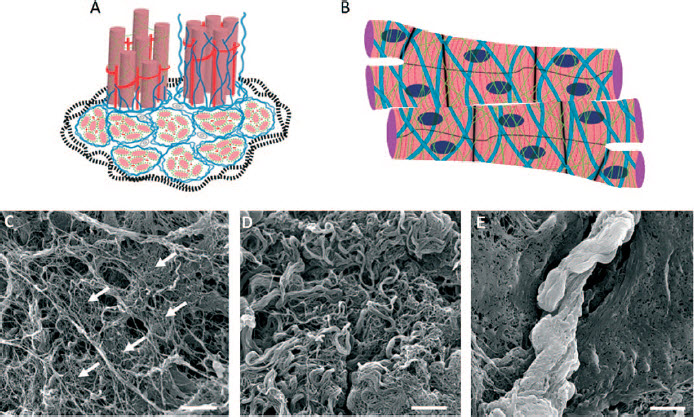
The proteoglycans, which are composed of glycosa- minoglycan (GAG) chains bound to a protein core, form a gel-like medium in which the fibrous proteins of the ECM are embedded. An increasing number of growth factors (GFs) have been found to associate with GAGs and proteins that compose the ECM. The GFs can thus be bound and stored in the ECM, protected from proteolysis and ready to be released following enzymatic activity or fluctuations in the local pH. On the other hand, GFs can also function in their ECM-bound state, by being ‘presented’ on the GAGs to their corresponding cell-receptors. 9,10,66
Collectively, the biochemical composition and mechanical properties of the ECM, its specific topography and associated signalling molecules work in ensemble to guide and maintain the formation, morphology, homo- eostasis and proper function of the supported tissue. Therefore, fabrication of scaffolds that mimic this natural cell microenvironment may greatly enhance the functionality of the resulting engineered patch.
Composite synthetic/natural scaffolds for mimicking the natural cell microenvironment
Materials for scaffold fabrication in TE may be classified into two main categories: synthetic materials and substances that are synthesised by living organisms. 67 Synthetic polymers are very attractive candidates for TE since they are readily available, relatively inexpensive, easily controlled in terms of their mechanical and chemical properties, can be non-toxic and non-immu- nogenic and may be designed for degradation under predefined conditions. Indeed, the use of synthetic polymers for scaffold fabrication has been explored since the late 80s of the last century, and the list of FDA-licenced biocompatible and biodegradable synthetic materials suitable for TE is continuously growing. The main downsides of these substances are that they lack the biological cues inherent in the natural ECM, and are thus commonly considered inferior to natural materials in terms of promoting cell binding, growth, differentiation, survival and tissue organisation. 68 In contrast, natural polymers, such as polysaccharides and ECM- derived polypeptides are considered more ‘cell friendly’ as they better imitate the characteristics of the natural ECM, and can provide cells with anchoring sites and signals that are essential for the formation of functional tissue. However, low availability, relatively high prices, batch to batch variability, poor physical and chemical stability, inadequate mechanical properties, risk of immunogenicity and limited processability are among the factors that restrict their use as standalone-com- ponents in fabrication of scaffolds for TE. 69,70 Ideally, one should aspire to generate a scaffold that is characterised by all the essential properties, whose combination will enable the successful fabrication of a matrix that faithfully imitates the natural cell-microenvironment of the tissue of interest.
Unfortunately, there is no single synthetic or natural substrate currently known that will answer all these requirements. Therefore, a combination of synthetic and natural substances is required in order to produce a composite with optimal qualities.
In this section of the article, we will focus on composite scaffolds that consist of both synthetic and natural components. The constituents may be pre-com- bined to yield a new starting material for the fabrication process, or alternatively, sequentially applied. Either way, the properties of the composite are expected to be superior to those of each individual component in terms of structural strength, biocompatibility and competence to support tissue formation.
Composite scaffolds with improved cell-binding capacity
The ability to promote cell attachment is a very basic and vital quality of any scaffold designated to be seeded with living, adherent cells. In the living body, cells interact with the ECM through distinct adhesion proteins, such as collagen, fibronectin and laminin.
71,72
Unfortunately, synthetic and natural bioengineered scaffolds that do not contain ECM components, lack the ability to promote cell adherence. Thus, several research groups have combined such scaffolds with ECM or ECM-like factors or motifs. For instance, Park et al. have used the synthetic biodegradable polymers poly(- lactic-co-glycolic acid) (PLGA) and poly( Scaffolds fabricated by the gas foaming process. A polymer is saturated with an inert gas by exposing to high gas pressure. Subsequent pressure reduction results in nucleation and expansion of gas bubbles within the polymer matrix, generating a porous structure a. Alternatively, ammonium bicarbonate particles are first mixed with a polymer solution. The solvent is then evaporated, and the composite is immersed in warm water or acidic aqueous solution. During particle dissolution, ammonia and carbon dioxide gases are released, generating gas bubbles, which turn the polymer into a porous structure b.
11–14
Using the gas foaming process, scaffolds made of biodegradable substances can be fabricated and used for tissue engineering applications. c SEM micrographs of the surface or cross-section of porous poly(L-lactic acid) [PLLA] scaffolds fabricated using the gas foaming method. Scale bar, 1 mm (reproduced with permission from Ref. 73)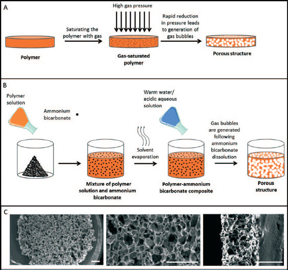
The mixture was processed by electrospinning (Fig. 5) into homogenous fibres with an average diameter of 380 nm. Upon hydration, the fibres swelled two- to threefold, and the scaffold turned into a stable fibre-laden hydrogel. The fibrous mats have been found to synergistically combine the beneficial characteristics of each individual component, namely: stability in aqueous environments even in the absence of cross-linking, mechanical strength and elasticity (from the PLGA), together with presentation of natural cell-adhesion ligands (from the elastin and gelatin). In vitro cell assays showed that after seeding, cardiac myoblasts and bone marrow stromal cells have adhered, migrated into the scaffold, grew in multilayers and displayed a spread morphology.
16
Scaffolds fabricated by the electrospinning technique. A polymer solution or melt is pumped through a capillary (such as a syringe needle) to which a high positive or negative electric potential is applied. As a result, the charge repulsion that is produced within the polymer solution overcomes the surface tension which held the drop at the tip of the capillary. A jet of polymer solution is ejected, while the solvent starts evaporating, forming fibres that travel towards and deposited on a grounded collector. A static, flat plate a or a rotating drum b are used to collect randomly oriented or aligned fibres, respectively.
11–14
When made from biocompatible and biodegradable substances, electrospun meshes could be utilised as scaffolds for tissue engineering applications. Bottom: SEM micrographs of randomly oriented c and aligned d electrospun albumin fibres (c and d reproduced with permission from Ref. 74)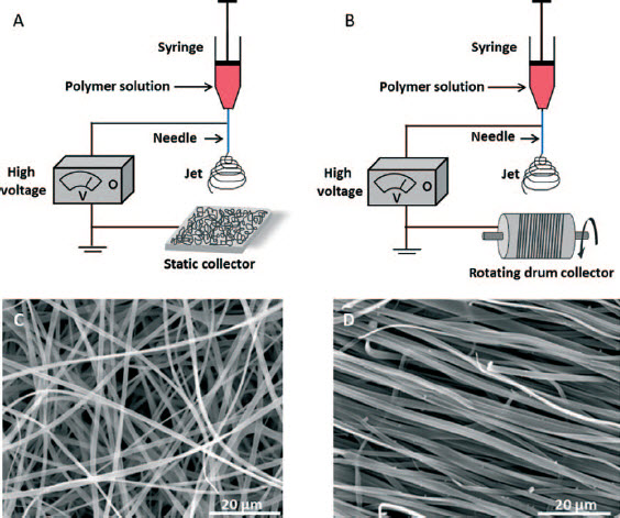
As discussed, cells interact with the ECM through adhesion proteins. More specifically, the interaction is mediated by distinct cell-adhesion motifs like RGD (in collagen, fibronectin and laminin), YIGSR (in laminin) and LDV (in fibronectin) that bind to cell-surface integrins or other receptors.
71,72
Engraftment of short peptides that contain such motifs may substitute the integration of full-length proteins, which may complicate the fabrication process or adversely affect the compatibility of the scaffold to its designated application. For example, Sapir et al. have decorated a scaffold made of alginate, an ionically cross-linkable, biocompatible polysaccharide isolated from brown algae, with two peptides. The first, G4RGDY contains the integrin-binding motif RGD. The second, G4SPP- RRARVTY is a heparin-binding peptide found in many ECM proteins, GFs and cytokines. This peptide has been shown to bind cell surfaces via syndecans, transmembrane heparin sulphate proteoglycans.
75,76
The peptides were covalently attached to alginate chains via carbodiimide chemistry, and the modified polysaccharide has been used to generate a matrix with a highly porous, well-interconnected pore structure. In comparison to unmodified alginate scaffolds or alginate scaffolds modified with scrambled peptides, cardiac cells that were seeded on the composites showed a significant improvement in attachment, spreading, viability, cyto- skeletal organisation, myocyte specific protein expression and tissue organisation.
17
Another example of using cell-adhesion motifs for improving the cell-binding capacity of synthetic scaffolds was introduced by Rai et al.
18
The group developed cardiac patches consisting of poly(glycerol sebacate) (PGS), a biocompatible elastomer with a Young's Modulus of 0.282 + 0.025 MPa and an elongation of 267 + 59.4%. Poly(glycerol seba- cate) has been widely used in TE of soft tissues, such as arteries, veins, hearts and neuronal networks.
77,78
By using the salt leaching method (Fig. 6), the authors fabricated porous PGS films that were sequentially treated with alkaline hydrolysis and acidification for incorporation of carboxylic groups on the polymer surface. Bioactive peptides harbouring the cell-adhesion motifs YIGSR and RGD of laminin and fibronectin, respectively, were then covalently bonded to the scaffold by carbodiimide chemistry. In vitro experiments showed that when cardiac progenitor cells were seeded within the functionalised matrix, adhesion, survival and growth of cardiac progenitor cells were improved when compared to the pristine PGS films.
18
Scaffolds fabricated by particulate leaching. a The method involves mixing of a polymer solution with water-soluble salt particles (porogen). The solvent is then evaporated and the polymer-porogen composite is immersed in water to leach out the salt, leaving a porous polymer structure.
11–14
b. SEM micrographs of biocompatible porous poly(e-caprolactone) (PCL) scaffolds fabricated by particulate leaching (reproduced with permission from Ref. 79)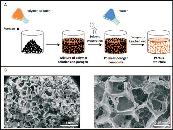
In addition to the requirement of supporting cell binding, the scaffold should also provide the patch with the appropriate physical qualities that are required for proper organisation and function of the engineered tissue.
Composite scaffolds with improved mechanical properties
Designated to support the growth and function of a contracting tissue, and to withstand implantation on the constantly beating myocardium, the mechanical properties of the scaffold should be carefully considered. Another advantage that can be obtained from using the combination of synthetic and natural substances is the ability to tune the mechanical properties of the fabricated matrix. Ideally, the latter should resemble those of the natural myocardium. For example, Kai et al. used a mixture of gelatin and the synthetic polymer poly(ε - caprolactone) (PCL), for electrospinning of fibres in random or aligned orientations. The resulting ECM-like meshes had small fibre diameters of around 250 nm with lower stiffness and better hydrophilicity when compared to electrospun PCL fibres. When fabricated in an aligned orientation, the gelatin-PCL nanofibrous scaffolds showed anisotropic mechanical properties that closely resemble those of the native myocardium. Importantly, the composite material improved cardiomyocyte attachment and induced a higher degree of cell alignment when compared to PCL scaffolds. 19 Another elegant way of generating a composite gelatinous scaffold that imitates the mechanical properties of the myocardium was presented by Kharaziha et al. and Ravichan- dran et al. The two groups have used different strategies to improve the mechanical properties of the rapidly degrading gelatin by combining it with PGS. In the study by Kharaziha and colleagues, gelatin and PGS were blended and electrospun in different ratios, and the resulting fibres were then cross-linked in order to stabilise the structure of the water-soluble gelatin. It was found that incorporation of PGS reduced the stiffness of the matrix, mimicking the elasticity of the myocardium. Moreover, when electrospun to form aligned architectures, the composite scaffold was shown to significantly improve the alignment, organisation and contractile function of seeded cardiomyocytes, in comparison to fibrous matrices that were composed of crosslinked gelatin. 20 An alternative strategy was adopted by Ravichandran et al. who generated a synthetic-natural hybrid scaffold by fabrication of PGS/gelatin core/shell electrospun fibres with an average diameter of 1 μm. In this case, the PGS was used as a core substance to mechanically support the ‘cell friendly’ gelatin shell, and provided the matrix with improved elasticity and ability to reversibly deform. Following stabilisation of the fibres by cross-linking with glutaraldehyde, a scaffold with mechanical properties that can resist the heart wall pressure was obtained. The authors reported that the composite matrix supported the adhesion of cardio- myocyte-mesenchymal stem cells (MSCs) coculture, which survived, migrated into the scaffold and expressed high levels of the cardiomyocyte-specific proteins tro- ponin-T and α-actinin. 21 In later studies, the group showed that the elastic PGS core can also be coated with other natural proteins that promote cell-biomaterial interactions, such as fibrinogen and collagen. 22,23 Either way, the combination of the synthetic polymer PGS with natural substances has resulted in scaffolds that are superior in their biological and mechanical properties when compared to any of their constituents individually.
Composite scaffolds that resemble the architecture of the natural cardiac ECM
The different chemical and physical qualities of natural and synthetic substances can also be utilised for the generation of complex structures, which resemble the structural hierarchy of the native myocardial tissue. For example, by co-electrospinning of PLGA and fibrin, a multi-scale scaffold composed of micrometric and nanometric fibres, respectively, could be obtained. In addition to the recapitulation of the cardiac ECM architecture, the tensile properties of the electrospun composite scaffold matched those of the human myocardium. The co-electrospinning of the micro and nanofibres also increased the pore size of the substrate and facilitated the infiltration of seeded MSCs, supporting their survival and proliferation. Interestingly, the hybrid scaffold was also found to provide a favourable microenvironment for the differentiation of 5-azacytidine-treated MSCs into a cardiac phenotype. 24
A unique approach for attaining patches that closely resemble the mechanical and topographical properties of the natural ECM is the fabrication of scaffolds that are based on decellularised native tissue. The latter are then further modified by combining with other synthetic or natural substances that were not derived from the tissue that ‘donated’ the ECM. For the purpose of this review, we refer to the decellularised tissue as a ‘natural’ constituent and the artificially applied modifying substances as the ‘synthetic’ component of the composite. The strategy of scaffold fabrication from decellularised tissue was adopted by Godier-Furnemont and colleagues. The researchers treated thin sheets of human cardiac explants with chemicals that extract the cellular component from the tissue, sparing the unique ultra-structural and mechanical properties of the native cardiac ECM. The decellularised matrices were then seeded with MSCs that have been treated with transforming growth factor beta (TGF-ß) and suspended in fibrin gel, which is known for its vasculogenic properties. It was found that implantation of the gel-matrix composite into infarcted rat model induced vascular network formation at the lesion site. Moreover, a recovery to baseline levels of left ventricular systolic dimensions and contractility was shown. These therapeutic effects were associated with migration of MSCs into the ischaemic myocardium, where their secretion of angiogenic factors further enhanced endogenous cell migration and revascularisa- tion.
25
In this regard, it is also worth noting that decel- lularised tissues can be further processed to generate injectable thermoresponsive ECM-based biomaterials that remain in a solution state at low temperatures and transform into gel upon incubation at 37°C. Cardiac patch fabricated by this approach can be delivered by a minimally invasive operation to the infarcted site.
80,81
Moreover, these biomaterials can be utilised as ‘bioinks’ for additive manufacturing processes, commonly known as ‘3D bioprinting’ (Fig. 7a-c). For instance, by using decellularised cardiac tissue-derived bioink in which myoblasts have been laden, Pati and colleagues have successfully generated 3D structures that supported the viability and functionality of the printed cells (Fig 7d). Moreover, gene expression analysis and immunohisto- logical studies showed that the maturation of myoblasts into myofibers was greater in these structures as compared to constructs made of pure collagen. The authors have also demonstrated the construction of hybrid porous structures in which PCL was fabricated as a supporting framework into which tissue-specific bioinks were deposited (Fig 7
e).
26
Recently, our group developed injectable thermoresponsive hydrogels based on decellularised omental tissue.
82,83
The hydrogels, which retained key features of the complex structure and composition of the ECM, were found to support the culture of encapsulated cardiomyocytes. Importantly, as the omentum can be easily and safely harvested from the patient, the biomaterial can serve as an autologous, non- immunogenic protective vehicle for the transported cells.
27
Our continuing research focuses on the implementation of omentum-based thermoresponsive hydrogels as bioinks for printing complex 3D scaffolds for cardiac TE applications. Engineering tissues or scaffolds by 3D printing. In this method, digital data are converted into physical 3D shapes. The process is executed by a computer-controlled robot that fabricates the object through a series of consecutive layers. A common 3D printing procedure is based on the direct-write technique in which a 3D printer a deposits a filament of material b, layer-by-layer, to yield a 3D structure c
84
(a-c, reproduced with permission from Ref. 85). When using biocompatible materials as ‘bioinks’ for additive manufacturing processes, complex 3D structures that support cell viability, functionality and tissue organisation can be generated. Bottom images show tissue constructs fabricated by 3D printing. A cardiac tissue construct printed with a bioink derived from a decellularised heart d. Adipose tissue construct printed with a decellularised fatty tissue supported by a poly(e-caprolactone) (PCL) framework e. Scale bar, 5 mm (dand e reproduced with permission from Ref. 26)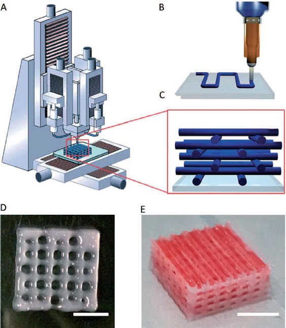
Composite scaffolds for presentation and release of growth factors
Growth factors represent the largest group among the bioactive molecules incorporated within composite scaffolds for TE applications. These are cell-secreted, soluble signalling polypeptide molecules, which bind to specific receptors on target cells and affect behaviours, such as survival, growth, migration and differen- tiation. 86 Certain GFs, commonly known as ‘angiogenic growth factors’, also regulate the development of a functional vascular system that provides the tissue with rapid oxygen, nutrients and waste exchange. 87
Growth factors can be stored in the ECM that protects them from degradation, assists in their presentation and constitutes a depot from which they can diffuse and act on responsive cells in the surrounding tissue. 9,10,66 Therefore, these molecules are considered promising candidates for integration into bioengineered, ECM mimicking scaffolds. Incorporation of GFs can broaden the capacity of the scaffolds to recruit cells, elicit cellular responses and influence tissue fate. Primarily, three main strategies have been used for generation of GF-incor- porating composite scaffolds: 1. immobilisation of GFs to the backbone of the polymeric scaffold; 2. constitutive, sustained release of free GFs that have been entrapped in the scaffold material; 3. triggered, ‘on- demand’ release of GFs from the scaffold.
Composite scaffolds with covalently immobilised GFs
Growth factor signalling can be prolonged when the factor is covalently tethered to the surface of the scaffold. 88 Therefore, Miyagi et al. have developed a collagenous scaffold to which the pro-angiogenic factor, vascular endothelial growth factor (VEGF), was covalently immobilised by carbodiimide chemistry. When the composite scaffold was used to replace a full thickness defect of the right ventricular free wall in a rat model, increased blood vessel density and improved tissue formation were evident. The authors attributed these results to the increase in recruitment, proliferation and survival of bone marrow cells, which, in turn, can also enhance the recruitment of endothelial progenitor cells to the graft. 28 More evidence for the therapeutic potential of composite scaffolds composed of VEGF- conjugated biodegradable materials came from the study of Wu et al. The authors described their development of an injectable, aliphatic polyester hydrogel made of [poly (δ-valerolactone) - block-poly (ethylene glycol)-block- poly (δ-valerolactone) (PVL-b-PEG-b-PVL)]. The temperature sensitive polymer dissolves in water at room temperature but turns into gel at 37°C. For the synthesis of a VEGF-conjugated hydrogel, VEGF was covalently linked to the polymer by carbodiimide activation. When evaluated for its effect on cardiac recovery following myocardial infarction, this composite hydrogel scaffold was found to increase angiogenesis at the infarcted site, and improve myocardial recovery when compared to a simple mixture of hydrogel and free, unconjugated VEGF. 29
Another intriguing strategy for constructing a scaffold that presents an immobilised form of a signalling biomolecule is to incorporate the active moiety into the building blocks of the fabricated matrix. Such an approach, in which the process of molecular selfassembly has been applied (Fig. 8), was presented in an elegant work conducted by Webber et al. The group designed peptide amphiphile (PA) molecules, consisting of a hydrophobic alkyl segment covalently linked to a peptide sequence, which self-assembled into cylindrical nanostructures that present VEGF-mimetic epitopes on their surface (VEGF-PA) (Fig. 9a). When hydrated and mixed with divalent ions, the nanofibers formed an entangled hydrogel. Vascular endothelial growth factormimetic signalling of the VEGF-PA was tested and verified in vitro, leading to VEGF receptor phosphorylation upon stimulation of endothelial cells. Consequently, the treatment led to the enhancement of cell proliferation, survival and migration, commonly associated with VEGF signalling. When tested on a hind- limb ischaemia model of CVD, VEGF-PA induced angiogenesis and significantly enhanced the recovery of the ischaemic tissue in comparison to treatments with saline or mutant PA. Interestingly, the angiogenic and therapeutic effect of VEGP-PA was also significantly higher when compared to that of the bioactive VEGF- peptide component of the amphiphile molecule. The authors have suggested that this observation may result from stabilisation and/or polyvalency of the VEGF- mimetic epitopes when in the context of the cylindrical nanofibers.
30
Scaffolds fabricated by molecular self-assembly. The process involves spontaneous organisation of molecules into stable, structurally defined ordered aggregates. Assembly of structures is mediated by non-covalent molecular forces and influenced by the properties of the molecules, their concentration and other environmental factors, such as temperature, pH, ionic strength and solvent composition.
12,13,89
For instance, peptide amphiphile (PA) molecules consist of a hydrophobic alkyl segment covalently linked to a peptide sequence a. These molecules have been shown to self-assemble into cylindrical nanostructures in acidic aqueous environment, with their alkyl tails packed in the centre of the cylinder, and the peptide segment exposed to the solution. The structures disassemble when pH is brought back to neutral b (a and b reproduced with permission from Ref. 90). When selfassembled structures are generated in aqueous solution, nanofiber hydrogels may form. The latter can be used for presentation and/or sustained release of bioactive molecules, as well as for facilitating cell culture and tissue regeneration.
91,92
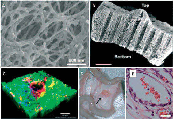
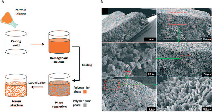
c SEM micrograph of PA nanofiber network (reproduced with permission from Ref. 93) Images from representative studies. a Entangled nanofiber gel networks composed of peptide amphiphiles (reproduced with permission from Ref. 30) b SEM micrograph of a laser-channelled alginate scaffold for promoting vessel formation. Scale bar, 1 mm c. CD31 stained endothelial cells (red) showing vessel formation in a fluorescently labelled alginate scaffold (green). Scale bar, 100 μm (b and c reproduced with permission from Ref. 35). d An alginate based cardiac patch, loaded with pro-survival and angiogenic factors. The patch was stitched to rat omentum to promote prevascularisation e. H&E staining of blood vessels inside the cardiac patch shown in d. The blood vessels are functional and anastomised to the host tissue as reflected by their red blood cell content. Scale bar, 20 μm (d and e reproduced with permission from Ref. 37) Scaffolds fabricated by thermally induced phase separation (TIPS). a The process is based on a reduction in thermal energy that induces the separation of a homogenous polymer solution into polymer-rich and polymer-poor phases. Lyophilisation is then used for solvent extraction, leaving a highly porous polymer structure.
11–14
By using the TIPS method, porous biocompatible and biodegradable structures can be fabricated and used as scaffolds for tissue regeneration. b SEM micrographs of micrometre and sub-micrometre structures of poly(propylene fumarate)-co-poly(L-lactic acid) porous scaffold fabricated using the TIPS method (Reproduced with permission from Ref. 104)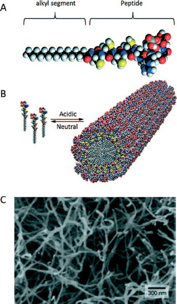
While VEGF promotes the early steps in the vascu- logenesis process, downstream events, such as vessel stabilisation and maturation, have been found to be mediated by angiopoietin-1 (Ang1). 87 By using carbo- diimide chemistry, Chiu et al. covalently immobilised those two factors together onto a porous collagen scaffold. When seeded with endothelial cells, the composite demonstrated improved angiogenic responses in terms of cell proliferation, metabolism and tube formation compared to cultivation on unmodified scaffolds. In addition, results from a chorioallantoic membrane (CAM) assay showed increased endothelial cells and blood vessel infiltration to the construct, also in comparison to scaffolds with independently immobilised VEGF or Ang1. Cell proliferation and metabolism were also improved in comparison to culturing on unmodified scaffolds in the presence of soluble GFs. This observation was interpreted by the authors as related to the increase in local concentration of the GFs on the scaffold, together with improved stability and effects of extended cell signalling that may associate with their covalent immobilisation. 31,32
Composite scaffolds for constitutive, sustained release of GFs
In order to provide a local, sustained supplementation of VEGF to the ischaemic area of the myocardium, Rufaihah et al. developed a hydrogel composed of cross-linked PEG-modified bovine fibrinogen. Vascular endothelial growth factor was loaded into the matrix by pre-mixing with the PEGylated fibrinogen solution before a UV-induced polymerisation step. When tested in vitro, VEGF was gradually and consistently released from the scaffold, inducing a higher degree of endothelial cell proliferation and migration when compared to pristine scaffolds. Upon injection into the infarcted area of model rat hearts, the composite hydrogel allowed a sustained release of the GFs, and induced the establishment of stable mature blood vessels. Together with providing a mechanical support to the damaged myocardium, the implantation of the matrix led to an increase in arterial density in both infarct and peri- infarct areas, reduced infarct size and improved cardiac function. 33
Similarly to the aforementioned Ang1, Platelet-derived growth factor (PDGF) BB is also known to encourage the maturation and stabilisation of newly formed blood vessels, a process that involves proliferation and recruitment of smooth muscle cells to nascent blood vessels. 94–96 It was found that optimal angiogenesis could be achieved when PDGF is sequentially given after VEGF administration. 97 Based on these findings, Hao et al. have generated a unique platform based on the sequential release of the two factors from alginate hydrogels. The different affinities of VEGF and PDGF to alginate resulted in a successive release of the factors from the hydrogel, with a delayed discharge of PDGF. When compared to administration of each factor alone, this dual delivery system promoted the formation of mature vessels, and improved cardiac function in a rat model of myocardial infarction. 34 Another example of incorporation and release of an angiogenic factor from a hydrogel scaffold was described in a study by Zieber and colleagues. The researchers implemented a unique biomimetic strategy, formulating an alginate scaffold with a fraction of alginate sulfate. 35 The rationale behind this composition was that alginate sulphate mimics the sulphated sequences in the natural GAGs heparin and heparin sulphate, that bind, protect and present many GFs and other proteins via specific, high-affinity electrostatic interactions. 98 The scaffold, in which microchannels were formed by using a CO2 laser engraving system, was loaded with the angiogenic basic fibroblast growth factor (bFGF). The authors showed that the sulphated alginate enabled high-affinity binding and sustained release of the factor, providing a continuous trigger for endothelial cells to form a vessel-like network around the fabricated channels (Fig 9b and c). 35
Whereas angiogenesis is a key process that is indispensable for cardiac tissue regeneration, additional GF- mediated physiological events should take place in order to establish a successful integration of a functional artificial tissue patch into the damaged area. For instance, Neuregulin-1 (NRG-1) is a GF that regulates different aspects of the myocardial tissue physiology, such as cardiovascular development and recovery after an ischaemic injury. By triggering the activation of MAPK and AKT signalling pathways, this factor regulates cellular behaviours, such as survival, migration, proliferation and differentiation. 99,100 Diaz- Herraez et al. have successfully incorporated NGF-1 into a PLGA particle scaffold prepared by the emulsion solvent evaporation method. In order to maximise the therapeutic effect of the system, adipose-derived stem cells, which are able to secrete other cytokines involved in cardiac regeneration, were also adhered to the particles. The authors showed that in this composite configuration, the scaffold was capable of delivering multiple GFs while being retained at the infarcted area for more than 2 weeks. 36 Similar to NGF-1, insulin-like growth factor-1 (IGF-1) regulates multiple cardiac functions, such as contractility, hypertrophy, metabolism and cell survival. This factor has been shown to prevent cardiomyocyte death following myocardial infarction. 101,102 In order to achieve mature vascular- isation of the cardiac patch before transplantation on the infarcted heart, Dvir and colleagues engineered an alginate matrix enriched with 10% alginate sulphate that enhanced its affinity to GFs and enabled their sustained release. The scaffold was loaded with the pro-survival and angiogenic factors IGF-1, VEGF and the stem-cell recruiting chemoattractant, stromal cell-derived factor (SDF-1). Upon seeding with cardiac cells and in vivo transplantation on rat omentum for 7 days, a functional vascular network developed within the patch (Fig. 9 d and e). Subsequently, the pre-vascularised construct was engrafted and successfully integrated into the infarct, improving cardiac function and preventing further de- terioration. 37 Nelson et al. have used a different strategy to protect and sustainably release IGF-1 and HGF (hepatocyte growth factor), another labile GF that plays a protective and reparative role in infarcted cardiac tissue. 103 The researchers used the thermally induced phase-separation technique (Fig. 10) to synthesise a biodegradable, 3D porous elastomeric poly(ester ure- thane)urea (PEUU) scaffold, in which the GFs were incorporated during preparation. The release kinetics of the loaded proteins was found to highly depend on the assay conditions. When incubated in PBS, an extended, complex triphasic release profile was observed without significant loss in the mass of the scaffold. In contrast, a relatively rapid, single phase release profile was evident upon the addition of polyurethane degradative enzyme (lipase), which simulates degradation of the scaffold. Although not tested in an in vivo setting, the bioactivity of the released GFs was confirmed by cell assays. 38
Triggered release of GFs from composite scaffolds
Composite scaffolds that constitutively release factors present an elegant way to attract, stimulate and/or direct cells towards functional tissue assembly. However, in nature, the release of ECM-tethered GFs is frequently triggered by local environmental factors, providing an ‘on-demand’ supplementation of soluble signalling molecules. One mechanism of triggerable release of ECM-bound VEGF involves local cellular enzymatic activity that either degrades the ECM or the ECM- binding domain within the GF itself. 105 Inspired by this natural feedback between cells, ECM and GFs, Zisch et al. presented a semi-synthetic scaffold, which was converted to a cleavable, matrix metalloproteinases (MMPs)-sensitive substrate. The composite construct was composed of vinyl sulphone-functionalised 4-arm PEG macromers, conjugated to VEGF variants or a peptide sequence that promotes cell adhesion. These structural blocks were cross-linked by thiols-containing peptides that comprise an MMP-cleavable sequence, generating 3D hydrogel networks. In spite of being bound to the matrix, the bioactivity of the VEGF molecules was found to be fully preserved. Importantly, the composite scaffold is capable of releasing the GF upon proteolytic activities executed by matrix-invading cells, namely: MMP-mediated matrix breakdown and cleavage of plasmin-sensitive sites in the native sequence of VEGF. 39 This concept of protease-dependent on- demand release of GFs was implemented in another study, in which a modified variant of VEGF was covalently coupled to a fibrin matrix. Only when cells started to invade and locally remodel the matrix, cell-associated proteolytic activity (e.g. MMPs or plasmin) cleaved the fibrin matrix and released the GF. 40 In both studies, cell- free experiments showed that the matrix-conjugated VEGF was protected from clearance and did not show diffusive burst release. Importantly, in vivo experiments revealed that cell-dependent proteolytic release of the GF induced local vessel formation more potently and with a more preferable morphology than upon treatment with a scaffold loaded with free, unbound native VEGF. 39,40
In another study, Kraehenbuehl et al. designed an MMP-sensitive scaffold for entrapment and release of thymosin ß4 (Tß4), a pro-survival peptide that acts as an angiogenic factor, chemoattractant, and a cardioprotective biomolecule. 106–108 The scaffold was synthesised of reactive branched vinylsulfone-PEG chains, functio- nalised with integrin-binding peptides and cross-linked with bifunctional peptides that serve as MMP substrates. The cross-linked chains formed a hydrogel, in which endothelial cells and Tß4 were co-encapsulated during the gelation process. Tß4 induced MMP secretion by the encapsulated endothelial cells, facilitating blood vessel sprouting from existing vasculature. Importantly, the secreted MMPs accelerated scaffold remodelling, which, in turn, enabled migration of the incorporated cells. This process may also facilitate recipient cell ingrowth upon in vivo transplantation. Moreover, MMP-induced scaffold degradation has also triggered the release of the encapsulated Tß4, which acted as a chemoattractant to host vascular cells surrounding the graft. 41 In a following study, endothelial and smooth muscle-like cells derived from human embryonic stem cells (hESC) were incorporated into the scaffold. The hydrogel assembled by in situ cross-linking at the infarcted area of rat myocardium. The authors showed that the gel, which degradation was accelerated by the elevated myocardial MMP levels following MI, promoted structural organisation of the host's endothelial cells. The composite was also found to preserve contractile performance, attenuate left ventricular dilation and decrease infarct size. These protective effects may, in part, be attributed to the structural support provided by the hydrogel itself and the cardioprotective effect of the released Tß4. However, the experimental data suggested that the improvement in cardiac function is mainly mediated by paracrine release of angiogenic and survival factors from the transplanted human cells. The viability of these cells was maintained by exposure to the portion of the pro-survival peptide that remained in the gel. 42
Another strategy for GF release is based on gelation of the biomaterial as a response to specific environmental conditions at the lesion site. For instance, Gerbern et al. developed a pH and temperature-responsive injectable hydrogel system composed of a random terpolymer of N-isopropylacrylamide (NIPAAm), propy- lacrylic acid (PAA), and butyl acrylate (BA). The copolymer was a liquid at room temperature and pH 7.4, but reversibly formed a gel at 37°C and in mildly acidic conditions (pH 6.8) found in the ischaemic myocardium. Experimental results showed that when injected into ischaemic myocardium, the bFGF supplemented polymer solidified and provided local retention of the factor. Basic fibroblast growth factor was retained at the site of injection with only minimal diffusion to distant regions of the heart and the amount of the recovered angiogenic factor after 1, 2 and 7 days was 10-fold higher in comparison to its administration in saline. Importantly, the localised, sustained release of bFGF significantly improved angiogenesis, blood flow and cardiac function of infarcted rats. 43
To conclude, the integration of GFs in bioengineered scaffolds, whether as immobilised molecules or in their free form that slowly releases to the surrounding, greatly enhances their capability to induce and maintain the formation of a functional tissue. Moreover, GF-inte- grated composite scaffolds better mimic the properties of the native ECM, which is naturally associated with such signalling molecules.
Indeed, many researchers focus their efforts on designing scaffolds that mimic the natural ECM and therefore provide the cultured cells with an essential biomimicking microenvironment. However, there are circumstances in which restoration and/or maintenance of proper tissue functions require modified scaffolds with qualities that cannot be found in the native ECM.
Composite scaffolds for enhancing electrical activity in the patch
The heart beats as a result of coordinated mechanical responses to electrical signals. The latter originate at the sinoatrial node and travel through excitable cardiac tissue, mediated by ion efflux through cell mem- branes. 109 In the case of MI, the scarred, infarcted area may disrupt the proper electrical signal flow in the heart. This may lead to dysfunctional contractions and inefficient blood supply. 110,111 It is thus desirable that an engineered cardiac patch will support the propagation of electrical signals in order to regain contractility of the damaged tissue. Unfortunately, during the process in which cardiomyocytes are isolated and seeded within a scaffold, their gap-junction proteins such as connexin 43 are internalised or lost. This results in a disruption of proper anisotropic transfer of electrical signal whose rapid restoration is crucial for proper development and function of myocardial tissue. 112 Moreover, the common biomaterials used for fabrication of porous matrices in cardiac TE possess poor conductivity. As a result, the isolating pore walls of the scaffold limit cell-cell interaction and delay electrical signal propagation. 48 To address this issue, several groups have developed composite scaffolds that promote efficient transfer of the electrical signal between the seeded cells.
Conductive polymer-incorporated composite scaffolds
One approach to enhance the conductivity of a scaffold is to incorporate conductive polymers into the fabricated matrix (for a recent review on conductive polymers in the context of TE, see Ref. 113). The biological benefits of enriching a non-conductive, biocompatible polymer (such as PCL) with conductive synthesised polyaniline (sPANi) were shown by Borriello et al. Ultrafine sPANi short fibres, 50–100 nm in diameter, were synthesised and blended with PCL to generate composite cast films or electrospun membranes. The authors showed that incorporation of sPANi significantly increased the conductivity of the composite material in a content- dependent manner, up to seven orders of magnitude. In order to evaluate the contribution of conductive sPANi incorporation into PCL-based scaffolds for TE applications, 5-azacytidine-treated hMSCs were seeded onto PCL or sPANi/PCL electrospun nanofiber meshes. The cells were then assayed for viability and differentiation towards cardiomyocyte-like cells. It was found that on days 3 and 5 post-seeding, the survival rate of cardio- myocyte-like cells was significantly higher when cells were cultured on sPANi/PCL substrates, indicating the potential of these conductive composite scaffolds to promote and support cardiac muscle regeneration. 44 A similar concept has also been applied by Hsiao et al. who used electrospinning of a polyaniline/PLGA blend to yield an aligned conductive nanofibrous mesh. This composite scaffold was shown to support cardiomyocyte adhesion, and transfer of electrical stimulation, leading to synchronised contraction between formed cell clus- ters. 45 Polyaniline has also been incorporated into PGS- based matrices, generating conductive composites with improved biocompatibility and mechanical properties. Moreover, the addition of polyaniline showed a pH buffering effect, restraining the localised drop in pH that is associated with the release of acidic degradation products during aqueous hydrolysis of PGS. 46,78
In another study performed by Kai et al., the conductive polymer polypyrrole (PPy) was blended with PCL and gelatin, and the mixture was electrospun to yield electrically conductive nanofibrous scaffolds. The authors reported that cardiomyocytes, seeded on these scaffolds, showed significantly higher cell adhesion and exhibited enhanced expression of connexin 43, when compared to seeding on a non-conductive PCL/gelatin substrate. 47
Incorporation of conducting nanomaterials
A different strategy for generating conductive scaffolds is to decorate them with conductive elements. Gold is among the most studied and applied metallic elements in TE and regenerative medicine. It is chemically inert, biocompatible, electrically conductive, and has a strong affinity to thiols and disulphides that facilitates its conjugation to biomolecules. Moreover, when processed into nanoparticles (AuNPs), it has a spectroscopic feature of displaying an intense surface plasmon reson- ance.
114,115
Dvir et al. have presented a new approach to overcome the poor conductivity of materials used for fabrication of scaffolds. In this study, 1 μm long gold nanowires (AuNWs) with an average diameter of 30 nm were synthesised by anisotropic gold seed elongation. The nanowires were then incorporated within porous alginate scaffolds, bridging the non-conducting pore walls and electrically connecting adjacent cells (Fig. 11a). Electrical measurements proved enhanced conductivity of the composite scaffold. In addition, the incorporation of AuNWs was also found to improve the mechanical properties of the composite. In comparison to the pristine alginate matrix, engineered cardiac tissues grown on the nanowired scaffolds were thicker, better aligned and contracted synchronously. Ca2+ imaging analysis also revealed that the nanowired scaffolds induced faster propagation of the electrical signal.
48
Images from representative studies. a and b the use of gold nanoparticles for improving conductivity and electrical communication between cells. a AuNWs assembled within the pore walls of an alginate macroporous scaffold (reproduced with permission from Ref. 48) b. AuNPs deposited on the surface of a poly(e-caprolactone) (PCL)-gelatin composite fibre. Scale bar, 300 nm (reproduced with permission from Ref. 50). c Carbon nanotubes embedded in a gelatin hydrogel improve the mechanical and electrical properties of the scaffold (reproduced with permission from Ref. 57). d Bright field optical micrograph of a folded electrospun fibre scaffolds integrated with a nanoelectronic network. The inset shows a photograph of the hybrid sheet before folding. Scale bars, 200 mm and 5 mm (inset) (reproduced with permission from Ref. 60)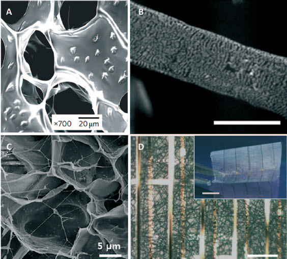
In another study, You et al. generated porous hydroxyethyl methacrylate (HEMA) hydrogels into which gold nanoparticles were introduced by in-gel synthesis. Along with conductivity, the resulting gels showed more physiologically relevant mechanical properties than polyaniline and PPy conductive biomaterials, and supported the adherence and viability of seeded cardiomyocytes. Moreover, expression of the gap-junc- tion protein connexin 43 was enhanced in cardiomyo- cytes that were seeded on the electrically conductive hydrogel, suggesting that conductive scaffolds may enhance cardiomyocyte function. 49
Gold nanoparticles have also been used for improving the electrical communication between adjacent cells when seeded on electrospun fibres. For instance, using a thin-film deposition technique, our group has incorporated AuNPs on the surface of PCL-gelatin electrospun fibres (Fig. 11b). In comparison to pristine scaffolds, cells that were seeded on the composite fibres showed a rapid formation of elongated and aligned cardiac tissue, capable of generating a strong contractile force. 50 In a later publication, we have demonstrated the same effect when AuNPs were incorporated on the surface of coiled PCL electrospun fibres that more reliably recapitulate the native architecture of the cardiac ECM. 51 AuNPs could also be deposited on other types of scaffolds. For example, it can convert a biological substance into a conductive composite material. This was demonstrated on fibrous decellularised omental matrices. 82,83 The composite scaffold, decorated with AuNPs, supported the formation of cardiac patches that generated stronger contraction forces while exhibiting lower excitation thresholds and faster calcium transients. Moreover, since this approach is based on autologous materials taken from the patient, it can minimise immune response and implant rejection. 52
Carbon nanostructure-incorporated composite scaffolds
While integration of metallic structures into poorly conductive substances has proved successful, other means of improving the conductivity of biocompatible scaffolds were proposed.
Carbon nanofibers (CNFs) and carbon nanotubes (CNTs) are cylindrical, high aspect-ratio nanostructures, made of graphene layers arranged as stacked cones, cups or plates (CNFs) or wrapped into perfect hollow cylinders (CNTs). The structures have extremely high electrical and thermal conductivity, together with extraordinary mechanical strength. These exceptional traits have made CNFs and CNTs applicable in various fields, including medicine, biotechnology, composite materials, microelectronics, textile, filtration, optics, energy storage and more. 116 These materials have also been applied to improve the mechanical and electrical properties of biocompatible scaffolds for TE appli- cations. 117 For instance, highly conductive CNFs have been used to confer the biocompatible polymer PLGA with the ability to transmit electric signals. As expected, the conductivity of the PLGA-CNF composites was increased as greater amounts of CNF were added to the blend. This composite was shown to promote the adhesion and growth ofhuman cardiomyocytes while hindering the growth of other non-electrogenic cells, such as endothelial cells and fibroblasts. It has also been demonstrated that applying continuous electrical stimulation elevated the density of the cardiac tissue cultured on the scaffold. 53–55
The great potential of CNFs and CNTs as conductive and enforcing additives to biomaterials for cardiac TE was further supported by later studies, where these carbon nanostructures were incorporated in various other porous scaffolds. For instance, CNFs were used as a doping material to develop a highly conductive chit- osan-based composite. The resulting scaffold was found to support the cultivation of cardiac cells, which demonstrated elevated metabolic activity, and high expression of cardiac genes involved in contraction and electrical coupling. 56 A different study showed that the incorporation of CNTs into a relatively mechanically weak gelatin hydrogel greatly enhanced its mechanical integrity, reduced the excitation threshold of seeded cardiomyocytes, and elevated spontaneous synchronous beating rates (Fig. 11c). Remarkably, the developing contractile tissue was also found to be protected from the effects of the gap-junction uncoupler heptanol and the cytotoxic agent doxorubicin. The authors have suggested that the defensive mechanism may be associated with CNTs ability to provide an alternative route for electrical signal propagation and by acting as free radical scavengers, respectively. 57
Aside from the expected enhancement in conductivity, incorporation of CNTs into biocompatible polymers may also improve other desirable properties of the fabricated scaffold. As an example, Kharaziha et al. have supplemented an electrospinning solution of PGS-gela- tin with bio-functionalised, methacrylated gelatin-coated CNTs. Upon electrospinning, aligned scaffolds of highly uniform, 170 nm diameter fibres were obtained, with 25 nm diameter CNTs, dispersed and aligned along the nanofibers axis. Interestingly, it was found that in addition to the improvement in conductivity, the composite fibres also showed a significant enhancement in their alignment and mechanical properties, such as toughness, tensile strength and elastic modulus. Apparently, the improved alignment was derived from the enhanced molecular orientation of the composite that may have resulted from the tendency of the CNTs to act as nucleating sites for the formation of oriented crystallites. On the other hand, the enhanced mechanical properties of the scaffold may have been associated with the orientation of the CNTs along the preferred axis of the fibres and their interaction with the polymeric matrix. In vitro cell assays showed that seeded cardiomyocytes that grew along the fibre direction presented increased retention, viability and contractility. 58
Conductive biomolecule-incorporated composite scaffolds
Another interesting strategy for improving the electric conductivity of a scaffold was presented by Kai et al.
The group chose melanin, a natural pigment characterised by good electrical conductivity and photoconductivity, as a filler for incorporation into electrospun poly(
Collectively, the results of the described studies stress the benefit of using electrically conductive composite scaffolds for cardiac TE. These scaffolds have been shown to enhance the expression of a cardiogenic phenotype, increase electrogenic activity and promote proper tissue assembly. Nonetheless, creative application of cutting-edge technologies allowed tissue engineers to take the art of scaffold fabrication another step forward.
Nanocomposites for sensing tissue function
As discussed earlier, a basic demand from a construct that is designated to serve as a scaffold for TE applications is the ability to recapitulate the properties of the natural ECM. Also discussed are modifications that provide the scaffold with properties that cannot be found in natural ECM, with the aim of maintaining a proper, native-like functionality of the supported tissue. Nonetheless, the generation of engineered tissues and organs that benefit from beyond-natural qualities has been a dream of scientists from the beginning of modern time. For instance, although providing the tissue with a proper microenvironment that supports its assembly, some crucial aspects regarding its functionality remained elusive. Among them are the actual status of the elec- trogenic activity and contractility of the patch. Taking up this challenge, Tian et al. have generated macro- porous, free-standing flexible scaffolds incorporating silicon nanowire field-effect transistors (FETs). These nanoelectronic scaffolds (nanoES) were then combined with synthetic (PLGA) or natural (collagen, alginate) ECM-like matrices, resulting in a 3D biocompatible composite scaffold with electrical sensory function (Fig. 11d). The hybrid mesh was then seeded with car- diomyocytes to yield a nanoelectronic-tissue construct. A high density of healthy tissue, intimately contacting the electronic elements, was observed upon culturing on the nanoES/PLGA substrate. Importantly, the monitoring capability of the system proved successful, recording regularly spaced spikes with characteristics consistent with extracellular electrical recordings from cardiomyocytes. The system also performed well in monitoring cellular responses to drugs and was capable of delivering simultaneous recordings from several nanowire FETs separated by up to 6.8 mm. 60
In conclusion, the ability to monitor the function of the patch is of great significance to tissue engineers and is essential for the success of the regenerative process. Considering the late advances in the fields of nanoelectronics and sensing devices, it is conceivable that in the near future, such hybrid scaffolds will gain more popularity and will offer more advanced features.
Final remarks
The amazing ability of the living body to heal and regenerate itself, together with the supportive care that is provided by modern medicine, is the most basic means that enable overcoming illness and trauma. Unfortunately, some of the most life-threatening diseases that are beyond nature's healing power are still waiting for an efficient medical solution. Among them, severe cardiac ischaemia that results in an acute myocardial infarction is of a special interest. Accordingly, many research groups worldwide are striving to develop an artificial, engineered cardiac patch that may substitute the need for complicated heart transplantations. By using state- of-the-art techniques and by harnessing the accumulating knowledge from multiple scientific disciplines, researchers are now able to fabricate composite ECM- like scaffolds that closely mimic the natural microenvironment of cardiac cells. In addition to the fine and complex topography of the matrix, these composites have also been modified to imitate the mechanical properties of the native myocardium, improve electric signal propagation, provide the cells with GFs and promote their interaction with the substrate. Moreover, the technology that enables the integration of sensing devices in living, engineered tissues is progressively developing. Results from studies discussed in this review demonstrated that application of such composite scaffolds in cardiac TE greatly improved pivotal physiological parameters of the artificial tissue patch. Together with recent advances in cellular biology and stem cell research, we foresee that bioengineered cardiac patches will be gradually applied as an approved treatment for MI patients. Needless to say, such a revolutionised therapy may save the lives of millions that desperately await a heart transplant, and improve the life quality of people who suffer from performance-limiting cardiac diseases.
Footnotes
Acknowledgement
TD acknowledges support from the Moxie Foundation, European Union FP7 program (Marie Curie, CIG), Alon Fellowship, Slezak foundation, and the Israeli Science Foundation.