
Abstract
In order to obtain a fast biomimetic coating on titanium substrates, calcium enriched silica films were prepared on pure titanium substrates by sol–gel method under the following conditions: pH = 3 and the molar ratio of tetraethyl orthosilicate (TEOS)/H2O/EtOH/Ca2+ = 1∶4∶11∶0·08. The titanium base calcium enriched silica film is soaked in a slightly supersaturated Ca/P solution, with the concentration of calcium ion and phosphate group being 1·5 times of that of simulated body fluid, for seven days, a uniform apatite layer is precipitated onto the surfaces of calcium enriched silica films, which is quite rapidly. The investigation adopted SEM, EDX, Fourier transform infrared spectroscope and XRD to indicate that the coating was carbonate hydroxyapatite. The results show that it is a feasible way to obtain carbonate hydroxyapatite through functionalising the pure titanium substrates by preparing calcium enriched silica films on it from a sol–gel method.
Introduction
Titanium and titanium alloys are widely used in biomedical and dental implants for their superior mechanical properties. They are biocompatible, but cannot be directly combined with human bones, only can be biologically fixed by mechanical locking through screws and bone cement. Loosening for long term after implantation has been reported. 1 In order to overcome this problem, various surface treatment techniques, such as plasma spraying, 2 Metal organic chemical vapour deposition and hydrothermal electrodeposition method have been employed to prepare hydroxyapatite (HA) bioactive coatings on pure titanium metal and titanium alloy substrates.3,4 A Ti/HA composite material is formed by introducing an HA coating onto the surface of titanium and titanium alloy, this material is considered as a novel material that can be used as bone substitute, with not only good mechanical properties of Ti but also excellent biological properties of HA.
Many researches have been dedicated to preparing Ti/HA composite materials in a biomimetic route recently. The so called biomimetic coating technique is to induce the deposition of apatite in simulated body fluid (SBF), 5 with ion concentrations nearly equal to those of human extracellular fluid. It is apparent that this method can be readily adapted to synthesising Ti/HA composite materials since it does not require complex equipment, the objects to be coated can be large or irregularly shaped. Above all, the process can be carried out with a low temperature, and can mimic the environment in vivo. The composition and structure of the formed layer will have some similarity to the tissue of the organism, and preparing Ti/HA composite materials in a biomimetic route will be an important direction of implantation research in the future. 2 But some reports show that the deposition of apatite needs one or more months, so the period of experiment could be a practical problem. 6
It has been reported that the formation of bone-like apatite on artificial materials in vivo is induced by functional groups, such as Si-OH, Ti-OH, Zr-OH, Nb-OH, Ta-OH, -COOH and -PO4H2.7–14 These groups have specific structures with negative charge, such as Si-O−, Ti-O−, Zr-O−, Nb-O−, Ta-O−, -COO−, -PO4H−, which interact with Ca2+ ions electrostatically and induce the formation of apatite via the formations of an amorphous calcium compound, e.g. calcium silicate, calcium titanate and amorphous calcium phosphate. 8 These fundamental findings provide methods for preparing new bioactive materials with different mechanical properties.
In the present paper, the authors describe the synthesis of carbonated hydroxyapatite coating (CHA) on calcium enriched silica films based on titanium substrate. The surface of titanium can be functionalised with Si-OH groups by calcium enriched silica films, which possess good biological properties. After being immersed in SBF for seven days, a great number of spherical CHA deposits spread all over the surface of the films.
Materials and experimental method
Preparation of silica films
The sample material was titanium (99·5 pure) in the form of foils with a thickness of 0·1 mm, purchased from Northwest Institute for Non-ferrous Metal Research (China). Before sol–gel treatment, titanium foils were cleaned ultrasonically in distilled water, acetone, ethanol (75 wt-) and distilled water for 10 min respectively, and then immersed in the mixture of HCl (18 wt-) and H2SO4 (48 wt-) solution at 70°C for 30 min. After being washed with distilled water and dried in air, the samples were coated with calcium enriched silica films by dipping coating method.
In the present study, calcium enriched silica films were prepared by a sol–gel method through controlling the hydrolysis and condensation process of tetraethyl orthosilicate (TEOS, Chinese Medicine Company of Shanghai Chemical Reagents) at 70°C for a period of time, using ethanol (EtOH) as a cosolvent, calcium nitrate tetra-hydrate [Ca(NO3)2.4H2O] as the source of calcium and hydrochloric acid as a catalyst followed after being dried at 70°C (the samples were placed in an empty vessel which was placed in a water bath) for 12 h and being sintered at 500°C (the temperature rises at a rate of 1°C min−1 under 200°C and 10°C min−1 over 200°C) for 1 h, followed by free cooling. Figure 1 shows the schematic illustration of the process followed for preparing calcium enriched silica films, the process is repeated for three times to obtain three layers.
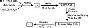
Schematic illustration of process followed for preparation of calcium enriched silica film
Different samples were prepared at pH = 3 with a TEOS/H2O/EtOH/Ca2+ ratio of 1∶4∶11∶(0·02–0·10) (molar ratio), the results show that sol prepared at pH = 3 with a TEOS/H2O/EtOH/Ca2+ ratio of 1∶4∶11∶0·08 (molar ratio) is suitable for coating calcium enriched silica films on titanium substrate after being aged for 6 h at 70°C. The sample obtained on this condition is described as SC, while other samples obtained are not uniform and have lots of cracks. The mechanical performance and growth of CHA will be affected. 15 Thus it is considered as the optimal condition, and the performance of SC is discussed in details.
Immersion in 1·5SBF
It has been found that the induction period for the formation of apatite decreases with increasing the concentration of calcium ion or phosphate ion in SBF, 16 so the authors use 1·5SBF (the 1·5SBF solution was prepared with the concentrations of calcium ions and phosphate ions 1·5 times of those of human blood plasma, the concentrations of other ions nearly equal to those of human blood plasma) as the solution for immersion. 1·5SBF was prepared as described in the literature. 17 Every titanium specimens (1×1 cm) coated with calcium enriched silica films was immersed in 50 mL 1·5SBF at 37°C, the solution was refreshed every two days, and buffered at pH 7·40 with tris-hydroxymethyl aminomethane [(CH2OH)3CNH3] and 1 mol L−1 HCl at room temperature. The specimens were taken out after one, three, five and seven days respectively, then washed with distilled water and dried in air.
Characteristics
The morphology of SC before and after the immersion was examined by scanning electron microscope (SEM, Philips XL30). The surface composition and structure of the specimens were examined by X-ray diffraction (XRD, Philips X'pert MPD), Fourier transform infrared spectroscope (FTIR, Nicolet Avatar 360 spectrometer) and energy dispersive X-ray microanalysis (EDX, Shimadzu EPM-810) respectively. The pH values of the 1·5SBF during the immersion were measured by a digital precision pH meter (828, Orion). The interfacial shear strength of SC after the immersion was measured with an adhesive strength test by a universal testing machine (LR5K, Lloyd) using a 2 kN load cell and a crosshead speed of 1·0 mm min−1. SC with 1×1 cm was glued to equal sized 316L stainless steel plates with epoxy resin, cured at 100°C for 2 h in an oven. For each testing material, five specimens were used, and the shear strength data was reported as the average value.
Results and discussion
Surface composition and structure of SC
Figures 2–4 show the XRD patterns, FTIR spectra and EDX spectra of SC before and after the immersion respectively. As shown in Fig. 2a, a weak peak of silica appears between 15 and 27° after it is calcinated for 1 h at 500°C, which shows that the silica has an amorphous structure. The absorption bands of -OH at 3200–3600 cm−1 and H-O-H at 1630 cm−1 could be observed as shown in Fig. 3a. The absorption bands of Si-O at 1080 cm−1,  at 790 cm−1 and Si-O-Si at 450 cm−1 could also be observed on the spectra.
18
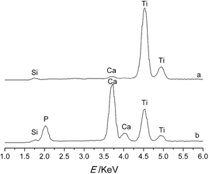
The EDX spectra of SC before the immersion shows the existence of silicon, calcium and titanium on the surface (Fig. 4a). It is reported that Ca(NO3)2 decomposes at 475°C.19 But the peak attributed to CaO (at 2θ = 37·36°) is not observed in Fig. 2a,
20
it is possibly because the amount of CaO is too small. Thus it can be concluded that the surface composition of SC are mainly amorphous SiO2, which possibly includes a small quantity of CaO.
at 790 cm−1 and Si-O-Si at 450 cm−1 could also be observed on the spectra.
18
The EDX spectra of SC before the immersion shows the existence of silicon, calcium and titanium on the surface (Fig. 4a). It is reported that Ca(NO3)2 decomposes at 475°C.19 But the peak attributed to CaO (at 2θ = 37·36°) is not observed in Fig. 2a,
20
it is possibly because the amount of CaO is too small. Thus it can be concluded that the surface composition of SC are mainly amorphous SiO2, which possibly includes a small quantity of CaO.
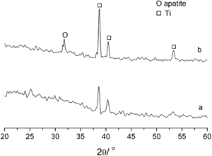
X-ray diffraction patterns of SC immersed in 1·5SBF solution for a zero day and b seven days
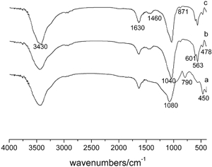
Fourier transform infrared spectroscope patterns of SC immersed in 1·5SBF solution for a zero day, b five days and c seven days
Energy dispersive X-ray microanalysis spectra of SC immersed in 1·5SBF solution for a zero day and b seven days
Bioactivity of SC
Generally, the evaluation of the bioactivity of biomaterials is conducted through immersing them in SBF, taking the period of inducement and growth of CHA into account. The shorter the period is, the better bioactivity they possess. 21
It can be seen from Fig. 2b that some weak diffraction peaks of apatite appear after SC is immersed in 1·5SBF for seven days which indicates that the grain size of the deposits is small and the deposits have weak crystallinity. It has been found that CaHPO4 is stable in a supersaturated solution at room temperature for pH<4·3. In contrast, HA is stable for pH>4·3. 22 In the present experiment, T = 37·0°C and pH = 7·40, thus it is likely to obtain HA due to thermodynamics reasons. The value of the diffraction peak at ∼31·8° is consistent with 211 crystal face of HA, according to the standard value of JCPDS (09-432). But no other crystal faces of HA are observed. The reason why other crystal faces of HA did not form was probably that the surface of functionalised materials could induce the formation of apatite nuclei in some specific orientation, therefore HA grew along some definite direction. The overlapping of crystal faces or the defects of crystal growth lead to the absence of other crystal faces of HA.
As shown in Fig. 3, after being immerse in 1·5SBF for five days, the absorption bands of  at 478, 563, 601 and 1040 cm−1 could be observed, reflecting the structure of apatite. With increasing the immersion time, the intensity of the absorption bands increases. The absorption bands of
  at 871 and 1420–1480 cm−1 could also be observed in the spectra,
23
indicating that carbonate ions have been incorporated in the HA lattice. With increasing the immersion time, the intensity of the absorption band of SiO2 at 450 cm−1 decreases gradually and disappears at 790 cm−1. The absorption band at ∼1080 cm−1 attributed to SiO2 might be overlapped by that of
at 871 and 1420–1480 cm−1 could also be observed in the spectra,
23
indicating that carbonate ions have been incorporated in the HA lattice. With increasing the immersion time, the intensity of the absorption band of SiO2 at 450 cm−1 decreases gradually and disappears at 790 cm−1. The absorption band at ∼1080 cm−1 attributed to SiO2 might be overlapped by that of  at ∼1040 cm−1 after the immersion. Possibly, as the immersion time being increased, the surface of the specimen is gradually covered with deposits, so the intensity of certain absorption band of SiO2 decreases or disappears.
As shown in Fig. 4b, after being immersed in 1·5SBF for seven days, apart from silicon and phosphorus, more calcium can be detected, while less titanium can be detected, indicating the formation of a denser and thicker Ca–P layer on the surface. The atomic ratio of Ca/P calculated from the EDX spectra of SC after being immersed in 1·5SBF for seven days is ∼2·20, higher than that of HA (1·67). A possible reason is that carbonate ions incorporated in the HA lattice take up some positions of phosphate ions.
Thus it is concluded that the surface deposits of SC are CHA. As mentioned above, the peaks of apatite in Fig. 2 are weak and wide, the possible reason is that carbonate ions are incorporated in the HA lattice, leading to structural disorder and a decrease in crystallinity. It is reported that, the mineral phase of hard tissue is a so called biological apatite, i.e. a non-stoichiometric HA, it contains several other ions, mainly carbonate (2·3–8) and other trace elements such as Mg2+, Na+, Fe2+,  , F− and Cl−.24 Consequently, CHA prepared by a biomimetic method is even more close to hard tissue than stoichiometric HA, so it is expected to possess better biocompatibility than stoichiometric HA.
Figure 5 shows the SEM micrographs of SC which is immersed in 1·5SBF solution for different periods of time. As shown in Fig. 5a, the surface of SC is uniform and porous, with small cracks, which may probably be caused by different horizontal shrinkage forces (due to the force existing in the surface of the films) and vertical shrinkage forces (arisen from the adhesive force between the films and the substrate) during the process of drying and heat treatment. After being immersed for one day (Fig. 5b), the size of pores and the cracks of the surface both become larger and there are some scattered spherical CHA on the surface of the films. Numberless leaves can be observed on the surface of spherical CHA in high magnification micrograph on the top left corner in Fig. 5b. During the immersion process, the size of pores expanded continuously. After being immersed for three days (Fig. 5c), more and more spherical CHA appears on the surface of the films and cover the cracks of the surface gradually, but they are still microspheres. After being immersed for five days (Fig. 5d), the number of spherical CHA is remarkably increased, the whole surface of SC is nearly completely covered with CHA. Spherical CHA become much larger than before. After being immersed for seven days (Fig. 5e), the number and the size of spherical CHA are both increased. The overlapping of deposition leads to the increase in density and thickness of CHA layer. Owing to the evaporation of water molecules from the specimens during the drying process, some cracks are brought to the CHA layer. Thus it is concluded that the surface of bio-inert titanium can be functionalised with Si-OH groups by calcium enriched silica films, and then will be able to induce the formation of apatite, indicating the good bioactivity.
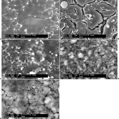
Images (SEM) of SC immersed in 1·5SBF solution for a zero day, b one day, c three days, d five days and e seven days
The above mentioned results show that SC is capable of inducing the formation of CHA in the short term. Despite the strong bonding between the CHA layer and bone structure, it has been recognised that the mechanical stability of the interface between the CHA layer and the titanium substrate could be a problem during surgical operation. The interfacial shear strength of SC with CHA layer after the immersion in 1·5SBF for seven days is ∼8·6 MPa. The relatively poor adhesion between SC and CHA layer is mainly arisen from the mismatch of the coefficients of thermal expansion between the titanium substrate and the CHA layer. The fracture surface of the titanium substrate is very smooth and clean, and all the silica film and CHA layer are adhered to the epoxy resin. It can be concluded that the fracture mechanism for SC is mainly the adhesive failure. 25 Therefore, further effort is needed to enhance the mechanical performance of the interface between the CHA coating and the titanium substrate.
pH variations of 1·5SBF and formation mechanism of CHA on surface of SC
During the immersion process, the 1·5SBF solution always kept clear, indicating that spherical CHA deposit on the surface of the films through inhomogeneous deposition. Figure 6 shows the variations of pH of 1·5SBF with immersion time. The pH value decreases from the initial 7·40 to 5·60 after being immersed for two days. The dissolution of apatite will be accelerated if the pH value of the solution decreases. 26 So it is necessary to refresh the solution every two days, keeping the concentrations of Ca2+ ions and phosphate ions, at the same time keeping the pH value favouring for the formation of apatite.
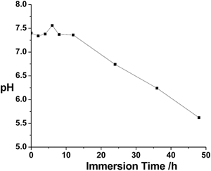
pH variations of 1·5SBF with immersion time
The OH− ions participate in the formation of apatite, hence it is presumed that the formation mechanism of CHA on SC is as follows.
Stage 1: Si-OH groups formed on surface of SC
At the beginning of the immersion, two reactions occur simultaneously. First, H2O attacks SiO2, which leads to the loss of soluble silica due to the breakage of Si-O-Si bonds and the formation of Si-OH (silanol) at the interface of SC and solution.16,21,27 The formation of polysilicic acid leads to an increase of H3O+ in the solution.16,21 At the ambient pH value of 6–9, the surface site is partially deprotonated and expressed as  , where H2O expresesses the explicitly hydrating water molecules.
28
Second, fractional CaO hydrolyse leading to the formation of alkalescent Ca(OH)2. The pH changes of the solution are not obvious, resulting from the opposite influences of the reactions mentioned above. The reactions are respectively expressed as follow
, where H2O expresesses the explicitly hydrating water molecules.
28
Second, fractional CaO hydrolyse leading to the formation of alkalescent Ca(OH)2. The pH changes of the solution are not obvious, resulting from the opposite influences of the reactions mentioned above. The reactions are respectively expressed as follow


Stage 2: SiO2 rich layer formed on surface of SC
A loose and porous SiO2 rich layer is formed on the surface of titanium due to the condensation and re-aggregation of Si-OH groups.16,21,27 The reaction is expressed as follow

Stage 3: Amorphous calcium phosphate layer formed on surface of SC
It has been reported that at the neutral pH of the solution, Si-OH groups of  are the active sites on silica surfaces for the nucleation of calcium phosphate.
28
There are three kinds of phosphate ions existing in the solution,
are the active sites on silica surfaces for the nucleation of calcium phosphate.
28
There are three kinds of phosphate ions existing in the solution,  ,
,  and
and  , but it is more favourable for
  to be absorbed on Ca2+ under the condition of near neutral pH environment, thus forming the crystal nucleus of
  .
28
The pathway is expressed as follow
.
28
The pathway is expressed as follow
Stage 4: Crystallisation and growth of CHA
With increasing the immersion time, the amorphous calcium phosphate gradually combines with OH− and  in the solution and forms into CHA, resulting in the continuously decrease of the pH value of solution. The apatite nuclei grows spontaneously in situ on the surface of SC into a dense and uniform surface layer by consuming Ca2+ and phosphate ions in 1·5SBF. Figure 7 shows the schematic illustration for the formation of CHA on the surface of SC. The decreasing trend of pH is slowed down, indicating that the formation rate of CHA is changed.
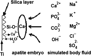
Schematic illustration for formation of CHA on surface of SC
In fact, stages 1 and 2 describe the dissolution and reprecipitation process of SiO2 respectively. The dissolution and reprecipitation of SiO2 are limited, an excessive quantity of soluble silica inhibits the formation of apatite, for the formation of apatite requires non-acidic conditions, and an excess of polysilicic acid around the surface or inside the pores will decrease the pH value to a level below the stability range of apatite. On the other hand, the surface of SC is covered gradually by apatite, so the dissolution and reprecipitation of SiO2 are delayed or inhibited. In addition, as seen from Fig. 5e, numberless spherical CHA deposits spread all over the surface of SC, indicating that Si-OH groups form uniformly on the surface of SC.
It has been reported that micropores and high surface area of bioactive materials can accelerate the nucleation and growth of bone-like apatite.27 The surface of SC is uniform and porous, abundant Si-OH groups on which are the active sites for the nucleation of apatite. In addition, concave surfaces are considered to be more advantageous to the nucleation of inorganic mineral phases than planar or convex surfaces in natural biomineralised systems, because enough concentrations of Ca2+ ions and phosphate ions round the materials is critical for the early induction of the formation of CHA. The liquid fluxion and the diffusion of ions will be blocked in the pores on the surface of SC, so higher concentrations of Ca2+ and phosphate ions will be kept there. At the same time, these positions are in favour of the nucleation of apatite. Furthermore, the size of pores on the surface of SC and the concentration of Ca2+ ions around the material increase, which is in favour of the nucleation and growth of CHA followed by the formation of CHA layer. It has been reported that it takes 15 days for pure silica coating based on Ti to induce the formation of apatite in vitro. 11 So the enrichment of calcium on the surface of SC is helpful for the formation of apatite.
Conclusions
Calcium enriched silica films were prepared on titanium substrate by a sol–gel method. The results show that sol prepared at pH = 3 with an TEOS/H2O/EtOH/Ca2+ ratio of 1∶4∶11∶0·08 (molar ratio) is suitable for coating calcium enriched silica films on titanium substrate after being aged for 6 h at 70°C. The surface of SC is uniform, porous, and nearly crackless after being calcinated for 1 h at 500°C. The surface composition of SC is mainly amorphous SiO2, including a small quantity of CaO.
After being immersed in 1·5SBF solution for seven days, the surface of bio-inert titanium can be functionalised with Si-OH groups by calcium enriched silica films, and then the formation of a dense and thick CHA layer can be induced, indicating the good bioactivity. Abundant Si-OH groups on the surface of SC are the active sites for the nucleation of apatite. The size of pores on the surface of SC and the concentration of Ca2+ around the material increase, which is in favour of the nucleation and growth of CHA followed by the formation of CHA layer. The results show that it is a feasible way to obtain CHA through functionalising the pure titanium substrate by preparing calcium enriched silica films on it from a sol–gel method.
The technology of preparing Ti/CHA composite materials by a biomimetic method is very simple and inexpensive, the process can be carried out at low temperature and mimic the environment in vivo. The composition and structure of the CHA layer may be somewhat similar to the tissue of the organism, which is by far more complex. The method presented has some advantages and is worthy of further study.
Footnotes
Acknowledgements
The authors would like to thank National Nature Science Foundation of China (grant no. 30600149), the Science Research Foundation of Ministry of Health & United Fujian Provincial Health and Education Project for Tackling the Key Research (project no. WKJ 2008-2-037), the Project of Fujian Education Department (project no. 209061, JA08030) and Fujian Provincial Department of Science and Technology (grant no. 2006I0015).