
Abstract
The microstructures of steels consisting of ferrite (steel 1) and bainite (steel 2) were characterised using atom probe tomography. The microstructural features, such as formation of Nb–C clusters in steel 1 fine Fe–C particles, were observed.
Introduction
The need for weight savings while maintaining, or improving, the safety aspects, has generated the development of new hot rolled, high strength steels, multiphase steels and ultrafine bainite steels. To understand the excellent mechanical properties of these steels, it is essential to have detailed information on the microstructure and the composition, particularly the distribution of the alloying elements. To analyse the composition distribution for such small features, the three-dimensional atom probe (3DAP) has advantage over other analysis techniques, such as TEM.
Atom probe tomography (APT) has been successfully used to study the microstructure of modern high strength steels by different research groups.1–3 For example, the solute distribution between phases in multiphase transformation induced plasticity steel was studied by APT,1 or APT evidence on the incomplete transformation phenomenon in nanobainitic steel was found.2
The present paper covers various applications of the APT technique.
Experimental
To demonstrate how the APT technique could be used for the nanoscale microstructural characterisation of high strength steels, two steels with composition of 0·05C–1·6Mn–0·3Si–0·05Nb–0·2Mo (wt-%) (steel 1) and 0·8C–1·5Si–1·98Mn–1·1Al–0·98Cr (wt-%) (steel 2) were chosen.
The thermomechanical processing of steel 1 was designed to produce ultrafine ferrite through deformation strain induced transformation4 and was performed using a computer controlled hot torsion deformation simulator. The specimens after austenitising at 1250°C for 5 min were cooled to 1100°C and held for 80 s, followed by two deformations of 0·4 with an interpass time of 20 s at a strain rate of 1 s−1. Following this, the specimens were cooled to 725°C, and a strain of 2 was applied. This was followed by quenching.
The steel 2 samples were austenitised at 1100°C for 30 min and then were isothermally heat treated at 200°C for 10 days following the work of Caballero and Bhadeshia.5
The microstructural characterisation was performed using TEM on a Philips microscope operated at 200 kV and APT.
A 3DAP is a combination of a field ion microscope (position sensitive detector) and a time of flight mass spectrometer.6 Through field evaporation, the surface atoms of the needle specimen are projected out one by one and layer by layer to the detector and captured. The recorded atoms are then reconstructed in three dimensions. This technique is very powerful in determining quantitatively the local composition distribution in three dimensions with atomic resolution, especially for nanoparticles, which are beyond the limit of electron microscopy.
The Monash Centre for Electron Microscopy at the Monash University has one Oxford nanoScience 3DAP with a state of the art delay line detector and a fast pulsing power supply. This 3DAP demonstrates high detection efficiency, superior mass resolution and excellent capability in encoding multiple ions arriving at the same time at the detector. The high performance of this tool makes it possible to analyse complex alloys that may contain four or more alloying elements.
Results and discussion
Steel 1 microstructure consisted of fine ferrite grains with high dislocation density, low grain boundary misorientations and an average grain size of 1±0·1 μm (Fig. 1a). The grain refinement was due to deformation strain induced transformation. This type of ferritic hot rolled steels with fine grain size can provide good local elongation and good stretch flangeability.7 However, the level of strength in these steels can only be achieved by additional precipitation hardening of the ferrite matrix with very fine precipitates.3 In the present research, the combined effect of Nb and Mo on the formation of carbides was studied.
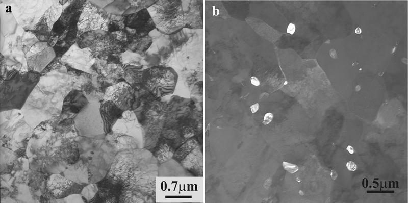
a bright field images of ferrite grains and b dark field images of particles formed in steel 1
The formation of three types of carbides was found through TEM: coarse round particles with an average diameter of 60±10 nm, coarse cubic particles with average size of 70±10 nm and very fine particles with the size from 2 to 5 nm (Fig. 1b). The coarse round particles are either undissolved during reheating or formed at high temperature, whereas the cubic particles are TiN and believed to form during casting. Analysing the energy dispersive X-ray spectroscopy spectrum and indexing the diffraction patterns, the round particles were found to be NbC and (MoNb)C with a cubic structure and lattice parameters of a = b = c = 0·44 nm and a = b = c = 0·436 respectively. However, the usage of TEM for fine particle characterisation in the ferrite matrix has a number of limitations. First, due to the coherent or semicoherent nature of these precipitates, it is necessary to use foils rather than replicas. The precipitates also have defined crystallographic relationships with the parent phase, and sometimes, imaging can require large rotations of the specimen, which, due to the magnetic nature of ferrite, can be an issue in many TEMs. This often leads to the appearance of precipitates in some grains and not in others.
The APT technique was used to study the nanoscale morphology and composition of precipitates. However, the disadvantage of this technique is that it represents a small area, and a number of samples are required to develop the complete ‘picture’. APT revealed not only the formation of nanoscale precipitates but also the presence of clusters. Most of the fine clusters and particles contained Nb and C (Fig. 2a). The precipitates had clearly visible planes of atoms that were different from the matrix crystallographic arrangement of atoms, while clusters could be identified as small segregations of solute atoms within the matrix phase without a clearly defined crystallographic structure. Standard methods, such as compositional profile across the cluster or particle (Fig. 2b) and selected atom maps, where the matrix ions are suppressed (Fig. 2c), were utilised. The nanoscale clusters/particles had a plate shape with an average thickness of 2·5±0·5 nm. They were parallel to each other and had a distance of ∼7·5 nm between them. APT allowed the determination of the composition, not only of precipitates but also of the nanoscale clusters. The composition of clusters varied from 17 to 20 at-%Nb and from 8 to 10 at-%C. However, the formation of Nb–C carbides with equilibrium compositions was also confirmed by APT. APT also enables the segregation of carbon to the grain boundary to be studied. One example of such segregation in steel 1 is shown in Fig. 2.
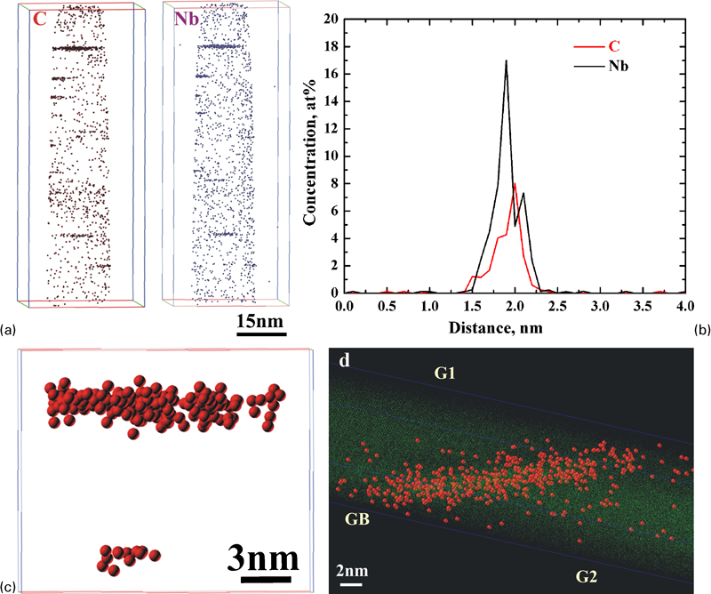
APT characterisation of steel 1
The ultrafine bainite steels have recently been developed by Caballero and Bhadeshia.5 The microstructure of the steel in the present study consisted of fine bainitic ferrite laths with an average thickness of 60±10 nm and retained austenite with an average thickness of 30±10 nm (Fig. 3a). The good strength–ductility balance in this steel is due to the microstructural refinement and formation of a high volume fraction of retained austenite (21±2%), which transforms to martensite during straining and provides the transformation induced plasticity effect. One of the main parameters that can dictate the stability of retained austenite during deformation is the carbon content. The carbon content determines the chemical driving force for the transformation of the retained austenite to martensite, the stress free transformation strain and the flow behaviour of retained austenite.8 However, most of the methods that have been used to analyse carbon content of retained austenite, such as X-ray diffraction, can provide the information on the average value, while the most important information for nanobainite steel research is the local carbon content of the retained austenite layer.
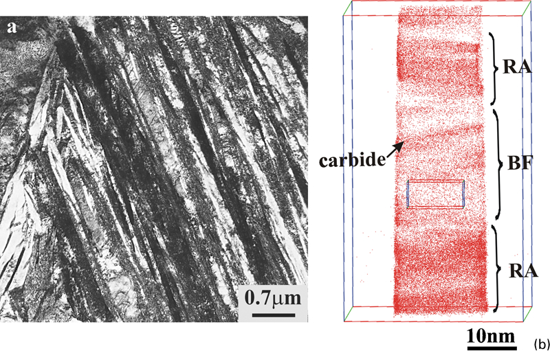
a TEM image and b carbon atom map of steel 2
Composition analysis of the phases of nanoscale bainitic steel using APT showed that the carbon level of the retained austenite varied from 1·9 to 6 at-%. Moreover, the bainitic ferrite was supersaturated with carbon, and carbon content of bainitic ferrite also varied from ∼0·3 to 0·7 at-%.
APT also showed the segregation of carbon to the dislocations in the bainitic ferrite and formation of Fe–C clusters and fine particles, which were not observed by TEM (Fig. 3b). The similar microstructural features have been observed by Caballero et al.9 The formation of Fe–C carbides was not expected due to the high Si level, which is used to suppress cementite formation during the isothermal transformation. Most of the particles were plate-like with the average thickness of 3·2±1 nm. All particles could be divided into three groups with average composition close to the compositions of Fe3C, Fe2·4C and Fe32C4. It appeared that the carbides were formed during the isothermal hold potentially by segregation of carbon to the dislocations in the bainitic ferrite.
Conclusions
The microstructural study of high strength steels using APT has shown that this technique is capable of providing very unique insights into nanostructure of steels, such as characterisation of clusters and fine particles, and segregation of carbon to the grain boundary.
Footnotes