
Abstract
Nickel nanowires were prepared by a magnetic field assisted method. An external magnetic field was applied to direct the growth of nanowires and to avoid overgrowing in diameter. The microstructure of the prepared Ni nanowires was examined by X-ray diffractometer, scanning electron microscopy and transmission electron microscopy. The characterised results indicated that the application of an external weak magnetic field resulted in the self-alignment of Ni nanoparticles with a preferential orientation and led to the formation of Ni nanowires of 95±10 nm in diameter. The present study could provide a facile route to prepare Ni nanowires in a large scale.
Introduction
As a typical one-dimensional (1D) nanostructure, nickel nanowires have attracted much attention owing to their unique properties,1,2 such as enhanced magnetic properties, 3 good catalytic activities, 4 etc. Many efforts have been focused on developing efficient routes to prepare Ni nanowires. 5 Currently, the common technique for preparing Ni nanowires is based on templates (named as template based method).6,7 However, most template based methods suffer from time consuming and complexity of multistep preparation of the templates and the residue separation after production, which have motivated investigations into template free methods. 8
The nanowire synthesis by template free methods from solutions often requires stringent growth conditions because favourable conditions should be created to promote 1D growth from the entire bulk liquid. 9 The findings that the magnetic field can significantly influence the movement of magnetic particles 10 lead to further studying on the growth and self-assembly behaviour of magnetic nanocrystallites under an external magnetic field. Thus, as a typical template free method, the use of magnetic field assistant in assembling magnetic nanoparticles (named as magnetic field assisted method) has been applied for the preparation of Co 11 or Ni 12 nanowires.
Compared to template based methods, the magnetic field assisted method for the fabrication 1D magnetic nanowires has expected advantages such as low cost, high efficiency, friendly environment and large scale production prospection. However, the magnetic field assisted method has a disadvantage that it is hard to control the size of nanowires, such as average diameter. For instance, 1D wires of metallic Ni with an average diameter of >100 nm [200, 12 250, 13 and 350 nm, 14 respectively] were prepared by magnetic field assisted methods. Generally, nanowires are defined as structures with at least one of their dimensions in the 1–100 nm range; typically, they are with diameter under 100 nm. Therefore, it is significant to develop simple and magnetic field assisted methods to prepare factual Ni nanowires with diameter in the range of 1–100 nm.
In the present study, we present a facile route to synthesise Ni nanowires via a solution method. To direct the growth of nanowires and to avoid overgrowing in diameter, a weak external magnetic field was applied. The synthesised Ni nanowires were characterised by X-ray diffraction (XRD), scanning electron microscopy (SEM) and transmission electron microscopy (TEM).
Experimental
All chemicals were of analytical reagent and used without further purification. In a typical process, 0.60 g nickel chloride (NiCl2·6H2O) (0.05 mol·L−1) and 0.74 g trisodium citrate (C6H5Na3O7·2H2O) (0.05 mol·L−1) were dissolved in 50 mL deionised water to form an absinthe green solution. The pH value of the solution was adjusted to 12.5 by 5 mol·L−1 NaOH solution. Hydrazine hydrate (N2H4·H2O) was added as reducing agent. The mole ratio of N2H4·H2O/NiCl2·6H2O was 2∶1. The obtained transparent navy blue solution was heated to 55°C by water bath and kept at 55°C for 25 min under an external magnetic field. The magnetic field intensity was ∼0.02 T provided by two same size NdFeB magnets whose poles were set oppositely with interval of 15 cm. The Ni samples were separated from the solution and washed with deionised water and finally dried in vacuum oven at 60°C for 12 h.
The morphology of the samples was observed by field emission scanning electron microscopy (FE-SEM, JSM-7500F, JEOL) operating at 10 kV accelerating voltage. The crystal structure was examined at room temperature by a DX-2000 XRD using Cu Kα radiation in the range of 2θ = 20–90° with a scanning rate of 3.6° min−1. Transmission electron microscopy micrographs, high resolution TEM images and selected area electron diffraction (SAED) patterns were obtained by employing a TEM (Tecnai G2 F20 S-TWIN, FEI) operating at 200 kV, and EDX analysis was performed with the spectrometer attached to the same TEM.
Results and discussion
The FE-SEM images of the prepared samples are shown in Fig. 1. Without a magnetic field applied, only spherical particles with an average diameter of ∼200 nm can be observed (Fig. 1a). In contrast, it is obvious that 1D Ni nanowires (Fig. 1b) of 95±10 nm in average diameter were obtained as a magnetic field (0.02 T) was applied. Particularly, the enlarged image (inset, Fig. 1b) clearly shows a 1D Ni nanowire with width fluctuation that is not a simple assemblage of particles. Intensive ultrasonication for 30 min (before drying) did not disintegrate the wires, which implies the mechanical stability of Ni nanowires and confirms that they are not merely a loose aggregate of spherical nanoparticles. Furthermore, no isolated spherical Ni particles were observed in Fig. 1b.
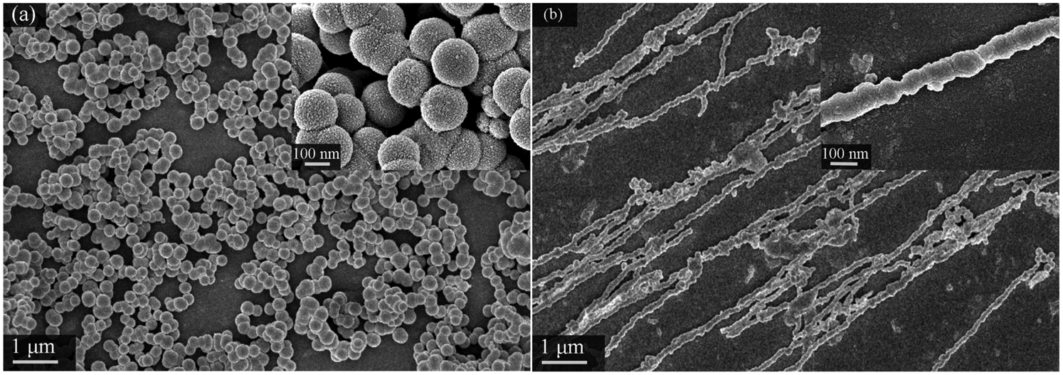
Images (FE-SEM) of Ni particles a prepared in absence of magnetic field and Ni nanowires and b prepared under external weak magnetic field: insets are local magnification of products
The microstructures of the Ni nanowires were further examined using TEM. A representative low magnification TEM image (Fig. 2a) shows more details of the prepared Ni nanowire. The wire has a necklace-like shape assembled by beads. Every bead is a nickel particle, which is confirmed by EDX characterisation (Fig. 2; inset, Fig. 2a). Two arrows point out the joints of one bead in the necklace, while the transverse and radial sizes of the bead are marked on the right in Fig. 2a. The wire possibly doubled its transverse size after the beads were assembled. A high magnification TEM image (Fig. 2b) shows the tip of the wire, which is rounded. From Fig. 2b, it can also be observed that two beads are bonded together tightly. The high resolution TEM image (Fig. 2c) reveals the good crystallinity of the nanowires that consist of crystallites. The SAED pattern (Fig. 1; inset, Fig. 2a) consists of diffraction rings of low intensity and a few spots of strong intensity simultaneously, which confirms that the wire is polycrystalline and the Ni crystallites have a preferred orientation in the wire. 15
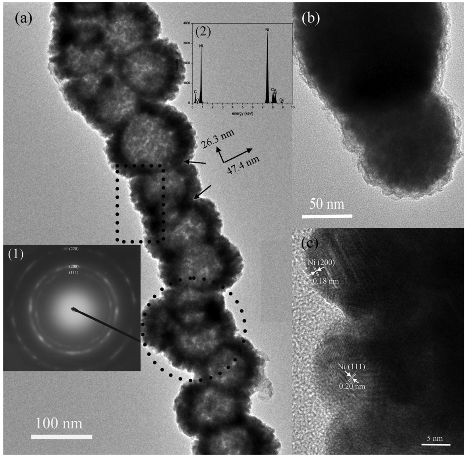
Micrographs showing a middle and b one tip of individual nanowire and c high resolution TEM image recorded from edge of wire in dashed rectangular area of a: insets of a show (1) SAED pattern and (2) EDX spectrum recorded from dashed circled area
Figure 3 shows an XRD pattern of the obtained product. It can be well indexed with the reflections of face centred cubic Ni (Powder Diffraction File standard cards, Joint Committee on Powder Diffraction Standards no. 65-2865, space group Fm-3m). The crystallite size is ∼11.4 nm, calculated by Scherrer's equation from the full width at half maximum of (111), (200) and (220) reflections. The size is less than R0 (where R0 is the critical size of single magnetic domain of spherical Ni particle, ∼82 nm). 16 Hence, each Ni crystallite might contain a single magnetic domain. Furthermore, the intensity ratio (∼3.9) of (110) peak to (200) peak is significantly larger than that (∼2.4) calculated by Powder Diffraction File standard card of face centred cubic Ni, indicating a preferential orientation of some Ni nanocrystallites with their easy magnetic axes [111] under the external magnetic field.
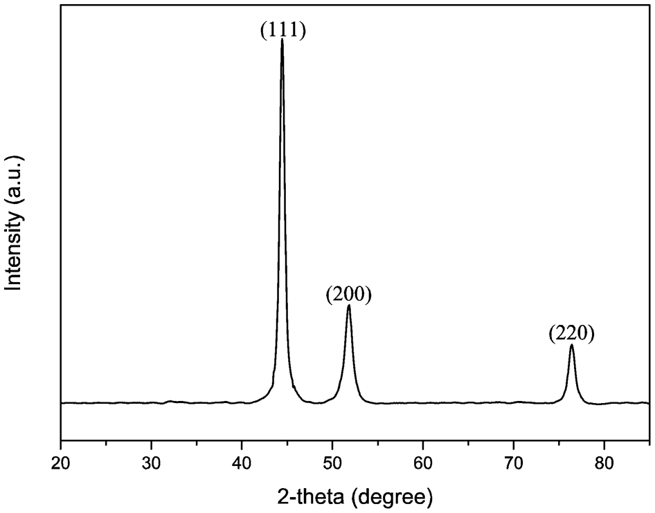
X-ray diffraction pattern of prepared Ni nanowires
Conclusions
In summary, we present a facile route to prepare Ni nanowires with appropriate diameter and well crystallised in an aqueous phase solution under magnetic field. The application of an external weak magnetic field resulted in the formation of Ni nanowires of ∼95±10 nm in diameter. The nanowires were self-aligned by Ni nanoparticles with a preferential orientation.
Footnotes
Acknowledgement
Financial support by the National Natural Science Foundation of China (grant nos. 50904046 and 51104103) is gratefully acknowledged.