
Abstract
Gold nanoparticles (AuNPs) hold promising applications in many fields such as electronics, optics and catalysis. In the past decades, there has been a growing interest for their application in medicine, in particular in nano-oncology as contrast agents, drug delivery vehicles or for diagnosis. Once injected intravenously and thanks to their small size, the AuNPs can circulate in the whole body via the blood stream and reach easily the tumour. However, what makes them very attractive for cancer treatment is their ability to distinguish healthy cells from cancer cells. While the current anticancer agents lack specific targeting, AuNPs, with their targeting efficiency, will enable the use of lower amount of drugs with all the positive aspects for the health of the patient. Additionally, their optical properties give them the ability to be used in imaging as an incredibly powerful contrast agent. For these reasons, they are believed to be one of the tools that, in the future, will enable to considerably increase the efficiency of cancer treatments by simultaneously imaging the tumour and treat it. They constitute an ideal theranostic drug delivery platform, in other words a unique combination of diagnostics and therapy. Many researches focus on the engineering of the nanoparticle surface in order to increase their biocompatibility and enable their further conjugation with bioactive ligands such as drugs, targeting or imaging agents for the design of multifunctional platforms. pH responsiveness, the ability to change properties with a change of proton concentration, is a remarkable asset for drug delivery carrier. Indeed, it has been demonstrated that cancer cells show very particular pHs in their environment: extracellular as well as intracellular. This characteristic has been exploited to create a more specific and efficient way to treat cancer. The present review focuses on the design of pH responsive AuNPs and particularly on the advantages and the potential applications of such hybrid nanomaterials in oncology.
This review was awarded a runner up prize in the 2015 Materials Literature Review Prize of the Institute of Materials, Minerals and Mining, run by the Editorial Board of MST. Sponsorship of the prize by TWI Ltd is gratefully acknowledged.
Gold nanoparticles in oncology
Current challenges in oncology
Cancer, the group of diseases which involves the rapid growth of abnormal cells, is still today a leading cause of death worldwide, and according to the World Health Organization, the number of deaths by cancer will continue to rise from 8.2 million in 2012 to over 13.1 million in 2030 †
World Health Organization (WHO). Fact sheet N 8297, updated February 2014. Retrieved from: http://www.who.int/mediacentre/factsheets/fs297/en.
Cancer Research UK. Retrieved from: http://www.cancerresearchuk.org/about-cancer/cancers-in-general/treatment/cancer-drugs/side-effects/
Another challenge for drug delivery systems in cancer therapy is the multidrug resistance. Patients who are initially responsive to the drug can develop drug resistant cells. Various strategies are adopted by the multiresistant cells: the loss of their surface receptors and the increase in their transporters pumps, leading to a decrease in the drug entry and the enhancement of the drug removal are among them.5,6 The development of novel delivery platforms able to transport and deliver the drug at its site of action in a specific manner but also able to overcome the cells resistance constitutes the main challenge in the research for cancer therapies today.
Nanotechnology is the engineering of materials conducted at the nanoscale. Thanks to the recent development of new equipments that enable to observe at such small scales, this branch of materials science has emerged and soon encountered numerous applications in various domains. Nanomaterials can be divided into three categories: one-dimensional (1D) structure such as nanowires, 2D structures such as thin films and 3D nanoparticles (NPs) such as quantum dots, silica NPs or magnetic NPs. 7
Nanoparticles appear today as the solution to the challenges in cancer treatment, as they offer many advantages. First, thanks to their small size, they can circulate easily in the whole body via the blood stream. Moreover, they can escape the immune system, and consequently, they show a relatively long circulation time; and more importantly, they are able to specifically target tumourous cells, leading to two positive aspects: the decrease in the required drug amount and thus its side effects, and the increase in efficiency. Second, they have proven to be able to cross the cellular membrane via receptor mediated endocytosis and to deliver the carried drug directly to the cytoplasm. 8 Last, NPs ameliorate the solubility of the therapeutic molecules, via conjugation or encapsulation, and thus, they are extending their in vivo stability and reducing their systemic toxicity. 9
The main nanomaterials under investigation for cancer diagnosis and tumour drug delivery purposes are polymeric NPs, liposomes, dendrimers, quantum dots, nanotubes and magnetic NPs. 10 This review focuses in particular on the AuNPs, which appear to be one of the most promising nanomaterials in oncology.
Advantages of AuNPs in oncology
Properties
AuNPs are usually red in colour and with a size between 5 and 100 nm. After intravenous injection and given their small size, they can reach the cells of interest passing through the vessels. They target the tumour cells according to two different strategies: ‘passive’ and ‘active’ targeting. In the passive targeting, AuNPs tend to accumulate close to the diseased tissues via the enhanced permeability and retention effect. Indeed, tumours usually show abnormalities in their vasculature such as hypervascularisation and extravasation. The concentration of the AuNPs can build-up there, as their size enables them to penetrate in the space between tumour cells and blood cells. On the other hand, they are unable to penetrate in the tightly closed normal blood vessels. The accumulation of the AuNPs depends on their size and molecular weight and is enhanced by the lack of renal clearance affecting the tumour tissues, which increases considerably the plasma half-life of the NPs. 11 Active targeting involves the functionalisation of the NPs with ligands able to target the tumour cells thanks to the interaction with specific cell receptors. 12 In that way, AuNPs can enhance the therapeutic index of the anticancer drug by transporting the drug directly to the tumour cells. 12
A highly interesting feature of AuNPs concern their unique optical properties and, in particular, the surface plasmon resonance effect. Thanks to their enhanced surface area, their scattering efficiency may be up to four to five times higher than their counterparts, 13 which explains why AuNPs have proved to be particularly attractive to enhance optical contrast in different imaging techniques such as dark field, photothermal interference contrast, atomic force microscopy or optical coherence tomography. 14
Properties of AuNPS and their associated advantages for application in nano-oncology
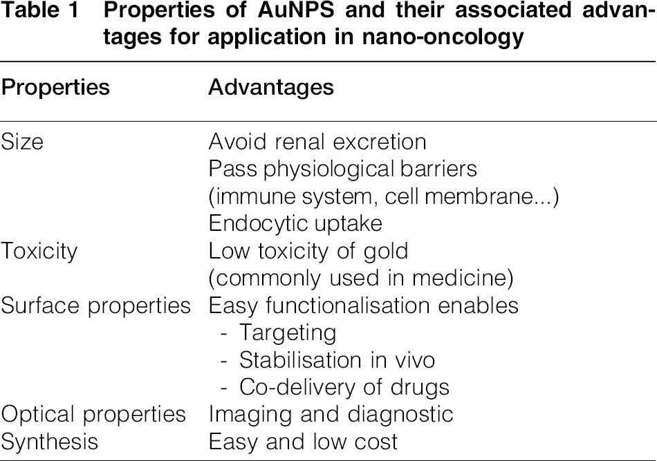
Another advantage of the AuNPs is their surface properties. The easy functionalisation of the surface enables the grafting of functional ligands and the loading of drugs (see section on ‘Surface engineering of AuNPs’). Finally, AuNPs have gained much attention in nanomedicine thanks to their easy synthesis,15–18 which allows a good control of size and dispersity. A low dispersity is absolutely crucial for good controlled drug delivery and biocompatibility. Indeed, apart from ornamental purposes, gold has also been used in medicine for centuries even if its mechanism of action is still not completely understood. The first known use of gold as a medicine appeared to be ∼2500 BC by the Chinese. Later, diseases such as skin ulcers and syphilis could be treated with the use of gold, and nowadays, it can be employed in the treatment of rheumatoid arthritis with the use of gold preparations (Solganol) or as antimicrobial agent. 19 The use of AuNPs in biomedical applications started in the 1970s with the discovery of immunogold labelling, and since then, AuNPs have considerably extended their range of applications. 16 All these properties and the range of applications mentioned above explain the increasing numbers of publications dedicated to AuNPs: >70 000 to date. 20
Therapy
AuNPs allow to deliver small molecular drugs but also large biomolecules such as peptides, proteins or nucleic acids. 21 Drugs can be loaded onto the AuNPs by covalent22–25 (disulphideor hydrazone bonding for instance) or non-covalent conjugations 26 (physical adsorption, ionic bonding) according to their nature. Covalent bindings are stronger than non-covalent ones, but the efficiency of the release is at stake.
The release of the drug can be triggered in various ways through external (temperature, pH or irradiation27–29) and internal stimuli (glutathione30,31). Photodynamic therapy combines light, photosensitizers and tissue oxygen to form reactive oxygen species in order to induce cells damages.32,33 Photothermal therapy, not to be confused with photodynamic therapy, uses the heat generated via electromagnetic radiation in order to induce damages to cells such as protein denaturation, mechanical membrane damage and cell fragmentation.34–42 Photothermal and chemotherapies are usually combined to enhance the cytotoxicity of the nanocarrier. Heat produced by a laser can also be used as a stimulus for AuNPs’ embedded thermoresponsive hydrogel phase transition and drug release. 43
Gold nanomaterials of specific shapes such as nanostars, nanocages or nanoshells have raised interests especially for their localised plasmon resonance. Gold nanorods (AuNRs), in particular, have been extensively studied. By modifying their aspect ratio, their absorbance can be shifted to the near infrared region (650–900 nm), where the background noise from the tissue is minimised, thus improving the quality of the image.13,14,16
AuNPs are one of the most studied nanomaterials in medicine thanks to their potential use as theranostic agents combining anticancer drug delivery and imaging of tumours.25,44,45 The work of Xiao et al. 25 described the design of multifunctional AuNRs covalently conjugated with doxorubicin (DOX) as therapeutic, targeting ligands and Cu chelators for PET imaging as a potential theranostic platform.
pH responsiveness in oncology
Concepts
How can pH responsiveness be useful in the treatment of cancer? At least two biological facts can help answer this question. First, it is known that tumour cells show a more acidic extracellular pH than healthy cells according to the so called ‘Warburg effect’. 46 The acidity comes from a higher rate of conversion of glucose into glycolic acid for the tumour cells. This property can be used for the design of targeting or pH triggered delivery systems. Second, the intracellular pH is not homogeneous. Biomolecules that have been internalised via receptor mediated endocytosis are initially entrapped into endosomes. In these compartments, the pH decreases progressively until complete degradation of the entrapped material in lysosomes (Fig. 1). Consequently, the drug needs to escape from the endosomes to the cytoplasm and/or to the nucleus, actual sites of therapeutic action, before they are inactivated by the lysosomes. As the pH is more acidic in late endosomes and lysosomes (pH 5–6.5) than in the cytoplasm (pH ∼7.4), some specific polymers have been designed to enhance the endosomal escape via pH responsiveness. 47
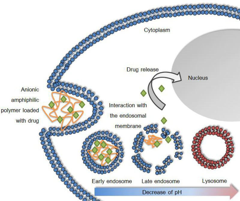
trafficking pathway followed by polymer/biomolecule conjugates internalised through receptor mediated endocytosis (reproduced from Yessine et al. 48 )
pH responsive nanosystems for cancer treatments
pH responsive polymers and peptides
Stimuli responsive polymers, also called ‘smart polymers’, are polymers that experience a change in their properties with a slight change of the physicochemical conditions. For instance, hydrophobicity, the phase or the conformation of these polymers, can be influenced by a change of pH, temperature or ionic strength of the environment. The two main families of ‘smart’ polymers are responsive to temperature and/or pH changes. Recently, they have gained an increasing attention in the medical field and especially in controlled drug delivery systems (DDSs). They can be applied in many ways such as dissolved in aqueous solutions, adsorbed, grafted, linked via hydrogen bonds and combined with different molecules such as proteins, peptides or nucleic acids, 49 but also NPs.
The main barrier to the use of stimuli responsive polymers in vivo remains the possible toxicity, as some polymers are not biodegradable and tend to accumulate. As an example, acrylic acid (AA) monomer is known to be neurotoxic; thus, the application of the widely used thermal responsive polymers such as poly (N-isopropylacrylamide) (PNIPAAm), poly (acrylic acid) (PAA), poly (methacrylic acid) (PMAA) or poly (α-propylacrylic acid) (PPAA) in medicine is at stake, particularly as clinical trials are expensive and usually focus on Food and Drug Administration (FDA) approved polymers (poly(lactic-co-glycolic acid) (PLGA), Poly(ethylene glycol) (PEG)). For these reasons, diagnostics applications are more likely to be approved, as there is no direct contact with the body. 49
The conformation and/or solubility of pH responsive polymers depends on the proton concentration in the environment. The pH responsiveness is usually given by the balance of hydrophilicity/hydrophobicity between the polymer backbone and its side chains. In general, the pH responsive polymers contain an ionisable weak acidic or basic group. The most common ones are PAA and poly(4-vinylpyridine) (PVP). 50 The pH responsive polymers have many applications in the medical field. As enteric coatings for oral drug tablets, they allow the release of the drug in the intestine rather than in the stomach, where the acidic pH could damage it. Other applications concern the previously mentioned ‘endosomal escape enhancement’ and also the hydrogel based DDSs. 49
pH responsive cationic polymers have been investigated for their application as vectors in drug delivery since they are able to efficiently disrupt the endosomal membrane. For instance, poly(ethyleneimine) 51 can damage the lipid membrane via the ‘proton sponge effect’: acting as a buffer, the polymer prevents endosomal acidification, so more protons enter in the endosome and more ions try to escape from it; as a result, the osmotic pressure increases until disruption of the endosome membrane. 52 Unfortunately, despite their efficiency to transfect negatively charged cell membranes via electrostatic interactions, the cationic polymers have proved to be cytotoxic and to bind with negatively charged serum proteins that inhibit their transfection efficiency and greatly limit their use in clinical trials. 53
Anionic polymers are less likely to form complexes with negatively charged serum proteins and thus to be inactivated than cationic polymers. Consequently, the aggregation of proteins and the toxicity can be limited. Some anionic polymers are able to mimic the strategy used by viruses to escape from the endosome via fusogenic peptides. The influenza virus, for instance, is first internalised by receptor mediated endocytosis, and then, the acidification activates the fusion peptide located at the N terminus of the protein by protonation. The resultant change of conformation takes the fusion peptide away from the viral surface (at least 10 nm), allowing the fusion with the endosomal membrane. The peptide will then adopt an α helical structure and insert into the lipid membrane, thus altering the membrane properties, and the content of the viral capsids will be released into the cytoplasm from the endosomes before they reach the lysosomes. 47 Gala and Ha2 are among the most common pH responsive fusogenic peptides. 54
Similarly, some anionic pH sensitive polymers are able to interact with the lipid membrane and escape from the endosomes by changing their conformation from coil to globule with a decrease in pH. These polymers contain weak polyacids, which upon ionisation will present a more extended conformation due to increased electrostatic interactions between the charged carboxylate groups. Other forces such as hydrophobic interactions, hydrogen bondings or Van der Waals forces may play a role in the conformational transition upon pH change. The pH at which the conformational change occurs depends on the balance between hydrophobic and hydrophilic groups and thus can be adjusted. Synthetic anionic polymers are preferred to viruses, as they are easier to produce at large scale, 55 but more obviously for safety reasons as the introduction of a viral vector into the body can induce severe reactions from the immune system. 52
Niren et al. 56 developed a pH responsive membrane disruptive anionic polymer able to target tumour cells and to enhance the endosomal escape. The polymer consisted of a hydrophilic membrane disruptive backbone and was grafted with PEG chains through acid degradable linkage for serum stability and with lactose for cell targeting. The polymer proved to successfully target hepatocytes and enhanced the release of rhodamine labelled oligonucleotides into the cytoplasm, where they diffused into the nucleus.
Chen et al.
53
preferred polymers based on
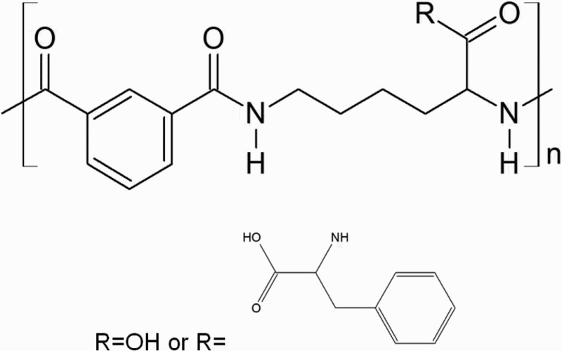
Repeat unit structures of poly(
pH responsive NPs
pH responsive NPs are usually composed of two elements: a core (the NP) and a corona (the grafted elements). Different strategies of pH responsiveness can be adopted: either the corona or the core is pH responsive according to the final aim. As described in the section on ‘Concepts’, cancer cells exhibit two different pH specificities: a more acidic extracellular pH and a gradual intracellular pH decrease. When the corona is pH responsive, usually via dissolution or charge change, the NP is generally designed for specific cancer cell targeting. On the other hand, when the core is pH responsive via conformational change, for instance the nanomaterial has been designed to respond to the intracellular gradual change of pH and aims at a fast drug release in the cytoplasm. 5 Some recent examples of pH responsive systems are described below.
Yang et al.
59
developed a pH triggered cytosolic drug delivery nanocarrier based on polymer micelles. In the study, the authors grafted phenylboronic acid modified cholesterol onto catechol pending monomethoxypoly(ethylene glycol)-poly(
Rosenholm et al. 59 investigated the internalisation of 300 nm polyethyleneimine (PEI) coated silica NPs. They demonstrated the successful internalisation of the NPs and the delivery of hydrophobic fluorophores to the cytoplasm. The release from the endosome was enhanced via the osmotic swelling promoted by the cationic pH responsive polymer PEI. Wan et al. 60 also chose to use PEI polymer for endosomal enhancement to coat porous carbon spheres via electrostatic interaction. The drug paclitaxel was loaded in the sphere pores, and folate acid was conjugated as a targeting ligand. The resultant NPs exhibited a relatively high drug loading (up to 51.37%) and an enhanced cellular uptake for folate receptor positive KB cells. Furthermore, in vivo studies showed that mice bearing hepatic tumour had a slower tumour growth rate and a lower tumour volume after injection with functionalised carbon spheres compared to the commercial Taxol. Akita et al. 61 designed a liposomal NP encapsulating DNA composed of SS cleavable bonds and pH activated lipid-like material (ssPalm) for simultaneous pH triggered release and endosomal escape. This material contained tertiary amines that helped the proton sponge effect in the cellular compartments (endosomes/lysosomes) and lead to membrane destabilisation but also a disulphide bond (S–S) cleavable in the cytoplasm, thanks to which DNA was delivered after destabilisation of the liposomal membrane. The authors reported a successful suppression of tumour growth in mice bearing renal cell carcinoma.
The difference of extracellular pH between tumour cells and healthy cells has been exploited for cells internalisation enhancement by Bae and co-workers.
62
Their nanocarrier used biotin as a targeting ligand in a mixed micelle system of which the core was composed of poly(
Huang et al. 63 prepared casein coated iron oxide NPs, these NPs are known in particular for their magnetic resonance imaging signal enhancement. DOX and indocyanine green, used as model drugs, were encapsulated in an inner layer of amphiphilic polymers. Thanks to the slow degradation of casein under gastric conditions, the drugs were preserved from acid degradation. Ex vivo experiments demonstrated the presence of drugs in the small intestine sacs, and in vivo imaging in mice showed the NPs were able to pass the stomach barrier. Such pH responsive NPs have a real potential for theranostic oral drug delivery.
Some examples of pH responsive NPs have been described (liposomes, polymeric micelles and magnetic, silica and carbon NPs), where the pH responsiveness was exploited for different purposes: cell internalisation, endosomal release or pH triggered release via acid cleavable bonds with promising in vitro and in vivo results. The next sections will focus on pH responsive AuNPs: their design and most recent applications.
Design of pH responsive AuNPs
Surface engineering of AuNPs
The surface modification of the AuNPs aims at different goals such as stabilising the particles in different media, adding some functionalities: targeting or imaging abilities and loading of drugs as previously mentioned (Fig. 3).
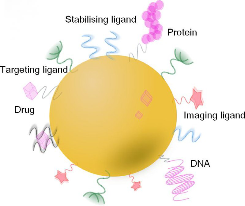
Multifunctional nature of AuNPs a active targeting ligands and surface modification with hydrophilic polymers like PEG b bioactive molecules (reproduced from Akhter et al. 13 )
Usually, AuNPs surface is already modified during their synthesis with some charged ligands to promote the electrostatic repulsion between particles and enable their stability. Nevertheless, this stability depends on environmental factors such as pH, ionic strength or temperature. In order to obtain stable dispersions for a large range of environmental conditions, a further surface modification with additional ligands (such as polymers) is often needed. In particular, polymers have many advantages as they increase the long term stability and solubility of the AuNPs, but also their amphiphilicity and compatibility.
The most employed strategy to functionalise AuNPs is the use of thiolated polymers since thiol has a strong interaction with gold. The thiolates can be directly functionalised during the synthesis of AuNPs using the Shiffrin–Brust synthesis, or via a subsequent bimolecular substitution of the thiolate ligands by functional thiols according to the ligand place exchange reaction 16 (Fig. 4). Other strategies for grafting include the click reaction introduced by Kolb et al. 64 in which a terminal alkyne reacts with an azide, 65 SN2 with alkylamines, coupling with amides or esters and polymerisations reactions via siloxanes formations for instance. 66
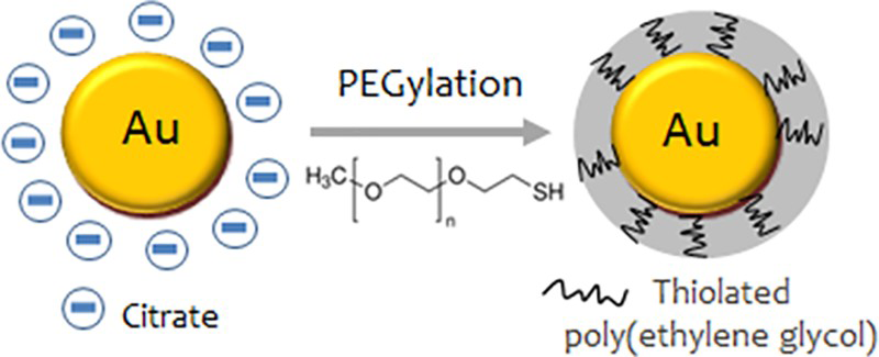
PEGylation of citrate capped AuNPs with a thiolated PEG
The most common strategy to improve the stability of AuNPS in biological media is the use of the PEG 67 and zwitterionic group.26,68 Upon the addition of PEG-SH to the gold colloids, the thiol groups will assemble to the Au and replace the original citrates. Once capped with PEG, particles are less likely to aggregate thanks to steric stabilisation, and their circulation time in the blood stream is improved. PEG is indeed one of the most common stabilisers because of its good solubility, antifouling properties and high degree of biocompatibility. However, the stability is strongly dependent on the size of the chains and the concentration of the polymer, 69 and these parameters need to be carefully considered. PEG is also used as a spacer to decrease the interaction between the ligand and the biological target. 70

Schematic illustration of PEGylated dox-AuNP@CaP (reproduced from Cha et al. 71 with permission from Springer Science+Business Media)
pH responsiveness and AuNPs
pH degradable outer layer
Cha et al. 71 encapsulated DOX in a calcium phosphate layer on PEGylated AuNPs. The mineralisation of the calcium phosphate layer occurred via successive addition of calcium and phosphate ions (Fig. 5). The resultant NPs were stable under physiological conditions, and in vitro experiments demonstrated low cytotoxicity. An enhanced drug release via dissolution of the calcium phosphate coating under lysosomal conditions was observed. Unfortunately, no in vitro study were carried out to confirm the successful internalisation. 71
Acid cleavable bonds
The use of boronic acid has been mentioned previously (see section on ‘pH responsive nanosystems for cancer treatments’). Another very common strategy used for pH triggered drug delivery is the use of hydrazone linkage. In order to deliver dox, Xiao et al. 25 conjugated the model drug to AuNRs via an hydrazine bond in a two-step reaction (Fig. 6). First, methoxy groups of methoxy thioglycolate (MTG) were substituted with anhydrous hydrazide by an ester amide exchange aminolysis reaction, and then, the resulting hydrazide modified AuNRs were conjugated with DOX. Under weak acidic condition, the hydrazide linkage (N–N) is cleaved. The results showed that the pH of the medium had a strong effect on the dox release: no release at pH 7.4 but 93 and 86% release in pH 5.3 respectively corresponding to the pHs of late and early endosomes. The drug delivery via acid cleavable hydrazone bonds is one of the most common strategies used to design pH responsive drug delivery carriers.25,72
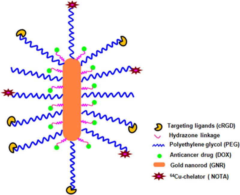
Schematic illustration of multifunctional AuNR-dox-cRGD nanocarriers for tumour targeted drug delivery and PET imaging (reproduced from Xiao et al. 25 )
Grafting with pH responsive polymers
Three main strategies can be employed to prepare polymer stabilised AuNPs: ‘grafting from’, ‘one-pot’ and post-functionalisation. Each of them presents their advantages and limitations. In the ‘grafting from’ technique, the NPs are first coated with an initiator and then the polymerisation takes place from the NP. Park et al. 73 prepared pH responsive AuNPs via the free radical polymerisation of isopropylacrylamide-co-acrylic acid (NIPAM-co-AA) on 40 nm AuNPs. Li et al. 50 chose PVP as a pH responsive polymer. PVP-AuNP conjugates have been reported in several publications. At low pH, the protonated pyridil groups of PVP prevent aggregation of NPs due to electrostatic repulsive forces, but with an increase in pH, the uncharged chains shrink and lead to particles aggregation. At acidic conditions, the pyridil groups are able to entrap negatively charged NPs. The NPS were obtained through surface initiated atom transfer radical polymerisation at ambient conditions. TEM images showed an important change of agglomeration state with the pH (around pH 3.2). However, even though the grafting density is usually high in this technique, the synthesis time is long and it requires the use of harmful solvents. 74
For the ‘one-pot’ synthesis, the AuNPs formation occurs with the end functionalised polymer chains acting as stabilisers.75,76 Cebrián et al. 76 synthesised PEI coated AuNPs for enhanced transfection efficiency using the polymer PEI as the reductant and stabiliser. The main advantage of this method is the control over the stoichiometry of the reagents; however, the final NPs are often very small, usually a few nanometres in size, and show a high polydispersity. 74
Post-functionalisation or ‘grafting to’ technique via trithiocarbonate or thiol chemistry is often used to prepare stabilised AuNPs, and the methods have been deeply studied and improved in the last decades. Zhang et al.
65
introduced PVP onto AuNPs via click chemistry: the polymer was first modified with an alkyne terminated ligand and then grafted to azide functionalised AuNPs. The resultant hybrid AuNPs exhibited a relatively high grafting density (6 chain/nm
2
) and a reversible pH transition at pH 5. Shim et al.
77
demonstrated the reversible conformational change of self-assembled monolayer of disulphide modified poly(
Physical adsorption with pH responsive polymers
Lastly, physical adsorption methods51,79,80 including encapsulation of AuNPs in the hydrophobic cores of amphiphilic polymers micelles or adsorption via electrostatic interactions have been investigated. Kang et al. 79 encapsulated AuNPs in polystyrene-b-poly(acrylic acid) (PS-b-PAA) micelles. More specifically, polymers and NPs were mixed in the presence of alkanethiol which rendered the surface of the NPs hydrophobic and direct them to the hydrophobic interior of the micelles. Bradley et al. 80 used a microgel network of pH and temperature responsive copolymer poly(NIPAM-co-DMAPMA) to encapsulate AuNPs. They asymmetrically adsorbed AuNPs onto microgel using an oil-in-water emulsion method and compared the pH responsive behaviour of these NPs with symmetrically adsorbed ones.
Applications
pH responsive NPs have been investigated for their use as sensors, catalysts, stabilisers or as intelligent building block to form one- to three-dimensional structures in a ‘bottom up’ fashion.74,78 Considering the tremendous interests for AuNPs in the medical field, pH responsiveness may provide an additional asset. Below are some examples of applications of pH responsive AuNPs in oncology.
Drug release
As previously mentioned, pH responsive polymers have been used to trigger the delivery of attached drugs.24,25,71,72,81 Gong's group designed AuNPs coated with a hydrophobic inner shell of poly(
Gold nanoparticles with a monolayer of doxorubicin-conjugated amphiphilic block copolymer for tumour-targeted drug delivery; reprinted from Prabaharan et al. 24 , Biomaterials, 30, 6065–6075, Copyright (2009), with permission from Elsevier
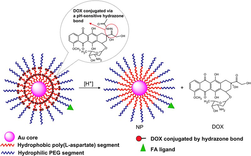
The same group also designed a pH responsive anticancer drug platform based on AuNPs functionalised with thiolated methoxy polyethylene glycol (MPEG-SH) and MTG, where the DOX is conjugated via a hydrazone linkage to the MTG segment. Epifluorescent microscopic images showed DOX fluorescence in the perinuclear region mainly and some fluorescence in the nuclei. These results also demonstrated a pH dependent release, an efficient endocytic uptake and cytoplasmic release. No comments were made on the endosomal escape mechanism. 72 Ruan et al. 82 also exploited the delivery of DOX via hydrazone bond. They investigated the in vitro and in vivo efficiency of their nanocarrier An-PEG-DOX-AuNPs, functionalised with angiopep2, an, a specific ligand use to target glioma cells. Despite a relatively low drug loading capacity of 9.7%, the resultant NP demonstrated in vitro pH dependent release. An-PEG-DOX-AuNPs increased the life expectancy of glioma bearing mice from 50 days for free dox to 65 days.
Monem et al. 83 developed an original way to combine pH responsive drug delivery and phototherapy by coating AuNRs with a layer of silica (SiO2) loaded with DOX. The authors claimed they have obtained a high loading efficiency thanks to the notable silica coating thickness (between 10 and 13 nm), but no further details were given. The AuNRs exhibited four times greater cytotoxicity to breast cancer cell lines than free DOX and an inhibition of the tumour growth of Ehrlich carcinoma in female mice in the first 4–5 days after injection of the AuNRs followed by near-infrared (NIR) irradiation. The cumulative effect of DOX release and phototherapy was more efficient than free DOX or NIR irradiation alone.
Membrane destabilising NPs
To our knowledge, no publication has reported the use of pH responsive AuNPs for cell targeting purposes. However, the development of pH responsive AuNPs allowing the control of the agglomeration state has been receiving great attention.50,74,77 Indeed, if the pH for transition can be tuned to match the extracellular pH of cancer cells, this could help in building up the concentration of NPs around the cancer cells and thus in improving the targeting efficiency.
Similarly, only few publications concern the functionalisation of NPs with pH responsive polymers aimed at enhancing the endosomal escape.74,84,83 Cebrián et al. 76 investigated the transfection efficiency of PEI coated AuNPs bound with DNA.
Two size of AuNPs-PEI were prepared ( < 10 and < 100 nm), and they demonstrated that, even if both NPs proved to be successfully internalised (Fig. 9), only small size hybrid NPs were able to release DNA from the endosomes. A possible explanation given by the authors was that small particles had a higher PEI loading capacity; this not only enables stronger interactions with DNA but also improves the electrostatic interaction between the polymer and the membrane. However, as already mentioned and despite its efficiency, the application of the positively charged PEI in DDSs is limited due to non-specific interactions with negatively charged serum proteins or negatively charged cell membranes and its toxicity. The use of biocompatible and anionic pH responsive polymers may provide a smart alternative for the design of a new generation of hybrid NPs in oncology.
Conclusions
Recently, many advances have been carried out in nano-oncology with the use of nanomaterials as carriers for delivery of drugs. Among the cancer treatments based on nanotechnology available on the market, Doxil was the first FDA approved nanodrug (1995).
85
Doxil has been mainly used to treat ovarian cancer for 20 years. In this system, the drug, DOX, is encapsulated in PEGylated nanoscale liposomes. Other liposome based nanotherapeutics have then been approved by the FDA such as AmBisome® (Gilead Sciences) and DepoDur® (Pacira Pharmaceuticals).
86
In 1994, PEG-
Wicki et al. 88 mentioned a total of 1575 nanomedicine formulations registered for clinical trials by December 2014, and 1381 of them are in the field of cancer therapy according to the registry maintained by clinicaltrials.gov. Nevertheless, this number is very low compared to the number of studies on NPs for cancer treatment as Web of Science yielded 57 944 publications available in December 2014. 87 This demonstrates that although NPs showed clear assets to improve the actual cancer treatments, they are still not accepted as a convincing replacement. Below, two examples of the most advanced clinical studies concerning AuNPs are presented. CytImmune sciences is currently investigating the development of PEGylated AuNPs (CYT-6091) for tumour necrosis factor alpha delivery for the treatment of solid tumours. Phase I of clinical trials demonstrated that the gold nanocarriers could be administrated at doses of tumour necrosis factor that were previously toxic and that the tumour cells could be specifically targeted. At the time of writing Phase II is ongoing. CYT-6091 constitutes nowadays the most advanced nanocarrier system based on AuNPs. 89 Another AuNP based cancer treatment is under phase I clinical trial: AuroLase® (Nanospectra Biosciences). Unlike CYT-6091, AuroLase® is not a DDS but a phototherapeutic. The treatment is based on silica gold nanoshell composed of a silica core and a gold layer. By changing the thickness of the gold layer, it is possible to tune the wavelength at which the light is absorbed and thus create specific photothermal device. Indeed, AuroLase® is aimed at the photothermal ablation of tumour via the use of laser. 88
Nanotechnology is a key field to improve cancer treatments. The specific properties of AuNPs make them ideal drug nanocarriers. Their advantages are numerous and non-negligible: long circulation time, protection of the drug and decrease in the systemic toxicity, specific targeting of the tumour, reduction in the side effects and simultaneous visual tracking of the therapy. The present review demonstrates that the pH responsiveness ability of AuNPscan provides a considerable interest in cancer therapy. Among their potential applications, pH responsive NPs can enhance the specific targeting of tumour cells via controlled assembly. Second, the change of their surface conformation or charge can help the internalisation of the nanotherapeutic. Furthermore, drug payloads can be released via pH triggering. Last, they allow cytoplasmic drug delivery thanks to endosomal membrane destabilisation. We strongly believe that pH responsiveness constitutes an advanced functionality for the current gold nanomaterials under investigation. Additionally, thanks to their easy functionalisation, it is possible to design multiresponsive systems where for instance pH and temperature (phototherapy) responsiveness are combined with imaging ability creating ideal theranostic agents.
Unfortunately, despite the elevated numbers of successful in vivo results for gold nanomaterials in oncology, clinical trials still focus on marketed NPs and no inorganic NPs have been commercialised due mainly to toxicity concerns. Deep investigations on biocompatibility, systemic toxicity, release kinetics and in vitro and in vivo behaviour need to be carried out. Indeed as of today, no inorganic NPs have been commercialised. Liposome based therapy is one of the most common approved nanotherapy. However, despite their ability to stabilise various payloads, liposomes are in general rapidly eliminated via renal clearance. It is also interesting to note that none of the above mentioned NPs systems that are currently commercialised or under clinical trials exhibit a pH responsive behaviour. Challenges in cancer treatments remain, but opportunities are more present than ever.
Footnotes
Acknowledgements
I would like to thank my PhD supervisor, Dr. Rongjun Chen in the Department of Chemical Engineering at Imperial College London who designed, supervised and supported this project, as well as commented and corrected this paper. I would also like to thank the Department of Chemical Engineering of Imperial College London for providing a full scholarship to my PhD research.